. 1998 Feb;18(2):880-6.
doi: 10.1128/MCB.18.2.880.

Regulation of Sos activity by intramolecular interactions

Affiliations

Regulation of Sos activity by intramolecular interactions

S Corbalan-Garcia et al. Mol Cell Biol. 1998 Feb.

Abstract

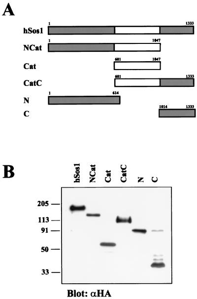
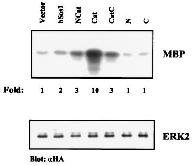
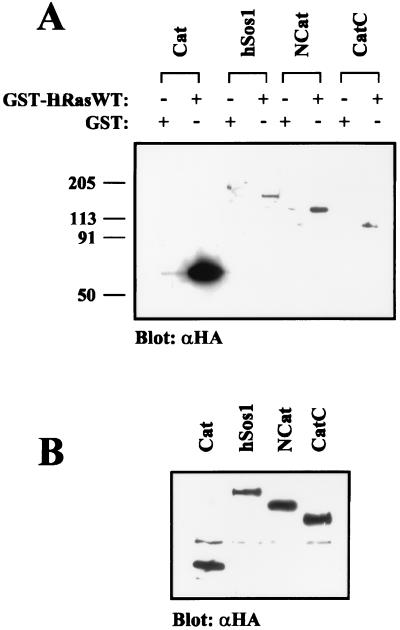
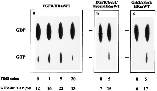
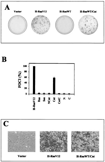
The guanine nucleotide exchange factor Sos mediates the coupling of receptor tyrosine kinases to Ras activation. To investigate the mechanisms that control Sos activity, we have analyzed the contribution of various domains to its catalytic activity. Using human Sos1 (hSos1) truncation mutants, we show that Sos proteins lacking either the amino or the carboxyl terminus domain, or both, display a guanine nucleotide exchange activity that is significantly higher compared with that of the full-length protein. These results demonstrate that both the amino and the carboxyl terminus domains of Sos are involved in the negative regulation of its catalytic activity. Furthermore, in vitro Ras binding experiments suggest that the amino and carboxyl terminus domains exert negative allosteric control on the interaction of the Sos catalytic domain with Ras. The guanine nucleotide exchange activity of hSos1 was not augmented by growth factor stimulation, indicating that Sos activity is constitutively maintained in a downregulated state. Deletion of both the amino and the carboxyl terminus domains was sufficient to activate the transforming potential of Sos. These findings suggest a novel negative regulatory role for the amino terminus domain of Sos and indicate a cooperation between the amino and the carboxyl terminus domains in the regulation of Sos activity.


Figures

FIG. 1
FIG. 1
Description of hSos1 truncation mutants. (A) Schematic representation of hSos1 constructs. Shading denotes noncatalytic regions of hSos1. All constructs contained an amino-terminal HA epitope tag. (B) Expression of hSos1 truncation mutants. COS1 cells were transfected with expression vectors encoding HA-tagged hSos1 constructs. Cell lysates were separated on SDS–7.5% PAGE gels and subsequently analyzed by immunoblotting with anti-HA antibody.
FIG. 2
FIG. 2
Activation of ERK MAP kinase by hSos1 truncation mutants. COS1 cells were transiently cotransfected with HA-tagged ERK2 (0.5 μg) and the indicated HA-tagged hSos1 constructs. ERK2 activation was measured in serum-starved cells by immune complex kinase assay using MBP as a substrate. (Upper) MBP phosphorylation as visualized by autoradiography. The amount of 32P incorporated into MBP was determined by phosphorimaging and is indicated under each lane in arbitrary units. Results shown are from a single representative experiment. Experiments were repeated three times with similar results. (Lower) Immunoblot with anti-HA antibody showing the level of expression of ERK2.
FIG. 3
FIG. 3
In vitro binding assay of hSos1 constructs to GST-HRasWT. (A) COS1 cells were cotransfected with the indicated hSos1 constructs. Following transfection, cells were grown in DMEM supplemented with 5% FCS for 24 h and serum starved for 24 h. Cell lysates were incubated with 2 μg of GST or 2 μg of nucleotide-free GST-HRasWT protein for 1 h at 4°C. Bound proteins were eluted in sample buffer and detected by immunoblotting with anti-HA antibody. (B) A proportion of cell lysates was separated on an SDS–7.5% polyacrylamide gel and was immunoblotted with anti-HA antibody.
FIG. 4
FIG. 4
Activation of Ras by hSos1 truncation mutants. (A) COS1 cells were cotransfected with HRasWT (0.2 μg) and the indicated hSos1 truncation mutants. Serum-starved cells were labeled with [32P]orthophosphate, and the guanine nucleotide content of HRasWT was analyzed as described in Materials and Methods. GTP and GDP markers are labeled on the right. (B) Quantitation of GTP/(GDP + GTP) percentages. Results are the averages of four independent experiments. Error bars represent the standard deviations.
FIG. 5
FIG. 5
Two-hybrid analysis of the interaction between the amino and carboxyl terminus domains of Sos. Yeast cells carrying a GAL4-LacZ reporter cassette were cotransformed with pairs of plasmids expressing proteins fused to the GAL4 binding domain or the GAL4 activation domain as indicated. The S. cerevisiae SNF1 and SNF4 fusion proteins were used as specificity controls. The interaction between the two fusion proteins is indicated by the induction of LacZ expression (dark color). Each patch represents an independent transformant.
FIG. 6
FIG. 6
Regulation of hSos1 activity by growth factor stimulation. (a) COS1 cells were cotransfected with 0.2 μg of HRasWT and 0.2 μg of EGF receptor. Cells were serum starved, labelled with [32P]orthophosphate and stimulated with 20 nM of EGF for 0, 1, 5, and 20 min. (b) COS1 cells were cotransfected with 0.2 μg of HRasWT, 0.2 μg of EGF receptor (EGFR); 0.2 μg of Grb2, and 0.005 μg of hSos1. Transfected cells were serum starved, labelled, and stimulated with EGF as described for panel a. (c) COS1 cells were transfected with 0.2 μg of HRasWT, 0.2 μg of Grb2, and 0.005 μg of hSos1. Transfected cells were serum starved, labelled with [32P]orthophosphate, and serum stimulated for 0 and 5 min. Guanine nucleotide content of HRasWT was analyzed as described in Materials and Methods. GTP and GDP markers are labeled on the left.
FIG. 7
FIG. 7
Focus-forming activity of hSos1 truncation mutants. (A) NIH 3T3 cells were transfected with 1 μg of HRasWT alone, 0.2 μg of HRasV12 as a positive control, and 1 μg of HRasWT and either 1 μg of hSos1 or 1 μg of the Cat domain. After 14 days, the dishes were stained with Giemsa to visualize the transformed foci. (B) Quantitation of the focus formation assay performed with all of the hSos1 constructs. The data are averages of three dishes and are representative of three independent assays. (C) The morphological appearance of NIH 3T3 cells transfected with vector alone, HRasV12, or HRasWT and the Cat domain.

References

    1. Aronheim A, Engelberg D, Li N, al-Alawi N, Schlessinger J, Karin M. Membrane targeting of the nucleotide exchange factor Sos is sufficient for activating the Ras signaling pathway. Cell. 1994;78:949–961. - PubMed
    1. Blattler D P, Garner F, Van Slyke K, Bradley A. Quantitative electrophoresis in polyacrylamide of 2-40% J Chromatogr. 1972;64:147–155.
    1. Boguski M S, McCormick F. Proteins regulating Ras and its relatives. Nature (London) 1993;366:643–654. - PubMed
    1. Buchsbaum R, Telliez J B, Goonesekera S, Feig L. The N-terminal pleckstrin, coiled-coil, and IQ domains of the exchange factor Ras-GRF act cooperatively to facilitate activation by calcium. Mol Cell Biol. 1996;16:4888–4896. - PMC - PubMed
    1. Buday L, Downward J. Epidermal growth factor regulates p21ras through the formation of a complex receptor, Grb2 adaptor protein, and Sos nucleotide exchange factor. Cell. 1993;48:611–620. - PubMed

Publication types

LinkOut - more resources