. 1998 Feb;18(2):936-43.
doi: 10.1128/MCB.18.2.936.

Two AAA family peroxins, PpPex1p and PpPex6p, interact with each other in an ATP-dependent manner and are associated with different subcellular membranous structures distinct from peroxisomes

Affiliations

Two AAA family peroxins, PpPex1p and PpPex6p, interact with each other in an ATP-dependent manner and are associated with different subcellular membranous structures distinct from peroxisomes

K N Faber et al. Mol Cell Biol. 1998 Feb.

Abstract

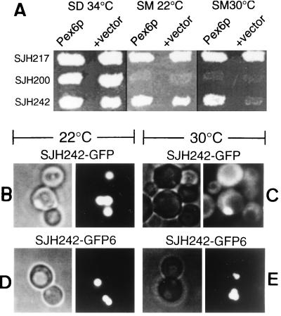
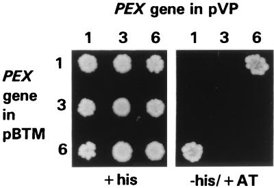
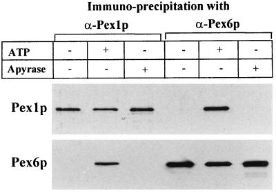
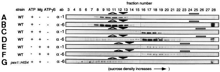
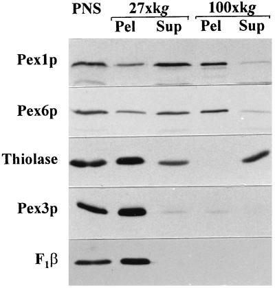
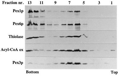
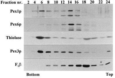
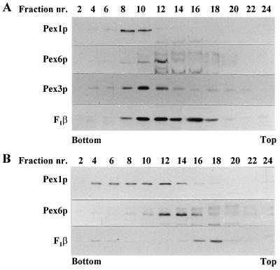
Two peroxins of the AAA family, PpPex1p and PpPex6p, are required for peroxisome biogenesis in the yeast Pichia pastoris. Cells from the corresponding deletion strains (Pp delta pex1 and Pp delta pex6) contain only small vesicular remnants of peroxisomes, the bulk of peroxisomal matrix proteins is mislocalized to the cytosol, and these cells cannot grow in peroxisome-requiring media (J. A. Heyman, E. Monosov, and S. Subramani, J. Cell Biol. 127:1259-1273, 1994; A. P. Spong and S. Subramani, J. Cell Biol. 123:535-548, 1993). We demonstrate that PpPex1p and PpPex6p interact in an ATP-dependent manner. Genetically, the interaction was observed in a suppressor screen with a strain harboring a temperature-sensitive allele of PpPEX1 and in the yeast two-hybrid system. Biochemially, these proteins were coimmunoprecipitated with antibodies raised against either of the proteins, but only in the presence of ATP. The protein complex formed under these conditions was 320 to 400 kDa in size, consistent with the formation of a heterodimeric PpPex1p-PpPex6p complex. Subcellular fractionation revealed PpPex1p and PpPex6p to be predominantly associated with membranous subcellular structures distinct from peroxisomes. Based on their behavior in subcellular fractionation experiments including flotation gradients and on the fact that these structures are also present in a Pp delta pex3 strain in which no morphologically detectable peroxisomal remnants have been observed, we propose that these structures are small vesicles. The identification of vesicle-associated peroxins is novel and implies a role for these vesicles in peroxisome biogenesis. We discuss the possible role of the ATP-dependent interaction between PpPex1p and PpPex6p in regulating peroxisome biogenesis events.


Figures

FIG. 1
FIG. 1
Overproduction of PpPex6p rescues the methanol utilization and peroxisome morphology defects of a Pppex1-ts strain. (A) Overexpression of PpPEX6 from plasmid pAS001 (29) in strains SJH217 (WT PpPEX1), SJH200 (Pppex1), and SJH242 (Pppex1-ts). Strains were replica plated onto SM or SD plates and grown for 3 days at the indicated temperature. (B to E) Fluorescence microscopy (right panels) of SJH242 synthesizing GFP-SKL (SJH242-GFP) from integrated plasmid pJAH127 grown at 22°C (B) and 30°C (C) and SJH242 overproducing PpPex6p and synthesizing GFP-SKL (SJH242-GFP6) from integrated plasmid pJAH125 grown at 22°C (D) and 30°C (E). Nomarski images are shown in the left panels.
FIG. 2
FIG. 2
PpPex1p and PpPex6p interact in the yeast two-hybrid system. Full-length clones coding for PpPex1p, PpPex3p, and PpPex6p were inserted into derivatives of plasmids pVP16 (transactivation domain) and pBTM116 (DNA-binding domain) (12). All combinations of pVP16 and pBTM116 plasmids containing the PpPEX genes were used to cotransform S. cerevisiae L40. Individual transformants were screened for expression of the chromosomal HIS4 marker gene (+his: media containing histidine [left panel]; −his/+AT: media lacking histidine and containing 25 mM 3-aminotriazole [right panel]).
FIG. 3
FIG. 3
Coimmunoprecipitation of PpPex1p and PpPex6p is ATP dependent. Shown are immunoblots (antibodies are indicated) of immunoprecipitates in which α-PpPex1p or α-PpPex6p was incubated with lysates from P. pastoris WT. The addition of ATP (0.5 mM) or apyrase (5 U/ml) to the immunoprecipitation buffer and wash buffer was as indicated.
FIG. 4
FIG. 4
PpPex1p and PpPex6p are present in a protein complex of approximately 320 to 400 kDa. Shown is a Western blot analysis of equal volumes of fractions from sucrose gradients loaded with lysates prepared from P. pastoris WT (A to F) or a Pppex1::HIS4 strain (G). ATP (0.5 mM) (A to D and G), 4 mM MgCl2 (C to G), or the nonhydrolyzable analog of ATP, ATPγS (E and F), was added to the lysis buffer, and the effect on the migration of PpPex1p and PpPex6p was determined. To assess protein separation within each gradient, the locations of dihydroxyacetone synthase (DHAS, 150-kDa dimer), catalase (242-kDa tetramer), and methanol oxidase (640-kDa octamer, but not correctly assembled in Pppex1 and Pppex6 cells) were determined by Western blot analysis. The two fractions containing peak levels of DHAS (open triangle), catalase (closed triangle), and methanol oxidase (bar) are indicated above each panel. Ab, antibody. α-1 and α-6, α-PpPex1p and α-PpPex6p antibodies, respectively.
FIG. 5
FIG. 5
PpPex1p and PpPex6p are enriched in the 100,000 × g pellet fraction. A PNS prepared from oleate-grown WT cells was subjected to differential centrifugation. Equal volumes of the PNS, 27,000 × g pellet (27×kg Pel) or supernatant (27×kg Sup), and 100,000 × g pellet (100×kg Pel) or supernatant (100×kg Sup) were loaded in each lane and analyzed by Western blot analysis with antibodies to the indicated proteins.
FIG. 6
FIG. 6
PpPex1p and PpPex6p are membrane associated. The 100,000 × g pellet fraction (obtained from the 27,000 × g supernatant fraction) prepared from oleate-grown WT cells was subjected to flotation gradient centrifugation. The gradient was drained from the top into 13 fractions (1, top; 13, bottom), and equal volumes of fractions were analyzed by Western blot analysis with antibodies to the indicated proteins. Very low amounts of thiolase, PpPex3p, and acyl coenzyme A oxidase (Acyl-CoA ox) were present in the 100,000 × g pellet fraction (relative to the 27,000 × g pellet fraction) used for flotation (Fig. 5). The Western blots analyzed for these proteins had to be overexposed, but clear signals were obtained due to the high quality of the antibodies. nr., number.
FIG. 7
FIG. 7
PpPex1p and PpPex6p are associated with subcellular structures distinct from peroxisomes. Thiolase, the F1β subunit of mitochondrial ATPase, PpPex3p, PpPex1p, and PpPex6p in fractions of a linear Nycodenz gradient loaded with a PNS fraction of oleate-grown WT cells were subjected to immunodetection. The gradient was drained from the bottom into 24 fractions (2, bottom; 24, top), and equal volumes of every second fraction were analyzed by Western blot analysis. nr., number.
FIG. 8
FIG. 8
PpPex1p and PpPex6p are present on different subcellular structures independent of the presence or absence of peroxisomes. A PNS fraction was prepared from glucose-grown P. pastoris WT cells (A) or PpΔpex3 cells (B) and loaded onto a linear Nycodenz gradient. Equal volumes of every second fraction of the gradient drained from the bottom (fraction 1) were analyzed for the presence of the indicated proteins by Western blot analysis. nr., number.

References

    1. Acharya U, Jacobs R, Peters J M, Watson N, Farquhar M G, Malhotra V. The formation of Golgi stacks from vesiculated Golgi membranes requires two distinct fusion events. Cell. 1995;82:895–904. - PubMed
    1. Albertini M, Rehling P, Erdmann R, Girzalsky W, Kiel J A K W, Veenhuis M, Kunau W-H. Pex14p, a peroxisomal membrane protein binding both receptors of the two PTS-dependent import pathways. Cell. 1997;89:83–92. - PubMed
    1. Arlt H, Tauer R, Feldmann H, Neupert W, Langer T. The YTA10-12 complex, an AAA protease with chaperone-like activity in the inner membrane of mitochondria. Cell. 1996;85:875–885. - PubMed
    1. Baerends R J S, Rasmussen S W, Hilbrands R E, van der Heide M, Faber K N, Reuvekamp P T W, Kiel J A K W, Cregg J M, van der Klei I J, Veenhuis M. The Hansenula polymorpha PER9 gene encodes a peroxisomal membrane protein essential for peroxisome assembly and integrity. J Biol Chem. 1996;271:8887–8894. - PubMed
    1. Beckers C J, Block M R, Glick B S, Rothman J E, Balch W E. Vesicular transport between the endoplasmic reticulum and the Golgi stack requires the NEM-sensitive fusion protein. Nature. 1989;339:397–398. - PubMed

Publication types

LinkOut - more resources