. 1998 Feb;18(2):944-52.
doi: 10.1128/MCB.18.2.944.

Mutation of Hip's carboxy-terminal region inhibits a transitional stage of progesterone receptor assembly

Affiliations

Mutation of Hip's carboxy-terminal region inhibits a transitional stage of progesterone receptor assembly

V Prapapanich et al. Mol Cell Biol. 1998 Feb.

Abstract

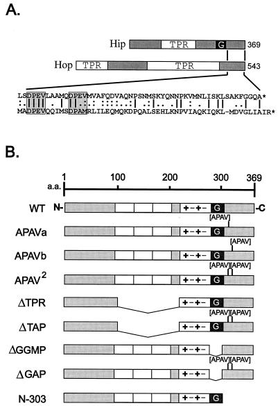
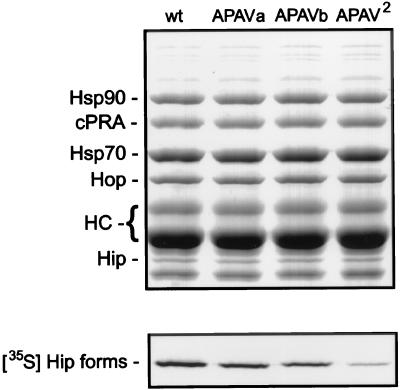
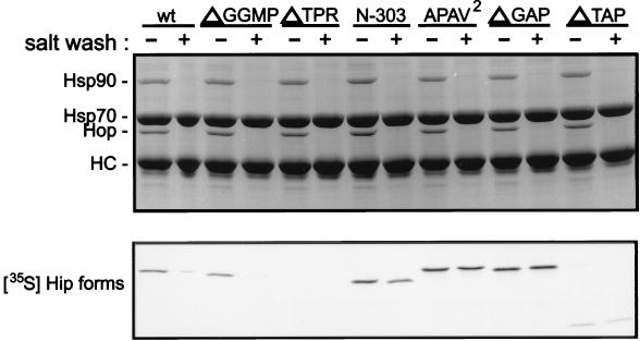
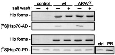
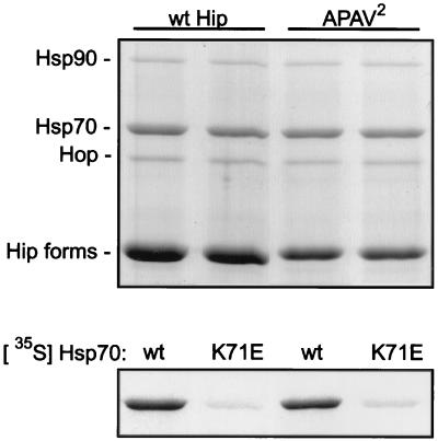
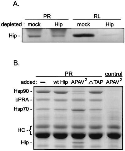
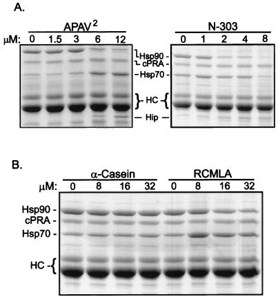
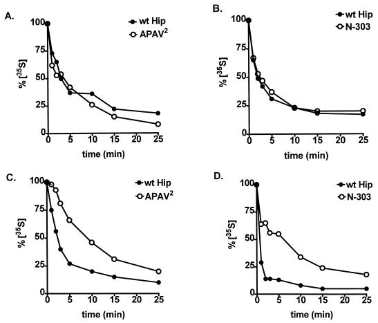
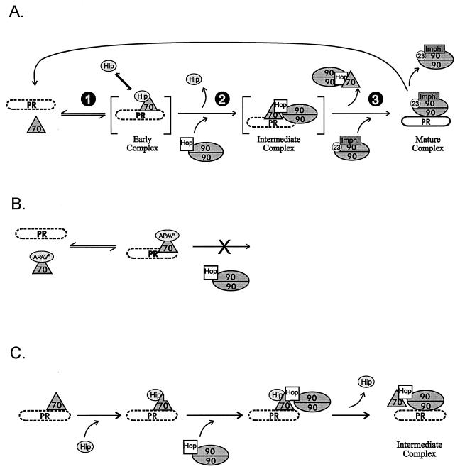
Steroid receptor complexes are assembled through an ordered, multistep pathway involving multiple components of the cytoplasmic chaperone machinery. Two of these components are Hsp70-binding proteins, Hip and Hop, that have some limited homology in their C-terminal regions, outside the sequences mapped for Hsp70 binding. Within this region of Hip is a DPEV sequence that occurs twice; in Hop, one DPEV sequence plus a partial second sequence occurs. In an effort to better understand Hip function as it relates to assembly of progesterone receptor complexes, the DPEV region of Hip was targeted for mutations. Each DPEV sequence was mutated to an APAV sequence, singly or in combination. The combined mutation, APAV2, was further combined with a deletion of Hip's tetratricopeptide repeat region that is required for Hsp70 binding or with a deletion of Hip's GGMP repeat. An additional mutant was prepared by truncation of Hip's DPEV-containing C terminus. By comparing interactions of various Hip forms with Hsp70, it was determined that mutation of the DPEV sequences created a dominant inhibitory form of Hip. The mutant Hip-Hsp70 complex was not prevented from interacting with progesterone receptor, but the mutant caused a dose-dependent inhibition of receptor assembly with Hsp90. The behavior of the Hip mutant is consistent with a model in which Hip and Hop are required to facilitate the transition from an early receptor complex with Hsp70 into later complexes containing Hsp90.


Figures

FIG. 1
FIG. 1
(A) Comparison of Hip and Hop sequences. Hsp70-binding proteins Hip and Hop both contain TPR domains. In Hip, the TPR domain plus surrounding highly charged sequences is required for Hsp70 binding. Also contained in Hip is a GGMP repeat domain (G) whose function is unknown. Hop contains two TPR regions; the N-terminal one is required for Hsp70 binding, and the central TPR binds to Hsp90. Little homology is shared between the highly degenerate TPR regions of Hip and Hop. The region of greatest similarity is near the C terminus of each protein (amino acid sequence alignment in exploded view at bottom). Most notable in this region are two DPEV sequences in Hip that align with a DPEV and DPAM in Hop (highlighted by shaded boxes). (B) Hip mutants. Each of the DPEV sequences in Hip was mutated to APAV, as illustrated. The mutation of both DPEV sequences (APAV2) was also combined with previously developed mutants in which the TPR or GGMP domains had been deleted. Another mutant from previous studies is the truncation mutant N-303, which lacks the DPEV-containing C terminus of Hip. TPR domains (open boxes), highly charged regions (+−+−), and a region of Hop homology (shaded box) at the C terminus are shown. a.a., amino acids; WT, wild type.
FIG. 2
FIG. 2
Association of radiolabeled APAV mutants with PR complexes. PR complexes were assembled in vitro by using RL supplemented with radiolabeled wtHip (wt) or the three APAV mutants. After assembly, components were separated by SDS-PAGE and visualized by Coomassie blue staining (upper panel). Proteins associated with recombinant chicken PR-A (cPRA) are indicated on the left. Also labeled are the heavy-chain bands (HC) from antibody used to immobilize PR-A for assembly reactions. The gel was dried and autoradiographed (lower panel) to reveal binding by radiolabeled Hip forms.
FIG. 3
FIG. 3
Coprecipitation of Hip forms with Hsp70. Hsp70 complexes were immunoprecipitated from RL supplemented with the radiolabeled Hip forms indicated above the lanes (wt, wtHip). An equimolar amount of each radiolabeled Hip form was added to separate mixtures for immunoprecipitation reactions. Immunoprecipitates were washed in buffer containing either no additional salt (−) or 0.5 M NaCl (+). Proteins in resin complexes were separated by SDS-PAGE and visualized by Coomassie blue staining (upper panel). Hsp70 and the coprecipitating proteins Hop and Hsp90 are indicated on the left. Hip is not evident in the stained gel due to its comigration with anti-Hsp70 BB70 heavy chain (HC). Coprecipitating radiolabeled Hip forms were detected by autoradiography of the dried gel (lower panel).
FIG. 4
FIG. 4
Association of wtHip and APAV2 with separate Hsp70 domains. For the main panels, recombinant wtHip (wt) or APAV2 was adsorbed to a covalently coupled immunoaffinity resin. The Hip resins or resin lacking bound Hip (control) were incubated in WB plus 5 mM ADP and 5 mM MgCl2. The buffer was further supplemented with the radiolabeled synthesis mixtures for the ATPase domain of Hsc70 (Hsc70-AD) or the peptide-binding domain (Hsc70-PD). Hip immunoprecipitates were divided and washed in buffer lacking (−) or containing (+) 0.5 M NaCl. As a positive control for Hsc70-PD binding to substrate, radiolabeled Hsc70-PD was incubated with immunoadsorbed PR or with immunoaffinity resin lacking PR (ctrl). After SDS-PAGE separation of components, the gels were Coomassie blue stained (Hip forms) and autoradiographed ([35S]Hsp70AD and [35S]Hsp70-PD).
FIG. 5
FIG. 5
Bulk interactions of Hip forms and their association with an Hsp70 mutant lacking ATPase activity. Recombinant wtHip or APAV2 was preadsorbed to a covalently coupled immunoaffinity resin. The resins were subsequently incubated in RL containing an ATP-regenerating system. Proteins on the resin complexes were separated by SDS-PAGE and Coomassie blue stained (upper panel). Stained bands representing coprecipitating Hsp70, Hop, and Hsp90 are indicated on the left. Radiolabeled Hsp70 forms were included in the bulk reaction mixtures, and the association of these forms with wtHip and APAV2 was resolved by autoradiography of the stained gel (lower panel). Either radiolabeled wild-type Hsp70 (wt) or an ATPase-deficient point mutant (K71E) was included in the reaction mixtures.
FIG. 6
FIG. 6
Hip involvement in PR assembly. (A) RL that was mock depleted or immunodepleted of endogenous Hip was used for assembly of PR complexes in vitro. The resulting PR complexes (PR lanes) and the total RL used for assembly reactions (RL lanes) were immunostained for the presence of Hip. (B) Purified recombinant Hip forms were added to RL at a 10-fold molar excess over endogenous Hip levels. The RL mixtures were used for PR assembly reactions, and the resulting PR complexes were separated by SDS-PAGE and visualized by Coomassie blue staining. The sample in the first lane contained no added protein, and the sample in the final lane (control) lacked PR in the assembly reaction mixture. Proteins associated with PR (recombinant chicken PR-A [cPRA]), along with the PR22 heavy chains (HC), are indicated on the left.
FIG. 7
FIG. 7
Dose dependence and specificity of PR assembly inhibition. (A) PR complexes were assembled in RL containing the concentrations of added APAV2 or N-303 indicated above the lanes. Protein components on the resulting resin complexes were separated by SDS-PAGE and Coomassie stained. (B) Similar PR reconstitutions were performed with Hip mutants replaced by the concentrations of the Hsp70 substrate α-casein or RCMLA indicated above the lanes.
FIG. 8
FIG. 8
Effects of mutant Hip forms on the dynamics of Hsp70 interactions. The rates of exchange of radiolabeled Hsp70 on PR complexes (A and B) and on Hip complexes (C and D) were compared in the presence of wtHip (A to D), APAV2 (A and C), or N-303 (B and D). As detailed in Materials and Methods, PR or Hip complexes were assembled in RL containing radiolabeled Hsp70 and then transferred to fresh RL lacking radioactivity. At the times indicated, aliquots were removed from the secondary assemblies. Each sample was separated by SDS-PAGE, Coomassie blue stained to monitor total protein levels, and autoradiographed to measure the binding of radioactive Hsp70. Bands were quantitated by densitometry, and radioactive bands were normalized to the amount of Coomassie blue-stained Hsp70 in each sample. The resulting values are plotted as a percentage of radiolabeled Hsp70 present at initiation of the secondary assembly reactions (0 min).
FIG. 9
FIG. 9
Inhibition of Hop binding to Hsp70 in the presence of misfolded substrates. Hop was immunoadsorbed from RL in the presence of 0.5 M NaCl to dissociate Hsp70 and Hsp90. Hop-resin (lane Hop) was then incubated with purified Hsp70 (lane Hsp70) or with Hsp70 preincubated with a 10-fold molar excess of α-casein, RCMLA, or BSA as indicated above the lanes. In the final lane, immunoaffinity resin lacking Hop was incubated with Hsp70. Migration positions for Hsp70, Hop, and the anti-Hop immunoglobulin heavy chain (HC) are indicated on the left.
FIG. 10
FIG. 10
(A) Normal assembly cycle. During cell-free assembly of PR complexes, an ordered pathway is followed. (1) Hsp70 and Hip bind at an early stage. (2) An intermediate complex containing Hsp70, Hop, and Hsp90 is formed. (3) The functionally mature complex contains Hsp90, p23, and any one of three immunophilins (Imph.). Hormone binding by PR is deficient (dashed outline of PR) until the mature complex is formed. The mature complex is not stable; if PR dissociates without binding hormone, it reenters the assembly cycle. (B) APAV2-arrested assembly. Hip mutant APAV2, or N-303, is permissive for Hsp70 binding and dissociation but prevents the coassociation of Hop-Hsp90 and Hsp70 on PR. (C) Hip-mediated transition in PR complexes. It is proposed that Hip normally facilitates binding of Hop to Hsp70 when Hsp70 is associated with PR, thus promoting progression of PR assembly to the intermediate complex containing Hsp90. The indirect association of Hsp90 with PR complexes may favor displacement of Hsp70 and direct binding of Hsp90 to PR.

References

    1. Barent, R. L., S. C. Nair, D. C. Carr, R. A. Rimerman, Y. Ruan, Y. Zhang, J. Brennan, and D. F. Smith. Analysis of FKBP51/FKBP52 chimeras and mutants for Hsp90 binding and association with progesterone receptor complexes. Mol. Endocrinol., in press. - PubMed
    1. Buchberger A, Theyssen H, Schroder H, McCarty J S, Virgallita G, Milkereit P, Reinstein J, Bukau B. Nucleotide-induced conformational changes in the ATPase and substrate binding domains of the DnaK chaperone provide evidence for interdomain communication. J Biol Chem. 1995;270:16903–16910. - PubMed
    1. Chang H C, Nathan D F, Lindquist S. In vivo analysis of the Hsp90 cochaperone Sti1 (p60) Mol Cell Biol. 1997;17:318–325. - PMC - PubMed
    1. Chen S, Prapapanich V, Rimerman R, Honore B, Smith D. Interactions of p60, a mediator of progesterone receptor assembly, with heat shock proteins hsp90 and hsp70. Mol Endocrinol. 1996;10:682–693. - PubMed
    1. Chen, S., V. Prapapanich, and D. F. Smith. Unpublished data.

Publication types

LinkOut - more resources