. 1998 Feb;18(2):967-77.
doi: 10.1128/MCB.18.2.967.

A dominant-negative inhibitor of CREB reveals that it is a general mediator of stimulus-dependent transcription of c-fos

Affiliations

A dominant-negative inhibitor of CREB reveals that it is a general mediator of stimulus-dependent transcription of c-fos

S Ahn et al. Mol Cell Biol. 1998 Feb.

Abstract

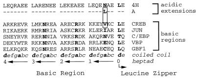
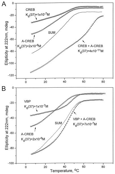
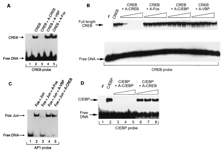
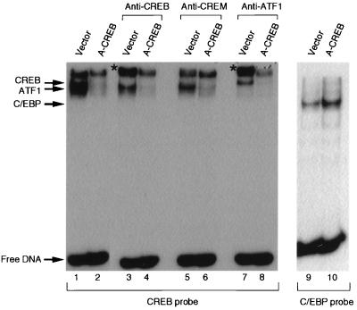
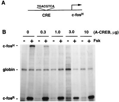
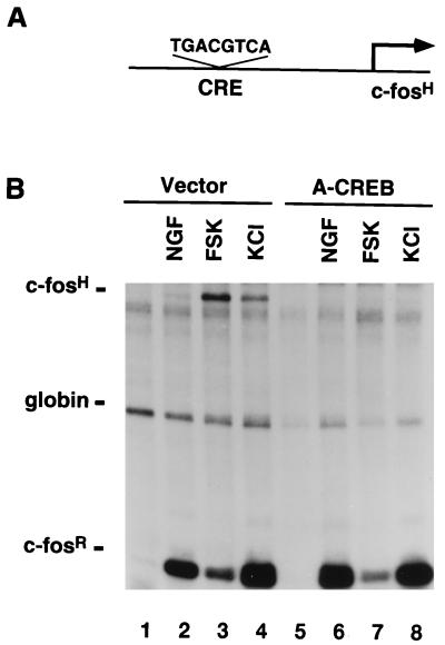
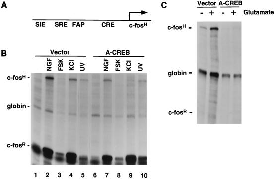
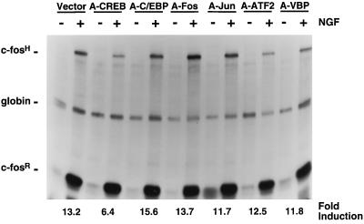
Several studies have characterized the upstream regulatory region of c-fos, and identified cis-acting elements termed the cyclic AMP (cAMP) response elements (CREs) that are critical for c-fos transcription in response to a variety of extracellular stimuli. Although several transcription factors can bind to CREs in vitro, the identity of the transcription factor(s) that activates the c-fos promoter via the CRE in vivo remains unclear. To help identify the trans-acting factors that regulate stimulus-dependent transcription of c-fos via the CREs, dominant-negative (D-N) inhibitor proteins that function by preventing DNA binding of B-ZIP proteins in a dimerization domain-dependent fashion were developed. A D-N inhibitor of CREB, termed A-CREB, was constructed by fusing a designed acidic amphipathic extension onto the N terminus of the CREB leucine zipper domain. The acidic extension of A-CREB interacts with the basic region of CREB forming a coiled-coil extension of the leucine zipper and thus prevents the basic region of wild-type CREB from binding to DNA. Other D-N inhibitors generated in a similar manner with the dimerization domains of Fos, Jun, C/EBP, ATF-2, or VBP did not block CREB DNA binding activity, nor did they inhibit transcriptional activation of a minimal promoter containing a single CRE in PC12 cells. A-CREB inhibited activation of CRE-mediated transcription evoked by three distinct stimuli: forskolin, which increases intracellular cAMP; membrane depolarization, which promotes Ca2+ influx; and nerve growth factor (NGF). A-CREB completely inhibited cAMP-mediated, but only partially inhibited Ca2+- and NGF-mediated, transcription of a reporter gene containing 750 bp of the native c-fos promoter. Moreover, glutamate induction of c-fos expression in primary cortical neurons was dependent on CREB. In contrast, induction of c-fos transcription by UV light was not inhibited by A-CREB. Lastly, A-CREB attenuated NGF induction of morphological differentiation in PC12 cells. These results suggest that CREB or its closely related family members are general mediators of stimulus-dependent transcription of c-fos and are required for at least some of the long-term actions of NGF.


Figures

FIG. 1
FIG. 1
Structure of A-CREB. Protein sequence of the acidic amphipathic extension that was fused with the CREB leucine zipper domain to produce A-CREB. The upper panel depicts the amino acid sequence of the acidic amphipathic extension (4H) and the single amino acid change in position a (N to L) needed to create the potent new acidic extension (A) that interacts with the CREB basic region. The lower panel shows the amino acid sequences of basic regions of CREB, Jun, C/EBP, VBP, and GBF1. The box encloses the potential interacting residues: Leu of the new acidic extension (A) and Val of the CREB basic region. The first Leu of the zipper, the invariant Asn and Arg of the basic region, and the position a amino acid which is critical for the efficacy of the new acidic extension, are in boldface. The coiled-coil nomenclature of the basic region extending from the leucine zipper is indicated below, with hydrophobic positions a and d in boldface. The numbering of the heptads in the acidic extension is indicated.
FIG. 2
FIG. 2
Thermal stability of A-CREB mixed with CREB or VBP. (A) CD thermal denaturation curves recorded at 222 nm for CREB (squares), A-CREB (diamonds), and a mixture of CREB and A-CREB (circles). The solid line labeled SUM is expected if CREB and A-CREB do not interact. The line through each data set is a fitted curve that was used to calculate Tm as described previously (33), and the calculated Kd(37) is shown. (B) CD thermal denaturation curves at 222 nm of VBP (squares), A-CREB (diamonds), and a mixture of VBP and A-CREB (circles). The solid line labeled SUM is expected if VBP and A-CREB do not interact. (C) A schematic representation of an A-CREB–CREB dimer. The left panel shows a CREB homodimer with unhelical basic regions. The right panel shows a heterodimer of CREB and A-CREB resulting in an α-helical formation of the basic region.
FIG. 2
FIG. 2
Thermal stability of A-CREB mixed with CREB or VBP. (A) CD thermal denaturation curves recorded at 222 nm for CREB (squares), A-CREB (diamonds), and a mixture of CREB and A-CREB (circles). The solid line labeled SUM is expected if CREB and A-CREB do not interact. The line through each data set is a fitted curve that was used to calculate Tm as described previously (33), and the calculated Kd(37) is shown. (B) CD thermal denaturation curves at 222 nm of VBP (squares), A-CREB (diamonds), and a mixture of VBP and A-CREB (circles). The solid line labeled SUM is expected if VBP and A-CREB do not interact. (C) A schematic representation of an A-CREB–CREB dimer. The left panel shows a CREB homodimer with unhelical basic regions. The right panel shows a heterodimer of CREB and A-CREB resulting in an α-helical formation of the basic region.
FIG. 3
FIG. 3
A-CREB inhibits DNA binding activity of CREB. The inhibition of CREB DNA binding activity is leucine zipper specific. (A) A-CREB, but not A-VBP or A-Fos, inhibits the DNA binding activity of the DNA binding domain of CREB. An EMSA was performed with a 25-bp double-stranded DNA oligonucleotide containing a consensus CRE and the DNA binding domain of CREB (5 × 10⁻⁶ M; lane 2). The binding reaction mixture contained 1.0 molar equivalent of A-CREB (lane 3), A-VBP (lane 4), or A-Fos (lane 5). (B) A-CREB, but not A-Fos, A-C/EBP, or A-VBP, inhibits the DNA binding activity of full-length CREB. Increasing concentrations (10−8 M, 3 × 10⁻⁷ M, 10−7 M, 3 × 10⁻⁶ M, and 10−6 M) of A-CREB, A-Fos, A-C/EBP, or A-VBP were added to the reaction mixture containing CREB (2 × 10⁻⁷ M), and an EMSA was performed as for panel A. (C) A-CREB does not inhibit a Fos-Jun complex from binding to the AP-1 site. An EMSA was performed with a probe containing an AP-1 site (lane 1). A Fos-Jun complex (5 × 10⁻⁷ M) binds DNA (lane 2), and 1 molar equivalent of A-Fos completely inhibits Fos-Jun DNA binding (lane 3). Neither A-VBP (lane 4) nor A-CREB (lane 5) inhibits AP-1 DNA binding activity. (D) A-CREB does not inhibit DNA binding activity of C/EBP. An EMSA was performed with a probe containing a C/EBP binding site (lane 1). C/EBP (10−8 M) was challenged with A-C/EBP or A-CREB at 1, 3, and 10 molar excesses. A-C/EBP completely inhibits C/EBP binding (lanes 3 to 5), while A-CREB has no effect on C/EBP DNA binding activity (lanes 6 to 8).
FIG. 4
FIG. 4
A-CREB inhibits CREB DNA binding activity in extracts of transfected cells. CREB DNA binding activity is inhibited in nuclear extracts prepared from HEK293-T cells that were transfected with an expression vector encoding A-CREB (lane 2) or the control vector (lane 1). For supershift analysis, antibodies against CREB (lanes 3 and 4), CREM (lanes 5 and 6), and ATF-1 (lanes 7 and 8) were added to the DNA binding reaction mixtures. A-CREB inhibited CREB and ATF-1 binding activity; the supershifted products are indicated by a star. C/EBP DNA binding activity is not inhibited in nuclear extracts from cells transfected with A-CREB (lanes 9 and 10).
FIG. 5
FIG. 5
A-CREB blocks cAMP-induced gene expression in a dose-dependent manner. (A) A schematic representation of the pAF42CRE reporter gene construct used in these experiments. (B) PC12 cells were transfected with the reporter plasmid pAF42CRE, an α-globin internal control expression vector (pSVα-1), and indicated amounts of A-CREB expression vector as described in Materials and Methods. The total DNA concentration was kept constant by including empty expression vector DNA. After 48 h of transfection, cells were stimulated with forskolin (Fsk) (10 μM, 1 h), and RNA was analyzed by an RPA. c-fosH, protected RNA fragment from the transfected human c-fos reporter gene; globin, fragment protected from the α-globin internal control plasmid; c-fosR, fragment protected by the endogenous rat c-fos mRNA.
FIG. 6
FIG. 6
A-CREB, but not other acidic leucine-zippers (A-ZIPs), inhibits cAMP activation of CRE-mediated gene expression. Transcriptional activation of pAF42CRE was evoked by forskolin. For this experiment, PC12 cells were transfected with pAF42CRE, α-globin expression vector, and expression vectors encoding the indicated A-ZIP proteins (3 μg each). Two days later, cells were treated with forskolin (Fsk; 10 μM, 1 h) or were left untreated and an RPA was performed. This experiment was performed two times with similar results.
FIG. 7
FIG. 7
A-CREB does not inhibit SRE-mediated gene expression. (A) A schematic representation of the pAF42SRE construct used in this experiment. (B) PC12 cells were transfected with the pAF42SRE reporter gene, an α-globin expression vector, and an empty vector or the A-CREB expression vector. Two days later, cells were treated with NGF (100 ng/ml, 45 min) or were left untreated and an RPA was performed. This experiment was performed three times with similar results.
FIG. 8
FIG. 8
CREB is critical for cAMP, Ca2+, and NGF induction of CRE-mediated gene expression. (A) A schematic representation of the pAF42CRE reporter construct. (B) Transcriptional activation of pAF42CRE by NGF, cAMP, or Ca2+. PC12 cells were transfected with the pAF42CRE reporter gene, an α-globin expression vector, and either an empty vector or the A-CREB expression vector (3 μg). Two days later, cells were either left unstimulated (lanes 1 and 5) or were stimulated with NGF (100 ng/ml, 45 min; lanes 2 and 6) or forskolin (FSK) (10 μM, 1 h; lanes 3 and 7) or were subjected to membrane depolarization with KCl (50 mM, 1 h; lanes 4 and 8) and an RPA was performed. A-CREB blocked NGF, forskolin, and KCl induction of pAF42CRE by 95, 100, and 98%, respectively. The quantification was done as described in Materials and Methods. This experiment was performed two times with similar results.
FIG. 9
FIG. 9
A-CREB inhibits NGF, cAMP, and Ca2+ induction of c-fos expression but not induction by UV light. (A) A schematic representation of the pF4 reporter construct. (B) Transcriptional activation of pF4 by various stimuli. PC12 cells were transfected with reporter gene pF4, an α-globin expression vector, and either an empty vector or the A-CREB expression vector. After 2 days, cells were either left unstimulated (lanes 1 and 6) or were stimulated with NGF (100 ng/ml, 45 min; lanes 2 and 7), forskolin (FSK) (10 μM, 1 h; lanes 3 and 8), KCl (50 mM, 1 h; lanes 4 and 9), or UV light (400 J/m2; lanes 5 and 10) and an RPA was performed. (C) Primary E19 rat cortical neurons were transfected as for panel B. Two days later, cells were stimulated with glutamate (10 μM, 1 h) and an RPA was performed. A-CREB blocked NGF, forskolin, KCl, and UV light induction of pF4 by 64, 99, 84, and 2%, respectively. The quantification was done as described in Materials and Methods. These experiments were performed three times with similar results.
FIG. 10
FIG. 10
A-CREB, but not other A-ZIPs, attenuates NGF induction of c-fos expression. For this experiment, PC12 cells were transfected with the pF4 reporter gene, α-globin, and either an empty vector or expression vectors encoding A-CREB, A-C/EBP, A-Fos, A-Jun, A-ATF-2, or A-VBP. After 2 days, cells were either left untreated or were treated with NGF (200 ng/ml, 45 min) and an RPA was performed. The quantification was done as described in Materials and Methods. This experiment was performed two times with similar results.
FIG. 11
FIG. 11
CREB contributes to NGF induction of morphological differentiation in PC12 cells. (A) PC12 cells were transfected with a β-galactosidase (β-Gal) plasmid and either an empty expression vector or the A-CREB expression vector. Then, cells were stimulated with NGF (100 ng/ml). After 3 days, cells were fixed and stained for β-Gal activity and the morphological differentiation of transfected cells was calculated as described in the Materials and Methods (the results shown are means ± the standard errors of the means; n = 8). (B) A-CREB inhibition of morphological differentiation is dose dependent, and the 50% inhibitory concentration is equivalent to that seen for A-CREB inhibition of CRE-mediated transcription (quantification of the results shown in Fig. 5B).

References

    1. Bading H, Ginty D D, Greenberg M E. Regulation of gene expression in hippocampal neurons by distinct calcium signaling pathway. Science. 1993;260:181–186. - PubMed
    1. Barton H, Muthusamy M, Chanyangam C, Fischer C, Cledenin C, Leiden J M. Defective thymocyte proliferation and IL-2 production in transgenic mice expressing a dominant-negative form of CREB. Nature. 1996;379:81–85. - PubMed
    1. Berkowitz L A, Riabowal K T, Gilman M Z. Multiple sequence elements of a single functional class are required for cyclic AMP responsiveness of the mouse c-fos promoter. Mol Cell Biol. 1989;9:4272–4281. - PMC - PubMed
    1. Blackstone C D, Moss S J, Martin L J, Price D L, Huganir R L. Biochemical characterization and localization of a non-NMDA receptor in rat brain. J Neurochem. 1992;58:1118–1126. - PubMed
    1. Blendy J A, Kaestner K H, Schmid W, Gass P, Schutz G. Targeting of the CREB gene leads to up-regulation of a novel CREB mRNA isoform. EMBO J. 1996;15:1098–1106. - PMC - PubMed

Publication types