. 1998 Feb;18(2):978-88.
doi: 10.1128/MCB.18.2.978.

Hepatitis B virus X-associated protein 2 is a subunit of the unliganded aryl hydrocarbon receptor core complex and exhibits transcriptional enhancer activity

Affiliations

Hepatitis B virus X-associated protein 2 is a subunit of the unliganded aryl hydrocarbon receptor core complex and exhibits transcriptional enhancer activity

B K Meyer et al. Mol Cell Biol. 1998 Feb.

Abstract

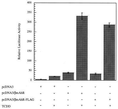
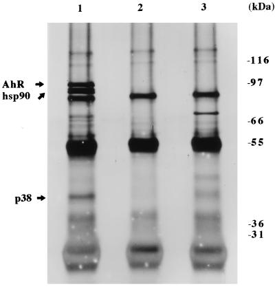
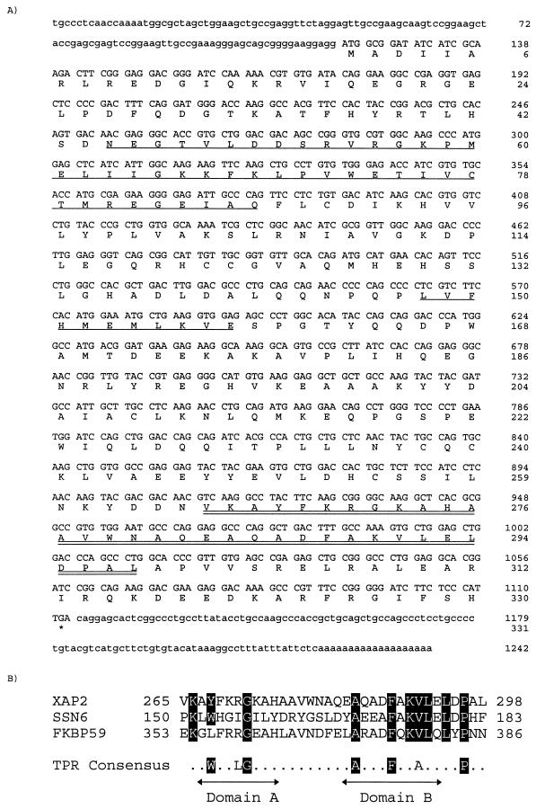
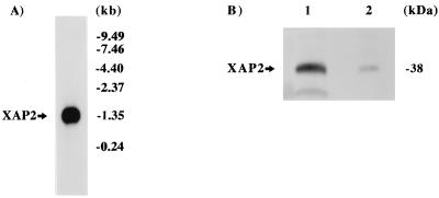
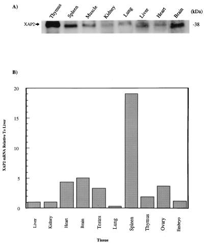
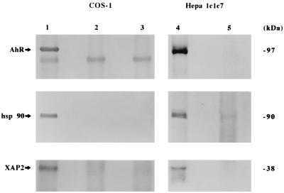
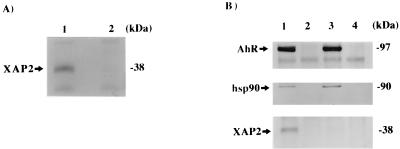
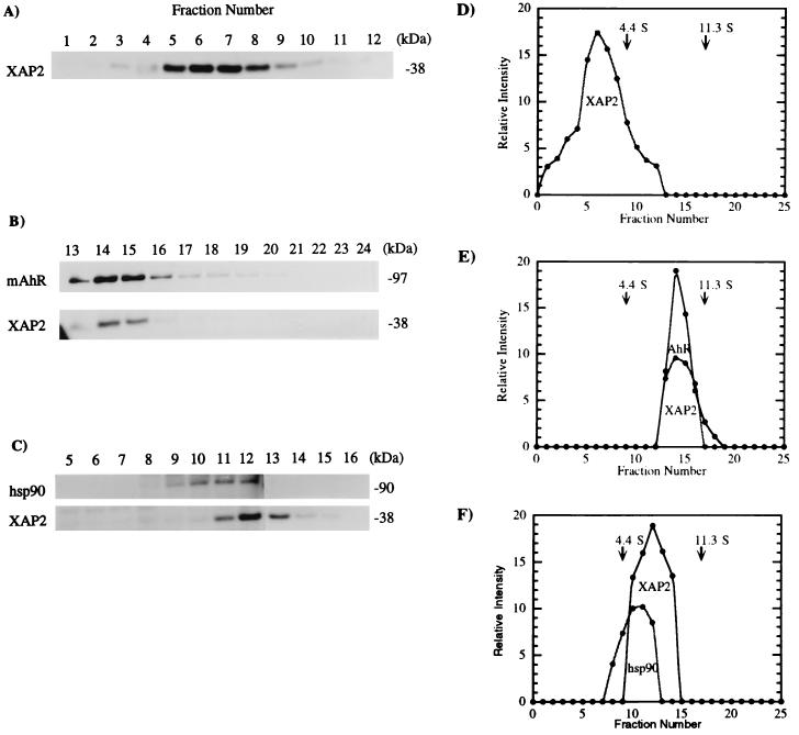
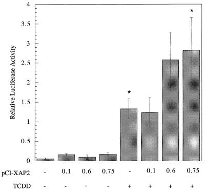
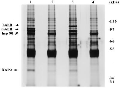
Prior to ligand activation, the unactivated aryl hydrocarbon receptor (AhR) exists in a heterotetrameric 9S core complex consisting of the AhR ligand-binding subunit, a dimer of hsp90, and an unknown subunit. Here we report the purification of an approximately 38-kDa protein (p38) from COS-1 cell cytosol that is a member of this complex by coprecipitation with a FLAG-tagged AhR. Internal amino acid sequence information was obtained, and p38 was identified as the hepatitis B virus X-associated protein 2 (XAP2). The simian ortholog of XAP2 was cloned from a COS-1 cDNA library; it codes for a 330-amino-acid protein containing regions of homology to the immunophilins FKBP12 and FKBP52. A tetratricopeptide repeat (TPR) domain in the carboxy-terminal region of XAP2 was similar to the third and fourth TPR domains of human FKBP52 and the Saccharomyces cerevisiae transcriptional modulator SSN6, respectively. Polyclonal antibodies raised against XAP2 recognized p38 in the unliganded AhR complex in COS-1 and Hepa 1c1c7 cells. It was ubiquitously expressed in murine tissues at the protein and mRNA levels. It was not required for the assembly of an AhR-hsp90 complex in vitro. Additionally, XAP2 did not directly associate with hsp90 upon in vitro translation, but was present in a 9S form when cotranslated in vitro with murine AhR. XAP2 enhanced the ability of endogenous murine and human AhR complexes to activate a dioxin-responsive element-luciferase reporter twofold, following transient expression of XAP2 in Hepa 1c1c7 and HeLa cells.


Figures

FIG. 1
FIG. 1
A transfected AhR is responsive to TCDD in COS-1 cells. COS-1 cells were transfected with pcDNA3, pcDNA3/βmAhR, or pcDNA3/βmAhR-FLAG in the presence of pGudLuc 6.1 and pcDNA3.1/lacZ/his plasmids. Relative luciferase activity was measured following induction by TCDD or carrier solvent. Each transfection was performed in triplicate.
FIG. 2
FIG. 2
The AhR exists in a heterotetrameric complex in COS-1 cells. COS-1 cells were transfected with 9 μg of pcDNA3/βmAhR-FLAG or pcDNA3; cytosolic fractions were isolated, immunoabsorbed with M2 affinity gel, and resolved by SDS-PAGE; and the gel was silver stained. Lanes: 1, pcDNA3/βmAhR-FLAG; 2, pcDNA3/βmAhR-FLAG with M2 affinity gel blocked with FLAG peptide; 3, pcDNA3.
FIG. 3
FIG. 3
Predicted coding sequence for simian XAP2. (A) Nucleotide and predicted amino acid sequence of simian XAP2. Singly underlined regions indicate homology to FKBP12; the doubly underlined region indicates a conserved TPR domain. (B) Alignment of XAP2 carboxy-terminal TPR, TPR3 of FKBP59, and TPR4 of SSN6. The consensus TPR was generated from CDC16, CDC23, CDC27, SSN6, and SK13 (28).
FIG. 4
FIG. 4
Presence of XAP2 in COS-1 cells. (A) Northern blot analysis of XAP2 mRNA. Poly(A)+ mRNA (4 μg) from COS-1 cells was probed with simian XAP2 cDNA. (B) Immunoblot analysis of XAP2. Lanes: 1, XAP2 translated in vitro; 2, 100 μg of COS-1 cytosol.
FIG. 5
FIG. 5
XAP2 is ubiquitously expressed in murine tissues. (A) Cytosolic extracts (100 μg) from the tissues indicated were resolved on SDS-PAGE, transferred to a PVDF membrane, and immunoblotted with XAP2 polyclonal antibodies. (B) QRT-PCR analysis of XAP2 mRNA in the murine tissues indicated. All values were normalized to XAP2 mRNA expressed in the liver.
FIG. 6
FIG. 6
XAP2 is a subunit stably associated with the unliganded AhR core complex in COS-1 and Hepa 1 cells. (A) COS-1 cells were transfected with 9 μg of pcDNA3/βmAhR-FLAG or pcDNA3, immunoabsorbed with M2 affinity gel, resolved on SDS-PAGE, blotted, and probed with antibodies raised against the AhR (RPT1), hsp90 (hsp 86/84), and XAP2. Lanes: 1, pcDNA3/βmAhR-FLAG; 2, pcDNA3/βmAhR-FLAG with M2 resin blocked with FLAG peptide; 3, pcDNA3 (cytosol from Hepa 1 cells was immunoabsorbed with polyclonal AhR antisera or rabbit immunoglobulin G, resolved on SDS-PAGE, and used for immunoblot analysis); 4, polyclonal AhR antisera; 5, rabbit IgG.
FIG. 7
FIG. 7
XAP2 is not required for assembly of the AhR with hsp90 in vitro. (A) Immunoblot analysis of XAP2 in RL. Lanes: 1, XAP2 transcribed and translated in vitro in RL; 2, RL alone. Each reaction was resolved by SDS-PAGE and used for immunoblot analysis with XAP2 polyclonal antibodies. (B) Immunoabsorption of AhR-hsp90-XAP2 complex. Lanes: 1, pcDNA3/βmAhR-FLAG cotranscribed and translated in vitro with XAP2 in RL; 2, same mixture as in lane 1 incubated with M2 affinity gel blocked with FLAG peptide; 3, pcDNA3/βmAhR-FLAG transcribed and translated in vitro in reticulocyte lysate; 4, same mixture as in lane 3 incubated with M2 affinity gel blocked with FLAG peptide. Each translation mixture was subjected to immunoabsorption with the M2 affinity gel, washed, resolved by SDS-PAGE, and used in an immunoblot analysis with the antibodies raised against the AhR (RPT1), hsp90 (hsp86/84), and XAP2.
FIG. 8
FIG. 8
XAP2 exists in a 0S to 4S complex in RL and shifts to 9S in the presence of mAhR. (A) pCI-XAP2 was transcribed and translated in rabbit RL with [35S]methionine and resolved on a sucrose density gradient, and fractions were collected, resolved by SDS-PAGE, and analyzed by autoradiography. (B) XAP2 was cotranscribed and translated with mAhR in RL by the same procedure. (C) Cytosol isolated from COS-1 cells was resolved on a sucrose density gradient, and fractions were collected, resolved by SDS-PAGE, blotted, and probed with antibodies against hsp90 (3B6p90) or XAP2 followed by incubation with [125I]GAM secondary antibody. (D to F) Quantitative assessments of panels A to C, respectively.
FIG. 9
FIG. 9
XAP2 acts a transcriptional enhancer in Hepa 1 cells. Hepa 1 cells were transfected with plasmids pGudLuc 6.1, pcDNA3.1/lacZ/his, and pCI-XAP2. The amount of pCI-XAP2 transfected is given in micrograms, and each transfection was brought to 0.75 μg with pCI vector. Relative luciferase activity was measured following induction by TCDD or carrier solvent. Each transfection was performed in triplicate. Asterisks indicate that a statistically significant difference was obtained (P < 0.05).
FIG. 10
FIG. 10
XAP2 is a member of the hAhR complex in COS-1 cells. COS-1 cells were transfected with 9 μg of pCI/hAhR-FLAG, pcDNA3/βmAhR-FLAG, or pcDNA3; cytosol was isolated, immunoabsorbed with M2 affinity gel, and resolved by SDS-PAGE; and the gel was silver stained. Lanes: 1, pcDNA3/βmAhR-FLAG; 2, pcDNA/βmAhR-FLAG with M2 affinity gel blocked with FLAG peptide; 3, pCI/hAhR-FLAG; 4, pCI/hAhR-FLAG with M2 affinity gel blocked with FLAG peptide.
FIG. 11
FIG. 11
XAP2 acts as a transcriptional enhancer in HeLa cells. HeLa cells were transfected with plasmids pGudLuc6.1, pcDNA3.1/lacZ/his, and pCI-XAP2. The amount of pCI-XAP2 transfected is given in micrograms and each transfection was brought to 3.0 μg with pCI vector. Relative luciferase activity was measured following induction by TCDD or carrier solvent. Each transfection was performed in triplicate. ∗, a statistically significant difference was obtained (P < 0.05).

References

    1. Altschul S F, Gish W, Miller W, Myers E W, Lipman D J. Basic local alignment search tool. J Mol Biol. 1990;215:403–410. - PubMed
    1. Aranyi P, Radanyi C, Renoir M, Devin J, Baulieu E-E. Covalent stabilization of the nontransformed chick oviduct cytosol progesterone receptor by chemical cross-linking. Biochemistry. 1988;27:1330–1336. - PubMed
    1. Bresnick E H, Dalman F C, Pratt W B. Direct stoichiometric evidence that the untransformed Mr 300 000, 9S, glucocorticoid receptor is a core unit derived from a larger heteromeric complex. Biochemistry. 1990;29:520–527. - PubMed
    1. Burbach K M, Poland A, Bradfield C A. Cloning of the Ah-receptor cDNA reveals a distinctive ligand-activated transcription factor. Proc Natl Acad Sci USA. 1992;89:8185–8189. - PMC - PubMed
    1. Carver L A, Bradfield C A. Ligand-dependent interaction of the aryl hydrocarbon receptor with a novel immunophilin homolog in vivo. J Biol Chem. 1997;272:11452–11456. - PubMed

Publication types

MeSH terms

Associated data