. 1998 Feb;18(2):1065-73.
doi: 10.1128/MCB.18.2.1065.

Epidermal growth factor induction of the c-jun promoter by a Rac pathway

Affiliations

Epidermal growth factor induction of the c-jun promoter by a Rac pathway

N Clarke et al. Mol Cell Biol. 1998 Feb.

Abstract

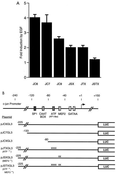
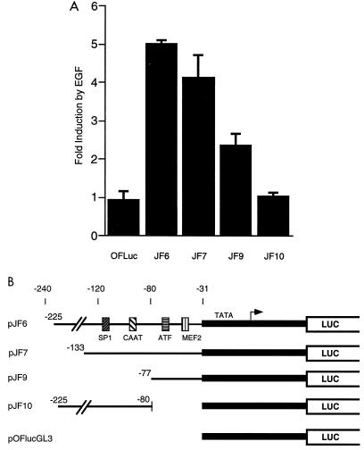
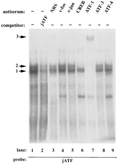
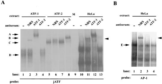
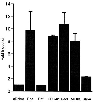
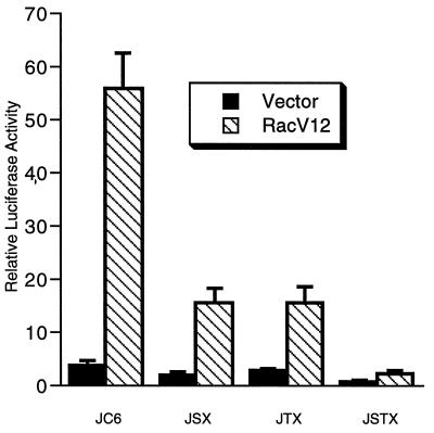
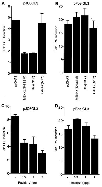
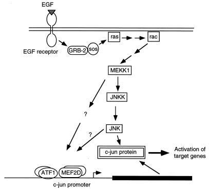
The c-jun proto-oncogene encodes a transcription factor which is activated by mitogens both transcriptionally and by phosphorylation by Jun N-terminal kinase (JNK). We have investigated the cellular signalling pathways involved in epidermal growth factor (EGF) induction of the c-jun promoter. We find that two sequence elements, which bind ATF1 and MEF2D transcription factors, are required in HeLa cells, although they are not sufficient for maximal induction. Activated forms of Ras, RacI, Cdc42Hs, and MEKK increased expression of the c-jun promoter, while dominant negative forms of Ras, RacI, and MEK kinase (MEKK) inhibited EGF induction. These and previously published results suggest that EGF activates the c-jun promoter by a Ras-to-Rac-to-MEKK pathway. This pathway is similar to that used for posttranslational activation of c-jun by JNK.


Figures

FIG. 1
FIG. 1
(A) MEF2 and ATF sites are required for EGF induction of the c-jun promoter. HeLa cells were transiently transfected with the c-jun pGL3-luciferase reporter plasmids, as indicated, and pCMV-β-galactosidase as an internal control. After transfection, the cells were serum starved and treated with or without EGF (100 ng/ml) for 3 h before preparing cell lysates for luciferase and β-galactosidase assays. The fold induction of luciferase activity in EGF-treated cells relative to untreated cells is shown. Values shown are the averages of at least two separate experiments done in duplicate ± standard errors of the means. (B) c-jun promoter constructs. The positions of binding sites for the transcription factors SP1, CTF, ATF, and MEF2 are indicated. The ATF site was previously referred to as an AP1-like element. The regions of the c-jun promoter in each construct are indicated. Point mutations in the ATF or MEF2 sites are indicated (x). LUC, luciferase.
FIG. 2
FIG. 2
(A) Upstream elements cooperate with ATF and MEF2 sites for EGF induction of the c-jun promoter. HeLa cells were transiently transfected with the indicated reporter plasmids (3 μg) and assayed for EGF induction of luciferase activity as described for Fig. 1. (B) Heterologous c-jun promoter constructs. The indicated fragments of the c-jun promoter were cloned upstream of a minimal c-fos promoter (shaded in grey) and a luciferase (LUC) reporter gene.
FIG. 3
FIG. 3
ATF1 and CREB from HeLa nuclear extracts bind to the c-Jun ATF site. Gel mobility assays were performed with HeLa cell nuclear extracts and a 32P-labeled double stranded oligonucleotide spanning the c-jun ATF site as a probe. A 50-fold molar excess of unlabeled oligonucleotide (jATF) was included as a specific competitor as indicated. Other nonspecific oligonucleotides had no effect (data not shown). The addition of antisera to the indicated proteins is shown. NRS, normal rabbit serum; −, no serum added. The arrows indicate the DNA-protein complexes obtained.
FIG. 4
FIG. 4
Antisera to ATF1, but not ATF2, specifically affect the c-Jun ATF complexes. (A) Gel mobility shift assays were performed with the c-jun ATF site probe and in vitro-translated ATF1 (lanes 1 to 4), in vitro-translated ATF2 (lanes 5 to 8), mock in vitro translation extract (lane 9), or HeLa cell nuclear extracts (lanes 10 to 13). Either no serum (−), nonimmune serum (NRS), or anti-ATF1 or anti-ATF2 serum was added as indicated. (B) HeLa nuclear extracts were assayed with an AP1 consensus site probe and the indicated sera. Arrows D and C mark complexes obtained with in vitro-translated ATF1 and ATF2. Antibody-supershifted complexes are marked with arrows A and B. Complex E shows the complex binding to the consensus AP1 site. The arrowheads to the right indicate a nonspecific complex obtained with nonimmune serum.
FIG. 5
FIG. 5
Activation of the c-jun promoter by activated forms of the small G proteins Ras, Rac, and Cdc42Hs. HeLa cells were transiently transfected with the c-jun–luciferase reporter pJC6GL3, pCMV-β-galactosidase as an internal control, and the following expression vectors (3 μg of each except for 2 μg for MEKK): pcDNA3 (empty vector control), Ras(V12), RafBXB, Cdc42Hs(V12), RacI(V12), RhoA(V14), or MEKK1. Cells were serum starved overnight prior to lysis for luciferase and β-galactosidase assays. The fold induction of luciferase activity is shown relative to activity in cells transfected with an empty expression vector (pcDNA3). The luciferase activities were normalized to the β-galactosidase activities except for assays with RasV12. With RasV12, the activity from pCMV-β-galactosidase was consistently induced fourfold, and this change was compensated for in normalizing the effect of Ras on pJC6GL3. The values shown are the averages of at least two separate experiments done in quadruplicate ± standard errors of the means.
FIG. 6
FIG. 6
Requirement of the MEF2 and ATF sites for Rac activation of the c-jun promoter. HeLa cells were transiently transfected with the indicated reporter genes, pCMV-β-galactosidase as an internal control, and either 2.5 μg of empty expression vector (pcDNA3) or racI(V12). After transfection, cells were serum starved in 0.2% newborn calf serum overnight and then lysed for luciferase and β-galactosidase assays. Relative luciferase activities normalized to the β-galactosidase activities are shown. The values shown are the averages of two separate experiments done in duplicate ± standard errors of the means.
FIG. 7
FIG. 7
Dominant negative Ras, RacI, and MEKK block EGF induction of the c-jun promoter. (A) HeLa cells were transiently transfected with pJC6GL3, pCMV-β-galactosidase, and 1 μg of the empty expression vector pcDNA3 or the indicated dominant negative vectors. After transfection, cells were serum starved for 30 h and then treated with or without EGF (100 ng/ml) for 3 h prior to lysis for luciferase and β-galactosidase assays. The fold induction in EGF-treated relative to untreated cells is shown. The results are the means of duplicates ± standard errors of the means. (B) HeLa cells were transiently transfected with the c-fos reporter plasmid pFos-GL3 (1 μg), pCMV-β-galactosidase (1.5 μg), and the indicated dominant negative vectors as for panel A except that the cells were treated with or without TPA (100 ng/ml) for 3 h. The fold induction by TPA relative to untreated cells is shown. (C) Increasing amounts of dominant negative RacI were transfected as for panel A with the c-jun reporter gene except that EGF was added at 200 ng/ml. (D) Increasing amounts of RacI(N17) were transfected with the c-fos reporter gene and assayed for the effect on TPA induction as for panel B.
FIG. 8
FIG. 8
Model of signalling pathways for EGF activation of c-jun. Components for signalling pathways to the c-jun promoter and for phosphorylation of c-Jun protein are shown and are described in the text. The pathway for induction of the c-jun promoter downstream of MEKK is unknown and may involve JNK and/or a separate pathway as indicated by the question marks. Two arrows between components indicate that the activation of the protein is not direct.

References

    1. Abate C, Luk D, Curran T. A ubiquitous nuclear protein stimulates the DNA-binding activity of fos and jun indirectly. Cell Growth Differ. 1990;1:455–462. - PubMed
    1. Angel P, Hattori K, Smeal T, Karin M. The jun proto-oncogene is positively autoregulated by its product, Jun/AP-1. Cell. 1988;55:875–885. - PubMed
    1. Angel P, Karin M. The role of Jun, Fos, and the AP-1 complex in cell-proliferation and transformation. Biochim Biophys Acta. 1991;1072:129–157. - PubMed
    1. Benbrook D M, Jones N C. Heterodimer formation between CREB and JUN proteins. Oncogene. 1990;5:295–302. - PubMed
    1. Bonni A, Ginty D D, Dudek H, Greenberg M E. Serine 133-phosphorylated CREB induces transcription via a cooperative mechanism that may confer specificity to neurotrophin signals. Mol Cell Neurosci. 1995;6:168–183. - PubMed

Publication types

MeSH terms

Substances