. 1998 Feb;18(2):1084-93.
doi: 10.1128/MCB.18.2.1084.

The promyelocytic leukemia gene product (PML) forms stable complexes with the retinoblastoma protein

Affiliations

The promyelocytic leukemia gene product (PML) forms stable complexes with the retinoblastoma protein

M Alcalay et al. Mol Cell Biol. 1998 Feb.

Abstract

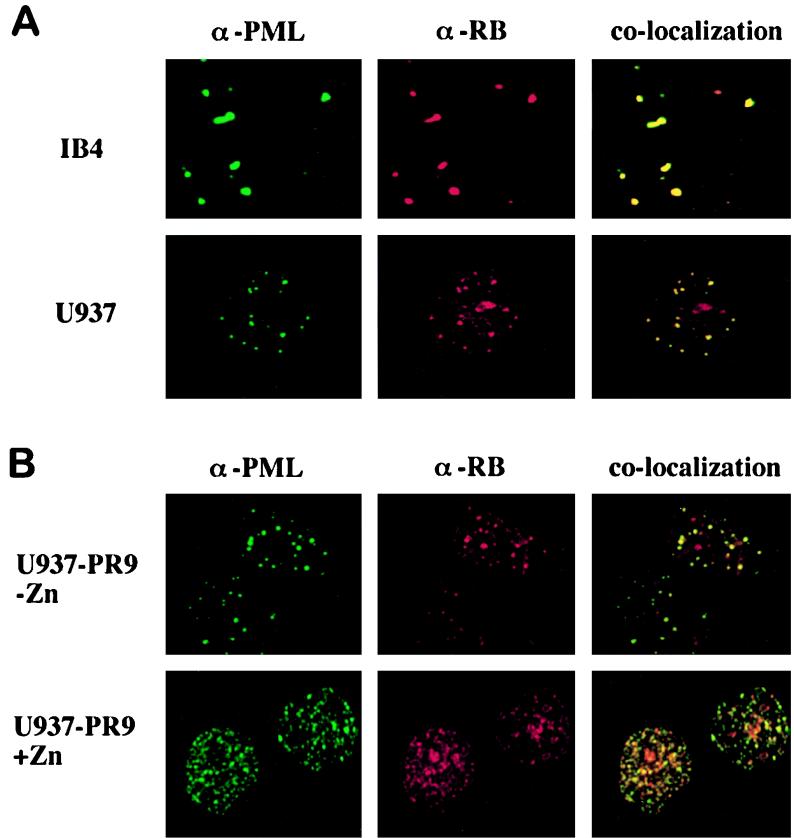
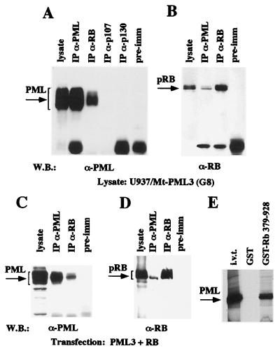
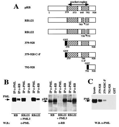
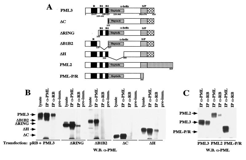
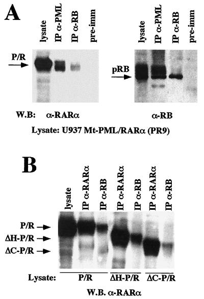
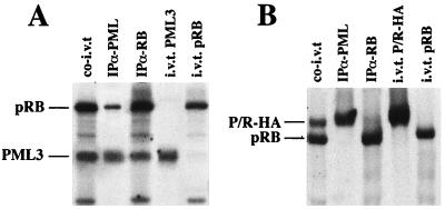
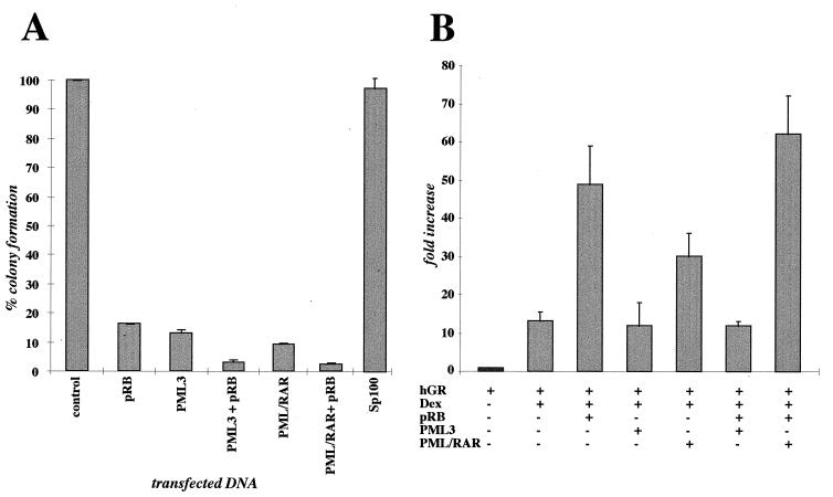
PML is a nuclear protein with growth-suppressive properties originally identified in the context of the PML-retinoic acid receptor alpha (RAR alpha) fusion protein of acute promyelocytic leukemia. PML localizes within distinct nuclear structures, called nuclear bodies, which are disrupted by the expression of PML-RAR alpha. We report that PML colocalizes with the nonphosphorylated fraction of the retinoblastoma protein (pRB) within nuclear bodies and that pRB is delocalized by PML-RAR alpha expression. Both PML and PML-RAR alpha form complexes with the nonphosphorylated form of pRB in vivo, and they interact with the pocket region of pRB. The regions of PML and PML-RAR alpha involved in pRB binding differ; in fact, the B boxes and the C-terminal region of PML, the latter of which is not present in PML-RAR alpha, are essential for the formation of stable complexes with pRB. Functionally, PML abolishes activation of glucocorticoid receptor-regulated transcription by pRB, whereas PML-RAR alpha further increases it. Our results suggest that PML may be part of transcription-regulatory complexes and that the oncogenic potential of the PML-RAR alpha protein may derive from the alteration of PML-regulated transcription.


Figures

FIG. 1
FIG. 1
pRB colocalizes with PML within nuclear bodies and is delocalized by PML-RARα expression. (A) Immunofluorescence experiments using an anti-pRB monoclonal antibody (α-RB; aRB1C1) revealed with a rhodamine-conjugated anti-mouse antibody and an anti-PML polyclonal antibody (α-PML; 2912A) revealed with an FITC-conjugated anti-rabbit antibody were performed with IB4 and U937 cells. Superimposition of PML and pRB staining is shown in the righthand panels, indicating colocalization of PML and pRB within NBs in both cell lines. (B) Similar experiments were performed with the U937 PR9 clone before and after induction of PML-RARα expression with ZnSO4. Superimposition of PML and pRB staining is shown in the righthand panels, revealing that pRB is delocalized into the PML-RARα microspeckles by the expression of the fusion protein. The anti-RB antibody aRB1C1 does not cross-react with PML-RARα, as revealed by Western blotting experiments and immunoprecipitations of in vitro-translated PML-RARα polypeptides (our unpublished results).
FIG. 2
FIG. 2
PML associates with the pRB complex. (A) PML coimmunoprecipitates with pRB but not with other pocket proteins. Cell lysates from the PML3-overexpressing U937 G8 clone were immunoprecipitated (IP) with anti-PML3 (α-PML) polyclonal (2417), anti-pRB (α-RB) monoclonal (XZ77), anti-p107 (α-p107) monoclonal (SD9), and anti-p130 (α-p130) polyclonal (C-20) antibodies and a preimmune (pre-imm) rabbit serum. The arrow on the left indicates PML3, revealed by Western blotting (W.B.) with anti-PML3 polyclonal antibody 2417. (B) PML coprecipitates with the nonphosphorylated pRB. Anti-PML immunoprecipitates from U937 (G8) cell lysates were analyzed by Western blotting with anti-pRB monoclonal antibody G3-245. The arrow on the left indicates pRB. (C and D) PML and pRB coimmunoprecipitate in transiently transfected cells. C33A cells were transiently transfected with the pCMV-PML3 and pCMV-pRB expression vectors, and lysates were immunoprecipitated with the anti-PML monoclonal (PG-M3) or the anti-pRB polyclonal (XZ77) antibody. Membranes were decorated with anti-PML3 antibody 2417 (C) or anti-pRB antibody G3-245 (D). Brackets on the left correspond to PML3 (C) and pRB (D). (E) In vitro binding analysis of bacterially expressed GST or GST-RB 379-928 (Fig. 3A) with in vitro-translated (i.v.t.) PML3 polypeptides.
FIG. 3
FIG. 3
The PML binding domain of pRB is within the pocket region. (A) Schematic representation of the pRB mutants used for the analysis. RBΔ21 and RBΔ22 represent deletion mutants lacking pRB exons 21 and 22, respectively, cloned in eukaryotic expression vector pCMV-Neo-BamHI for in vivo association studies. 379-928, 379-928 C-F, and 792-928 are different portions of the pRB C-terminal region expressed in bacteria as GST fusion proteins used for in vitro binding experiments (40, 52). (B) In vivo analysis of the association between PML and pRB deletion mutants. Coimmunoprecipitation experiments were performed with the antisera indicated above the blots (abbreviations are as defined in the legend to Fig. 2), using lysates from C33A cells transiently transfected with pCMV-PML3 and either the pCMV-RB, pCMV-RBΔ21, or pCMV-RBΔ22 expression vector. Left, analysis with the anti-PML3 antibody 2417; right, analysis with anti-pRB antibody G3-245. (C) In vitro binding analysis of GST-RB fusion proteins with a lysate from U937 (G8) cells overexpressing PML3. The GST-pRB proteins are shown in panel A; the procedure is described in Materials and Methods. The membrane was decorated with the anti-PML3 antibody 2417.
FIG. 4
FIG. 4
Determination of the PML regions involved in the association with pRB. (A) Schematic representation of the PML mutants (ΔH, ΔC, ΔRING, ΔB1B2, and PML-PR) and the two PML isoforms (PML2 and PML3) used in this study. The major PML domains are shown above or within the maps; the relevant amino acid boundaries are shown below. (B) The PML N terminus is involved in pRB binding. Coimmunoprecipitation experiments were performed with lysates from C33A cells transfected with pRB and either the PML3, ΔH, ΔC, ΔRING, or ΔB1B2 expression vector. The membrane was decorated with an anti-PML3 antibody directed against the C terminus (see the legend to Fig. 2 for abbreviations). (C) Relevance of the PML C terminus in pRB binding. Coimmunoprecipitation experiments were performed as described in Materials and Methods, using lysates from U937 clones expressing the PML3 and PML2 isoforms, which have different C termini, and the PML-P/R mutant, which bears a C-terminal deletion. The membrane was decorated with anti-PML antibody PG-M3, directed against the PML N terminus. Specific bands are indicated by arrows on the left.
FIG. 5
FIG. 5
PML-RARα associates with pRB. (A) Coimmunoprecipitation of pRB and PML-RARα was performed as described in Materials and Methods, using the lysate from a U937 clone expressing PML-RARα (PR9) (see the legend to Fig. 2 for abbreviations). Left, Western blot analysis of anti-PML (PG-M3) and anti-pRB (XZ77) immunoprecipitates with the anti-RARα anti-F antibody; right, Western blot analysis with the anti-pRB antibody G3-245. (B) PML-RARα mutants bearing deletions analogous to those described for PML in Fig. 3 within the PML moiety of PML-RARα (ΔC-PR and ΔH-PR [30]) were assayed for the capacity to bind pRB. Coimmunoprecipitation experiments were performed with lysates from U937 cell clones expressing the indicated PML-RARα mutants.
FIG. 6
FIG. 6
Reconstitution of the PML-pRB, but not of the PML-RARα-pRB, complex in rabbit reticulocyte lysates. pRB was in vitro translated together with PML3 (A) or P/R-HA (B), using rabbit reticulocyte lysates. The resulting products were immunoprecipitated with either anti-PML or anti-pRB antibodies, as indicated above the lanes (see the legend to Fig. 2 for abbreviations). A fraction of the co-in vitro translation product was loaded as a control (co-i.v.t.). In vitro translation products of the single proteins are loaded at the right of each panel, to exclude comigration of nonspecific bands.
FIG. 7
FIG. 7
Functional relevance of the PML-pRB association. (A) PML inhibits colony formation with a pRB-independent mechanism. C33A cells, which lack functional pRB, were transfected with equimolar quantities of the expression vectors indicated below the graph; G418-resistant colonies were scored after 12 to 15 days of selection. Results are shown as percentage of colonies with respect to C33A cells transfected with an empty vector (control). The results represent the average of four separate experiments, each performed in triplicate. (B) Effect of PML3 and PML-RARα on pRB regulation of GR-mediated transcription in HeLa cells. The data are expressed as fold activation of plasmid MTV-LTR-CAT with respect to the basal control value. Results are the average of four separate experiments, each performed in triplicate. hGR, human GR; Dex, dexamethasone.

Similar articles

Cited by

References

    1. Alcalay M, Zangrilli D, Pandolfi P P, Longo L, Mencarelli A, Giacomucci A, Rocchi M, Biondi A, Rambaldi A, Lo Coco F, Diverio D, Donti E, Grignani F, Pelicci P G. Translocation breakpoint of acute promyelocytic leukemia lies within the retinoic acid receptor alpha locus. Proc Natl Acad Sci USA. 1991;88:1977–1981. - PMC - PubMed
    1. Ascoli C A, Maul G G. Identification of a novel nuclear domain. J Cell Biol. 1991;112:785–795. - PMC - PubMed
    1. Baker S J, Markowitz S, Fearon E R, Willson K V, Vogelstein B. Suppression of human colorectal carcinoma cell growth by wild-type p53. Science. 1990;249:912–915. - PubMed
    1. Boddy M N, Howe K, Etkin L D, Solomon E, Freemont P S. PIC 1, a novel ubiquitin-like protein which interacts with the PML component of a multiprotein complex that is disrupted in acute promyelocytic leukemia. Oncogene. 1996;13:971–982. - PubMed
    1. Borden K L, Boddy M N, Lally J, O’Reilly N J, Martin S, Howe K, Solomon E, Freemont P S. The solution structure of the RING finger domain of the promyelocytic leukemia proto-oncoprotein PML. Proc Natl Acad Sci USA. 1995;93:1602–1606. - PMC - PubMed

Publication types

Substances

LinkOut - more resources