. 1998 Feb;18(2):1115-24.
doi: 10.1128/MCB.18.2.1115.

Invading the yeast nucleus: a nuclear localization signal at the C terminus of Ty1 integrase is required for transposition in vivo

Affiliations

Invading the yeast nucleus: a nuclear localization signal at the C terminus of Ty1 integrase is required for transposition in vivo

M A Kenna et al. Mol Cell Biol. 1998 Feb.

Abstract

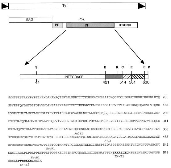
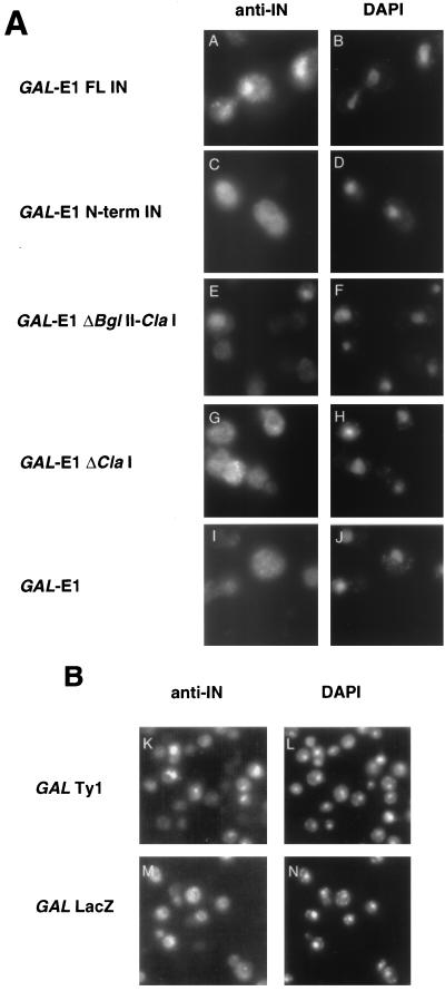
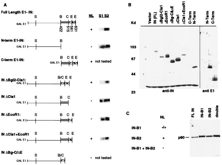
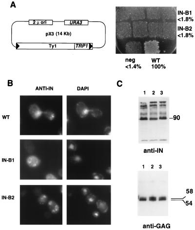
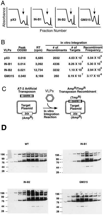
Retrotransposon Ty1 faces a formidable cell barrier during transposition--the yeast nuclear membrane which remains intact throughout the cell cycle. We investigated the mechanism by which transposition intermediates are transported from the cytoplasm (the presumed site of Ty1 DNA synthesis) to the nucleus, where they are integrated into the genome. Ty1 integrase has a nuclear localization signal (NLS) at its C terminus. Both full-length integrase and a C-terminal fragment localize to the nucleus. C-terminal deletion mutants in Ty1 integrase were used to map the putative NLS to the last 74 amino acid residues of integrase. Mutations in basic segments within this region decreased retrotransposition at least 50-fold in vivo. Furthermore, these mutant integrase proteins failed to localize to the nucleus. Production of virus-like particles, reverse transcriptase activity, and complete in vitro Ty1 integration resembled wild-type levels, consistent with failure of the mutant integrases to enter the nucleus.


Figures

FIG. 1
FIG. 1
Ty1 and IN structure. The structure of the Ty1 retrotransposon is diagrammed. Black triangles indicate the long-terminal-repeat regions. The GAG and POL ORFs and the element-encoded proteins derived from POL are indicated. The complete sequence of Ty1 IN is shown. Restriction site abbreviations: S, SalI; B, BglII; C, ClaI; E, EcoRI; K, KpnI; P, PvuII. The amino acid number is indicated beneath each restriction site. Key restriction sites are indicated above the corresponding amino acid sequence. IN NLS mutations, B1 and B2, made in the GAL-Ty1 plasmid are in boldface type. The numbers to the right of the sequence represent the last amino acid residue on that line of sequence.
FIG. 2
FIG. 2
Ty1 IN localizes to the nucleus. Anti-IN antibody was used on cells induced for the production of E1-IN from the GAL1 promoter (A) or on cells induced for Ty1 transposition (B, panels K and L [panels M and N show GAL-LacZ negative controls]). FITC-stained panels are on the left; DAPI-stained panels are on the right. Constructs tested for IN localization are indicated to the left of the micrographs. FL, full length.
FIG. 3
FIG. 3
Analysis of E1-IN fusion proteins. (A) C-terminal deletion constructs map the NLS region within the last 74 amino acids of IN. Constructs tested are diagrammed, with restriction sites indicated; abbreviations for these are defined in the legend to Fig. 1. Numbers shown below restriction sites in full-length E1-IN represent amino acid residues. Nuclear localization (NL) is indicated as present (+) or absent (−); these results are from the indirect immunofluorescence experiments with anti-IN antibody described in the legend to Fig. 2, except for that for the C-terminal construct that necessitated the use of anti-E1 (see text). Cell fractionation experiments (described in Materials and Methods) were performed on strains bearing the E1-IN truncation constructs. Bands corresponding to the indicated E1-IN protein are shown to the right of the NL data. (B) Immunoblot analysis of the truncated IN proteins. Whole-cell extracts were electrophoresed on an SDS–8% polyacrylamide gel. Proteins were detected with either MAb 8B11 (1:5,000) or anti-E1 (1:500). Molecular mass markers are shown at the left in kilodaltons. MAb 8B11 also recognizes a non-Ty1 band of ∼30 kDa. Since the C-terminal truncation does not contain the epitope for MAb 8B11, this protein extract, along with the N-terminal one, was probed with anti-E1. (C) Similar immunoblot analysis of the substitution mutant GAL-E1-IN constructs B1 (NSKKRS→AAGSAA) and B2 (RSKKRI→AAGSAA). NL is indicated as present (+), partial (−/+), or absent (−).
FIG. 4
FIG. 4
Two sites required for nuclear localization and transposition. (A) (Left) The pX3 GAL-Ty1-TRP1 plasmid (58) was used as the parent for making the IN B1 and B2 mutants (residues underlined in amino acid sequence of Fig. 1 were replaced with AAGSAA). Cells containing these pX3 derivatives were assayed for Ty1-TRP1 transposition. (Right) Transposition results for pX3 (wild-type IN [WT]) and mutants B1, B2, and RT (the negative control [neg]) are shown. Percentages of wild-type transposition frequency are indicated. (B) Indirect immunofluorescence. Anti-IN (MAb 8B11) panels are shown on the left (stained with FITC); DAPI staining is shown on the right. (C) (Top) Immunoblot with anti-IN (MAb 8B11) of whole-cell extracts prepared from wild-type (lane 1), IN B1 (lane 2), and IN B2 (lane 3) strains. The apparent molecular mass of fully processed IN is indicated on the right in kilodaltons. (Bottom) Immunoblot with R1-F (anti-Gag antibody). The apparent molecular masses of unprocessed and processed Gag proteins are indicated on the right in kilodaltons. Lanes are as described for the top panel.
FIG. 5
FIG. 5
Analysis of VLPs from wild-type and NLS mutant Ty1 strains. (A) Sucrose gradient fractionation profiles showing VLP peaks. The top of the gradient (20% sucrose) is presented at the left side of the x axis; the bottom of the gradient (70% sucrose) is at the right. The y axis represents absorbance at 254 nm. It is important to note that VLPs could be isolated from wild-type and NLS mutant Ty1 strains (small peaks under vertical arrows). The first peak at the top of the gradient is bulk protein, and the last peak at the right is the extreme bottom of the gradient. (B) Biochemical analysis of VLPs. The peak fraction from each VLP preparation was examined for RT and integration activities in vitro. All of the preparations showed normal RT activity. The activities of positive control avian myeloblastosis virus RT and the negative control, no enzyme (data not shown), were 93,500 and 465 cpm, respectively. VLPs isolated from the wild-type strain (pX3), or the NLS mutant strains (IN B1 and B2) supported normal levels of integration, whereas the negative control IN mutant strain (GM315) was 16-fold less active. OD280, optical density at 280 nm. (C) In vitro integration assay (25). The artificial transposon AT-2 serves as a substrate for Ty1 IN and is integrated into the target plasmid in vitro when VLPs are added. AT-2 carries the dhfr gene which confers trimethoprim resistance and thus serves as a selectable marker when the transposition reaction mixture is transformed into Escherichia coli. The target plasmid contains the bla gene conferring ampicillin resistance, while transposon recombinants carry both markers. The number of recombinants (Ampr Tmpr colonies) divided by the total number of targets recovered (Ampr colonies) provides the recombinant frequency. (D) Immunoblots of nine peak fractions from each gradient probed with MAb 8B11, showing similar yields of encapsidated IN protein in the four preparations. Molecular masses are indicated on the left in kilodaltons. WT, wild type; GM315, negative control IN mutant strain.

Similar articles

Cited by

References

    1. Adams A E, Pringle J R. Staining of actin with fluorochrome-conjugated phalloidin. Methods Enzymol. 1991;194:729–731. - PubMed
    1. Aitchison J D, Rout M P, Marelli M, Blobel G, Wozniak R W. Two novel related yeast nucleoporins, Nup170p and Nup157p: complementation with the vertebrate homologue Nup155p and functional interactions with the yeast nuclear pore-membrane protein Pom152p. J Cell Biol. 1995;131:1133–1148. - PMC - PubMed
    1. Bassett, D. Personal communication.
    1. Belcourt M F, Farabaugh P J. Ribosomal frameshifting in the yeast retrotransposon Ty: tRNAs induce slippage on a 7 nucleotide minimal site. Cell. 1990;62:339–352. - PMC - PubMed
    1. Boeke J D, Eichinger D J, Castrillon D, Fink G R. The Saccharomyces cerevisiae genome contains functional and nonfunctional copies of transposon Ty1. Mol Cell Biol. 1988;8:1432–1442. - PMC - PubMed

Publication types

MeSH terms