

Review
doi: 10.1073/pnas.95.3.831.
Frontoparietal cortical networks for directing attention and the eye to visual locations: identical, independent, or overlapping neural systems?
Affiliations
- PMID: 9448248
- PMCID: PMC33805
- DOI: 10.1073/pnas.95.3.831
Item in Clipboard
Review
Frontoparietal cortical networks for directing attention and the eye to visual locations: identical, independent, or overlapping neural systems?
Proc Natl Acad Sci U S A.
.
Abstract
Functional anatomical and single-unit recording studies indicate that a set of neural signals in parietal and frontal cortex mediates the covert allocation of attention to visual locations, as originally proposed by psychological studies. This frontoparietal network is the source of a location bias that interacts with extrastriate regions of the ventral visual system during object analysis to enhance visual processing. The frontoparietal network is not exclusively related to visual attention, but may coincide or overlap with regions involved in oculomotor processing. The relationship between attention and eye movement processes is discussed at the psychological, functional anatomical, and cellular level of analysis.
Figures
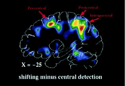
Sagittal PET section, 25 mm left of
midline, of group-averaged subtraction image between shifting-attention
and central-detection tasks.
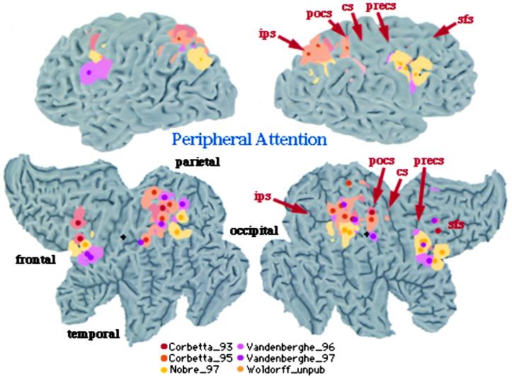
3D rendering and 2D flattened surface of
the Visible Man Brain, with the left hemisphere on the left. Lobes are
indicated in 2D surface. Sulci are indicated as follows: sfs, superior
frontal sulcus (s.); precs, precentral s.; cs, central s.; pocs,
postcentral s.; ips, intraparietal s. Foci of activation during
shifting attention [red (16), yellow (17), and orange (53)] and tonic
attention [pink (24), violet (25), and light orange (Woldorff
et al., unpublished data)].
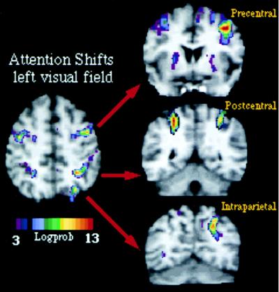
MPrage anatomical and fMRI activity in single
subject during shifting attention in left visual field. Transverse
section, z = 52. Coronal sections along precentral
(precs), postcentral (pocs), and intraparietal sulcus (ips).
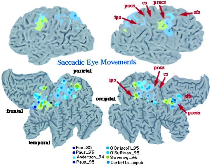
Visible Man Brain as in Fig. 2. Foci for saccadic
eye movements.
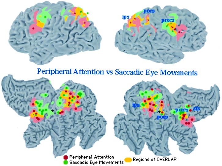
Visible Man Brain as in Fig. 2. Foci for
attention from Fig. 2 (red) and eye movement from Fig. 4 (green). Areas
of overlap are in yellow.
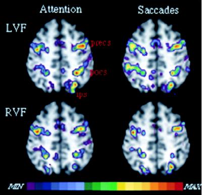
Anatomical MRI and fMRI activity in single
subject for shifting attention and saccadic eye movements in left (LVF)
and right (RVF) hemispheres. Slices at z = 52 mm.
References
-
- Ullman S. Cognition. 1984;18:97–159. - PubMed
-
- Eriksen C W, Hoffman J E. Percept Psychophys. 1972;12:201–204.
-
- Posner M I. Q J Exp Psychol. 1980;32:3–25. - PubMed
-
- Berlucchi G, Tassinari G, Marzi C A, Di Stefano M. Neuropsychologia. 1989;27:201–221. - PubMed
-
- Jonides J. In: Voluntary vs. Automatic Control over the Mind’s Eye’s Movement. Posner M I, Marin O, editors. Hillsdale, NJ: Lawrence Erlbaum Associates; 1981. pp. 187–205.
Publication types
MeSH terms
Grants and funding
LinkOut - more resources
Full Text Sources
Other Literature Sources
