Review
. 1998 Feb 3;95(3):839-46.
doi: 10.1073/pnas.95.3.839.

Neural components of topographical representation

Affiliations
Review

Neural components of topographical representation

G K Aguirre et al. Proc Natl Acad Sci U S A. .

Abstract

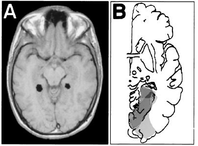
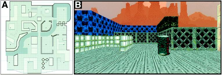
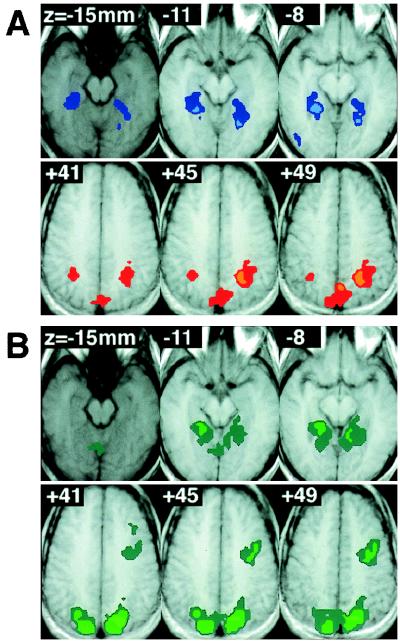
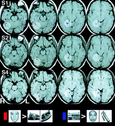
Studies of patients with focal brain damage suggest that topographical representation is subserved by dissociable neural subcomponents. This article offers a condensed review of the literature of "topographical disorientation" and describes several functional MRI studies designed to test hypotheses generated by that review. Three hypotheses are considered: (i) The parahippocampal cortex is critically involved in the acquisition of exocentric spatial information in humans; (ii) separable, posterior, dorsal, and ventral cortical regions subserve the perception and long term representation of position and identity, respectively, of landmarks; and (iii) there is a distinct area of the ventral occipitotemporal cortex that responds maximally to building stimuli and may play a role in the perception of salient landmarks. We conclude with a discussion of the inferential limitations of neuroimaging and lesion studies. It is proposed that combining these two approaches allows for inferences regarding the computational involvement of a neuroanatomical substrate in a given cognitive process although neither method can strictly support this conclusion alone.


Figures

Figure 1
Figure 1
Stimuli from an fMRI study of topographical learning. (A) Subject view. Actual stimuli were in full color and were several times wider in the horizontal plane than pictured. (B) Aerial view of maze. (C) Representative subject rendition of maze.
Figure 2
Figure 2
(A) Single axial slice showing voxels with significant fMRI signal changes during putative topographical learning in one subject. Right and left are reversed. Signal changes were observed, among other areas, bilaterally within the parahippocampal gyrus. (B) Common lesion site in four patients with anterograde topographical disorientation (8). Areas of lesion overlap are indicated by progressively darker shades of gray. The common lesion site is in the right parahippocampus.
Figure 3
Figure 3
Virtual reality town in which subjects were trained before scanning. (A) Aerial view. Subjectively, the town was ≈140 meters in width. (B) View of one location within the town. Each location was designated as such by the presence of a marker in the ground.
Figure 4
Figure 4
Sites of replicated significant activity across subjects. Right and left are reversed. (A) Shown are three inferior and three superior axial slices through a brain in standard space. In shades of blue are those voxels where multiple subjects had significantly greater signal during the appearance task compared with the position task; in shades of red are those voxels with greater signal during the position task compared with the appearance task. (B) In shades of green are shown those voxels that had significantly greater activity in both the appearance and position tasks compared with the baseline task over multiple subjects.
Figure 5
Figure 5
Putative “face” and “building” areas in three subjects. Shown are 4-mm axial slices in standard space, arranged from inferior to superior. Superimposed in red are those voxels that responded with significantly greater signal to the presentation of faces than to the presentation of buildings or objects. In blue are those areas with a greater response to building than to faces or objects. The circles of lighter contrast are intended to illustrate the anatomical correspondence of the suprathreshold voxels across subjects.

References

    1. O’Keefe J, Nadel L. The Hippocampus as a Cognitive Map. Oxford: Oxford Univ. Press; 1978.
    1. Brewer B, Pears J. In: Spatial Representation. Eilan N, McCarthy R, Brewer B, editors. Oxford: Blackwell; 1993. pp. 25–30.
    1. Heft H. Environ Psychol Nonverbal Behav. 1979;3:172–185.
    1. Thorndyke P W, Hayes R B. Cognit Psychol. 1982;14:560–589. - PubMed
    1. Magliano J P, Cohen R, Allen G L, Rodrigue J R. J Environ Psychol. 1995;15:65–75.

Publication types

LinkOut - more resources