Review
. 1998 Feb 3;95(3):883-90.
doi: 10.1073/pnas.95.3.883.

A neural system for human visual working memory

Affiliations
Review

A neural system for human visual working memory

L G Ungerleider et al. Proc Natl Acad Sci U S A. .

Abstract

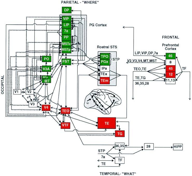
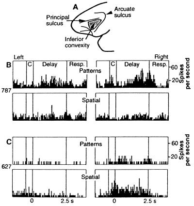
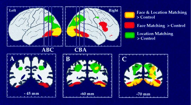
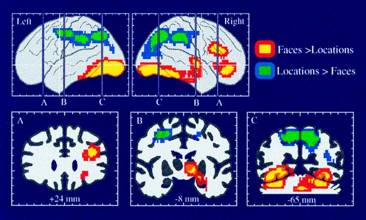
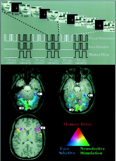
Working memory is the process of actively maintaining a representation of information for a brief period of time so that it is available for use. In monkeys, visual working memory involves the concerted activity of a distributed neural system, including posterior areas in visual cortex and anterior areas in prefrontal cortex. Within visual cortex, ventral stream areas are selectively involved in object vision, whereas dorsal stream areas are selectively involved in spatial vision. This domain specificity appears to extend forward into prefrontal cortex, with ventrolateral areas involved mainly in working memory for objects and dorsolateral areas involved mainly in working memory for spatial locations. The organization of this distributed neural system for working memory in monkeys appears to be conserved in humans, though some differences between the two species exist. In humans, as compared with monkeys, areas specialized for object vision in the ventral stream have a more inferior location in temporal cortex, whereas areas specialized for spatial vision in the dorsal stream have a more superior location in parietal cortex. Displacement of both sets of visual areas away from the posterior perisylvian cortex may be related to the emergence of language over the course of brain evolution. Whereas areas specialized for object working memory in humans and monkeys are similarly located in ventrolateral prefrontal cortex, those specialized for spatial working memory occupy a more superior and posterior location within dorsal prefrontal cortex in humans than in monkeys. As in posterior cortex, this displacement in frontal cortex also may be related to the emergence of new areas to serve distinctively human cognitive abilities.


Figures

Figure 1
Figure 1
Visual processing pathways in monkeys. Areas in the dorsal stream, having primarily visuospatial functions, are shown in green, and areas in the ventral stream, having primary object recognition functions, are shown in red. Lines connecting the areas indicate known anatomical connections, with heavy arrowheads indicating feed-forward connections from lower-order areas to higher-order ones and open arrowheads indicating feedback connections from higher-order areas to lower-order ones. Solid lines indicate connections from both central and peripheral visual field representations, and dotted lines indicate connections restricted to peripheral field representations. Shaded region on lateral view of the brain indicates extent of cortex included in the diagram. (From ref. ; for further details of the visual areas, see ref. .)
Figure 2
Figure 2
Location and response properties of the monkey prefrontal cortex. (A) Monkey prefrontal cortex is defined as the region extending forward from the fundus of the rostral bank of the arcuate sulcus to the frontal pole. Cortex within the principal sulcus is BA 46, whereas the cortex below it on the inferior prefrontal convexity (shaded region) is BA 12. (B) Responses of a single inferior convexity neuron with pattern-specific working memory activity. (Upper) Delay period activity specific to a pattern on pattern delayed response trials. (Lower) Lack of response on spatial delayed response trials. (C) Responses of a single neuron in the principal sulcus with spatial-specific working memory activity. (Upper) Lack of response on pattern delayed response trials. (Lower) Delay activity specific to right spatial cues on spatial delayed response trials. [Reproduced with permission from ref. (Copyright 1993, The American Association for the Advancement of Science.)]
Figure 3
Figure 3
The dorsal and ventral visual processing streams in human cortex, as demonstrated in a PET study of location and face perception. Areas shown in green had significantly increased rCBF during the location matching but not during the face matching task, as compared with rCBF during a sensorimotor control task. Areas shown in red had significantly increased rCBF during the face matching but not during the location matching task, as compared with rCBF during the control task. Areas shown in yellow had significantly increased rCBF during both face and location matching tasks. Maximum location intensities are shown on the lateral views of the left and right hemispheres. Coronal sections are taken at the anterior-posterior levels indicated on the lateral views. (Adapted from ref. .)
Figure 4
Figure 4
Selective activation of the dorsal and ventral visual processing pathways and of dorsal and ventral prefrontal areas, as demonstrated in a PET study of spatial location and face working memory. Areas shown in blue and green had significantly greater rCBF during the spatial, as compared with the face, working memory task. Areas shown in yellow and red had significantly greater rCBF during the face, as compared with the spatial, working memory task. The two working memory tasks used identical stimuli. (Adapted from ref. .)
Figure 5
Figure 5
Design and results of an fMRI study of working memory for faces (61). (Upper) Design of the task. For each series of fMRI scans, subjects performed 3½ baseline-activation task cycles, each consisting of two sensorimotor control trials followed by two working memory trials. During the memory task, subjects saw a picture of a face, a delay, and then another picture of a face. Subjects were asked to hold an image of the first face in mind during the delay and to respond with a left or right button press to indicate whether the second face matched the first. During the control task, subjects simply looked at the scrambled pictures and then pressed both buttons when the second scrambled picture appeared. Three time series are shown that represent the different cognitive components of the task: a transient, nonselective response to visual stimuli; a transient, selective response to faces; and sustained activity during memory delays. These time series (smoothed and delayed by convolution with a model of the hemodynamic response) were used as regressors in a multiple regression analysis of the time course of activation in each area. (Lower) Results from a single subject overlaid onto that subject’s anatomical images. Activations are color-coded according to the relative sizes of the three regression coefficients described above. Areas that responded transiently and nonselectively to any visual stimulus, such as posterior occipital cortex (a), are shown in green. Areas that responded transiently and showed a selective response to faces over scrambled faces, such as fusiform gyrus (b), are shown in blue. Areas that showed sustained activation during the memory delay after the stimulus was removed from view, such as inferior frontal cortex (c), are shown in red. Areas that showed a combination of these types of responses are shown in a blend of colors. (From ref. .)

References

    1. Fox P T, Raichle M E. Proc Natl Acad Sci USA. 1986;83:1140–1144. - PMC - PubMed
    1. Bandettini P A, Wong E C, Hinks R S, Tikofsky R S, Hyde J S. Magn Reson Med. 1992;25:390–397. - PubMed
    1. Kwong K K, Belliveau J W, Chesler D A, Goldberg I E, Weisskoff R M, Poncelet B P, Kennedy D N, Hoppel B E, Cohen M S, Turner R. Proc Natl Acad Sci USA. 1992;89:5675–5679. - PMC - PubMed
    1. Ogawa S, Tank D W, Menon R, Ellermann J M, Kim S G, Merkle H, Ugurbil K. Proc Natl Acad Sci USA. 1992;89:5951–5955. - PMC - PubMed
    1. Desimone R, Ungerleider L G. In: Handbook of Neuropsychology. Boller F, Grafman J, editors. Vol. 2. Amsterdam: Elsevier; 1989. pp. 267–300.

MeSH terms