

Functional neuroimaging studies of encoding, priming, and explicit memory retrieval
- PMID: 9448256
- PMCID: PMC33813
- DOI: 10.1073/pnas.95.3.891
Functional neuroimaging studies of encoding, priming, and explicit memory retrieval
Abstract
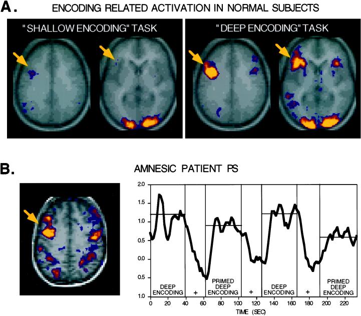
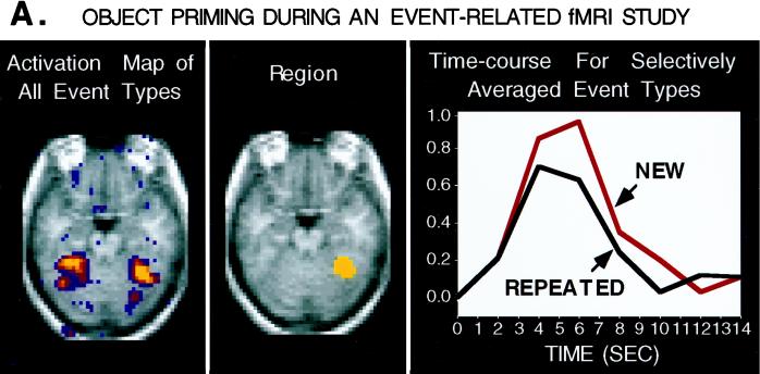
Human functional neuroimaging techniques provide a powerful means of linking neural level descriptions of brain function and cognition. The exploration of the functional anatomy underlying human memory comprises a prime example. Three highly reliable findings linking memory-related cognitive processes to brain activity are discussed. First, priming is accompanied by reductions in the amount of neural activation relative to naive or unprimed task performance. These reductions can be shown to be both anatomically and functionally specific and are found for both perceptual and conceptual task components. Second, verbal encoding, allowing subsequent conscious retrieval, is associated with activation of higher order brain regions including areas within the left inferior and dorsal prefrontal cortex. These areas also are activated by working memory and effortful word generation tasks, suggesting that these tasks, often discussed as separable, might rely on interdependent processes. Finally, explicit (intentional) retrieval shares much of the same functional anatomy as the encoding and word generation tasks but is associated with the recruitment of additional brain areas, including the anterior prefrontal cortex (right > left). These findings illustrate how neuroimaging techniques can be used to study memory processes and can both complement and extend data derived through other means. More recently developed methods, such as event-related functional MRI, will continue this progress and may provide additional new directions for research.
Figures
References
-
- Posner M I, Raichle M E. Images of Mind. New York: Scientific American Books; 1994.
-
- Tulving E. Elements of Episodic Memory. New York: Oxford Univ. Press; 1983.
-
- Gluck M A, Myers C E. Annu Rev Psychol. 1997;48:481–514. - PubMed
-
- Polster M R, Nadel L, Schacter D L. J Cognit Neurosci. 1991;3:95–116. - PubMed
-
- Raichle M E. In: The Handbook of Physiology: Section 1. The Nervous System: Vol. V. Higher Functions of the Brain: Part 1. Plum F, Mountcastle V, editors. Bethesda, MD: Am. Physiol. Assoc.; 1987. pp. 643–674.
Publication types
MeSH terms
Grants and funding
LinkOut - more resources
Full Text Sources
Medical
