Review
. 1998 Feb 3;95(3):906-13.
doi: 10.1073/pnas.95.3.906.

The role of left prefrontal cortex in language and memory

Affiliations
Review

The role of left prefrontal cortex in language and memory

J D Gabrieli et al. Proc Natl Acad Sci U S A. .

Abstract

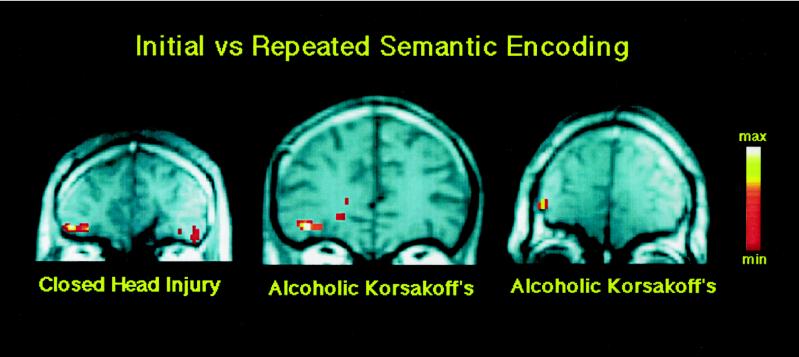
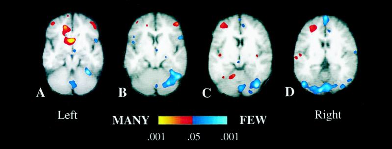
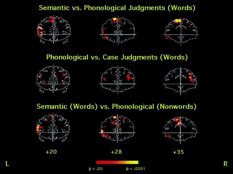
This article reviews attempts to characterize the mental operations mediated by left inferior prefrontal cortex, especially the anterior and inferior portion of the gyrus, with the functional neuroimaging techniques of positron emission tomography and functional magnetic resonance imaging. Activations in this region occur during semantic, relative to nonsemantic, tasks for the generation of words to semantic cues or the classification of words or pictures into semantic categories. This activation appears in the right prefrontal cortex of people known to be atypically right-hemisphere dominant for language. In this region, activations are associated with meaningful encoding that leads to superior explicit memory for stimuli and deactivations with implicit semantic memory (repetition priming) for words and pictures. New findings are reported showing that patients with global amnesia show deactivations in the same region associated with repetition priming, that activation in this region reflects selection of a response from among numerous relative to few alternatives, and that activations in a portion of this region are associated specifically with semantic relative to phonological processing. It is hypothesized that activations in left inferior prefrontal cortex reflect a domain-specific semantic working memory capacity that is invoked more for semantic than nonsemantic analyses regardless of stimulus modality, more for initial than for repeated semantic analysis of a word or picture, more when a response must be selected from among many than few legitimate alternatives, and that yields superior later explicit memory for experiences.


Figures

Figure 1
Figure 1
Coronal view of prefrontal cortex in three amnesic patients with two different etiologies of amnesia. The slices, from left to right, are 39, 35, and 42 mm anterior to the anterior commissure. Individual activations are overlaid on T1-weighted anatomic sections. Each patient shows greater activation in the left inferior prefrontal cortex (corresponding to Brodmann area 47) for the initial relative to the repeated semantic processing of words. The individual activations correspond to the anterior extent of regions that show semantic activation in Fig. 3. The right side of the brain is depicted on the right side of Figs. 1–3.
Figure 2
Figure 2
Averaged fMRI activation over six subjects depicted on averaged T1-weighted oblique axial sections (obtained at a 25-degree angle from the AC-PC line). (Left to Right) The sections depict increasingly superior planes that are 6 mm thick and separated by a 1-mm gap. Regions depicted in red–yellow represent areas that exhibited increased activation when subjects completed stems that had many, relative to few, completions (MANY condition), whereas regions in blue–light blue represent areas that exhibited increases during completion of stems with few, relative to many, completions (FEW condition). Major areas of increased activation during the MANY condition include the left middle frontal gyrus (Brodmann area 9/10, C and D), the right middle frontal gyrus (Brodmann area 10, A and B), the left cingulate gyrus (Brodmann areas 24/32, A), the left caudate nucleus (A), the left postcentral gyrus (Brodmann area 43, D), and the left anterior quandragular lobule of the cerebellum (C). Regions of increased activation during the FEW condition include several portions of the cerebellar vermis (Larsell’s lobules VI, VII, and VIII; A, C, and D), right cerebellar hemisphere (HVI and superior HVIIA, B and C), the right inferior frontal gyrus (Brodmann area 47, not depicted in figure), along the midline in the superior frontal gyrus (Brodmann area 8, D), and bilaterally in the fusiform gyrus (Brodmann area 37/19 and right Brodmann area 37, B–D).
Figure 3
Figure 3
Averaged fMRI activation in the prefrontal cortex over eight subjects for three coronal slice locations (20, 28, and 35 mm anterior to the anterior commissure). (Top) Areas that were more active for semantic processing (abstract/concrete judgment) than for phonological processing (syllable judgment). (Middle) Regions that were more active for phonological processing than for perceptual processing (letter case judgment). (Bottom) Areas that were activated for semantic processing of words compared with phonological processing of pronounceable nonwords. Left inferior frontal cortex (Brodmann areas 45 and 47) is located just above and below the left end of the horizontal line. Activation in left inferior cortex is prominent when semantic judgments are made (Top and Bottom) but not when phonological judgments are made (Middle).

References

    1. Gabrieli, J. D. E. (1998) Annu. Rev. Psychol. 49, in press. - PubMed
    1. Engel S A, Rumelhart D E, Wandell B A, Lee A T, Glover G H, Chichilnisky E J, Shadlen M N. Nature (London) 1994;369:525. - PubMed
    1. Engel S A, Zhang X, Wandell B. Nature. 1997;388:68–71. - PubMed
    1. Tootell R B H, Dale A M, Sereno M I, Malach R. Trends Neurosci. 1996;19:481–489. - PubMed
    1. Petersen S E, Fox P T, Posner M I, Mintun M, Raichle M E. Nature (London) 1988;331:585–589. - PubMed

Publication types