

Neuroimaging studies of word reading
- PMID: 9448259
- PMCID: PMC33816
- DOI: 10.1073/pnas.95.3.914
Neuroimaging studies of word reading
Abstract
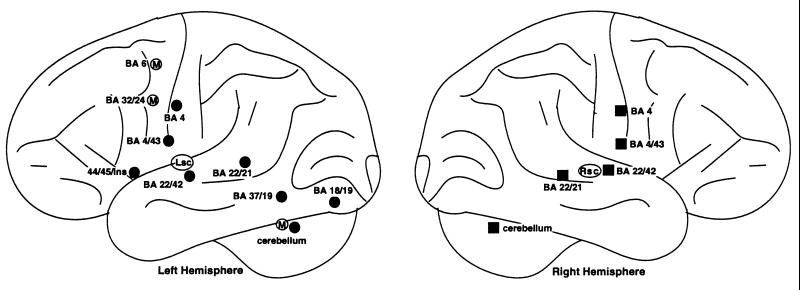
This review discusses how neuroimaging can contribute to our understanding of a fundamental aspect of skilled reading: the ability to pronounce a visually presented word. One contribution of neuroimaging is that it provides a tool for localizing brain regions that are active during word reading. To assess the extent to which similar results are obtained across studies, a quantitative review of nine neuroimaging investigations of word reading was conducted. Across these studies, the results converge to reveal a set of areas active during word reading, including left-lateralized regions in occipital and occipitotemporal cortex, the left frontal operculum, bilateral regions within the cerebellum, primary motor cortex, and the superior and middle temporal cortex, and medial regions in the supplementary motor area and anterior cingulate. Beyond localization, the challenge is to use neuroimaging as a tool for understanding how reading is accomplished. Central to this challenge will be the integration of neuroimaging results with information from other methodologies. To illustrate this point, this review will highlight the importance of spelling-to-sound consistency in the transformation from orthographic (word form) to phonological (word sound) representations, and then explore results from three neuroimaging studies in which the spelling-to-sound consistency of the stimuli was deliberately varied. Emphasis is placed on the pattern of activation observed within the left frontal cortex, because the results provide an example of the issues and benefits involved in relating neuroimaging results to behavioral results in normal and brain damaged subjects, and to theoretical models of reading.
Figures
References
-
- Geschwind N. Sci Am. 1972;226:76–83. - PubMed
-
- Price C J, Moore C J, Frackowiak R S J. Neuroimage. 1996;3:40–52. - PubMed
-
- Price C J, Wise R J S, Warburton E A, Moore C J, Howard D, Patterson K, Frackowiak R S J, Friston K J. Brain. 1996;119:919–931. - PubMed
-
- Rumsey J M, Horwitz B, Donohue C, Nace K, Maisog J M, Andreason P. Brain. 1997;120:739–759. - PubMed
-
- Petersen S E, Fox P T, Posner M I, Mintun M, Raichle M E. J Cognit Neurosci. 1989;1:153–170. - PubMed
Publication types
MeSH terms
Grants and funding
LinkOut - more resources
Full Text Sources
