

Structure of the elongating ribosome: arrangement of the two tRNAs before and after translocation
- PMID: 9448265
- PMCID: PMC18634
- DOI: 10.1073/pnas.95.3.945
Structure of the elongating ribosome: arrangement of the two tRNAs before and after translocation
Abstract
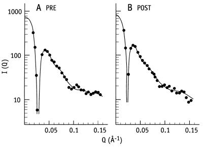
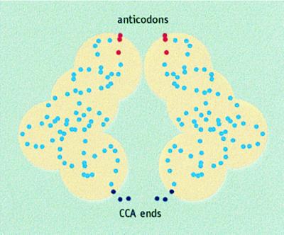
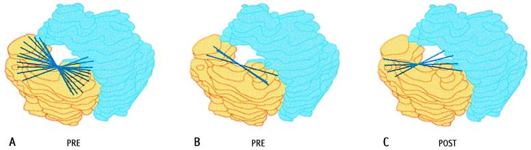
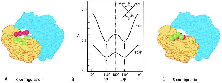
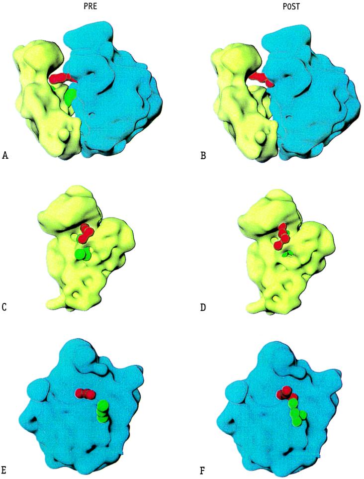
The ribosome uses tRNAs to translate the genetic information into the amino acid sequence of proteins. The mass ratio of a tRNA to the ribosome is in the order of 1:100; because of this unfavorable value it was not possible until now to determine the location of tRNAs within the ribosome by neutron-scattering techniques. However, the new technique of proton-spin contrast-variation improves the signal-to-noise ratio by more than one order of magnitude, thus enabling the direct determination of protonated tRNAs within a deuterated ribosome for the first time. Here we analyze a pair of ribosomal complexes being either in the pre- or post-translocational states that represent the main states of the elongating ribosome. Both complexes were derived from one preparation. The orientation of both tRNAs within the ribosome and their mutual arrangement are determined by using an electron microscopy model for the Escherichia coli ribosome and the tRNA structure. The mass center of gravity of the (tRNA)2mRNA complex moves within the ribosome by 12 +/- 4 A in the course of translocation as previously reported. The main results of the present analysis are that the mutual arrangement of the two tRNAs does not change on translocation and that the angle between them is, depending on the model used, 110 degrees +/- 10 degrees before and after translocation. The translocational movement of the constant tRNA complex within the ribosome can be described as a displacement toward the head of the 30S subunit combined with a rotational movement by about 18 degrees.
Figures
Similar articles
-
The elongating ribosome: structural and functional aspects.Biochem Cell Biol. 1995 Nov-Dec;73(11-12):1011-21. doi: 10.1139/o95-108. Biochem Cell Biol. 1995. PMID: 8722016 Review.
-
Direct localization of the tRNAs within the elongating ribosome by means of neutron scattering (proton-spin contrast-variation).J Mol Biol. 1997 Feb 21;266(2):343-56. doi: 10.1006/jmbi.1996.0788. J Mol Biol. 1997. PMID: 9047368
-
Structure of ratcheted ribosomes with tRNAs in hybrid states.Proc Natl Acad Sci U S A. 2008 Nov 4;105(44):16924-7. doi: 10.1073/pnas.0809587105. Epub 2008 Oct 29. Proc Natl Acad Sci U S A. 2008. PMID: 18971332 Free PMC article.
-
The ribosomal elongation cycle and the movement of tRNAs across the ribosome.Prog Nucleic Acid Res Mol Biol. 1998;59:177-204. doi: 10.1016/s0079-6603(08)61032-6. Prog Nucleic Acid Res Mol Biol. 1998. PMID: 9427843 Review.
-
Visualization of two transfer RNAs trapped in transit during elongation factor G-mediated translocation.Proc Natl Acad Sci U S A. 2013 Dec 24;110(52):20964-9. doi: 10.1073/pnas.1320387110. Epub 2013 Dec 9. Proc Natl Acad Sci U S A. 2013. PMID: 24324168 Free PMC article.
Cited by
-
Visualization of tRNA movements on the Escherichia coli 70S ribosome during the elongation cycle.J Cell Biol. 2000 Aug 7;150(3):447-60. doi: 10.1083/jcb.150.3.447. J Cell Biol. 2000. PMID: 10931859 Free PMC article.
-
Calculation of the relative geometry of tRNAs in the ribosome from directed hydroxyl-radical probing data.RNA. 2000 Feb;6(2):220-32. doi: 10.1017/s1355838200992112. RNA. 2000. PMID: 10688361 Free PMC article.
-
Design of typical genes for heterologous gene expression.Sci Rep. 2022 Jun 10;12(1):9625. doi: 10.1038/s41598-022-13089-1. Sci Rep. 2022. PMID: 35688911 Free PMC article.
-
Neutrons for biologists: a beginner's guide, or why you should consider using neutrons.J R Soc Interface. 2009 Oct 6;6 Suppl 5(Suppl 5):S567-73. doi: 10.1098/rsif.2009.0156.focus. Epub 2009 Aug 5. J R Soc Interface. 2009. PMID: 19656821 Free PMC article. Review.
-
Universal pattern and diverse strengths of successive synonymous codon bias in three domains of life, particularly among prokaryotic genomes.DNA Res. 2012 Dec;19(6):477-85. doi: 10.1093/dnares/dss027. Epub 2012 Nov 6. DNA Res. 2012. PMID: 23132389 Free PMC article.
References
-
- Matheson, A. T., Davies, J. A., Dennis, P. P. & Hill, W. E., eds. (1995) Biochem. Cell Biol. 73, 739–1227.
-
- Wower J, Sylvers L A, Rosen K V, Hixson S S, Zimmermann R A. In: The Translational Apparatus-Structure, Function, Regulation, Evolution. Nierhaus K H, Franceschi F, Subramanian A, Erdmann V, Wittmann-Liebold B, editors. New York: Plenum; 1993. pp. 455–464.
-
- Brimacombe R. Eur J Biochem. 1995;230:365–383. - PubMed
-
- Agrawal R K, Penczek P, Grassucci R A, Li Y, Leith A, Nierhaus K H, Frank J. Science. 1996;271:1000–1002. - PubMed
Publication types
MeSH terms
Substances
LinkOut - more resources
Full Text Sources
