. 1998 Feb 3;95(3):963-8.
doi: 10.1073/pnas.95.3.963.

Nlk is a murine protein kinase related to Erk/MAP kinases and localized in the nucleus

Affiliations

Nlk is a murine protein kinase related to Erk/MAP kinases and localized in the nucleus

B K Brott et al. Proc Natl Acad Sci U S A. .

Abstract

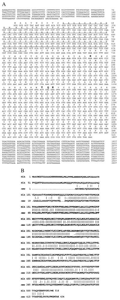
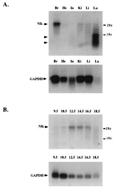
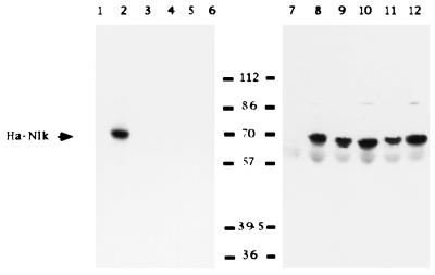
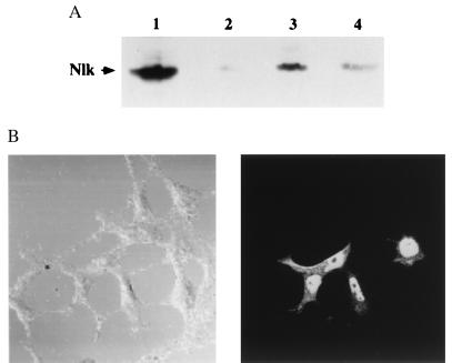
Extracellular-signal regulated kinases/microtubule-associated protein kinases (Erk/MAPKs) and cyclin-directed kinases (Cdks) are key regulators of many aspects of cell growth and division, as well as apoptosis. We have cloned a kinase, Nlk, that is a murine homolog of the Drosophila nemo (nmo) gene. The Nlk amino acid sequence is 54. 5% similar and 41.7% identical to murine Erk-2, and 49.6% similar and 38.4% identical to human Cdc2. It possesses an extended amino-terminal domain that is very rich in glutamine, alanine, proline, and histidine. This region bears similarity to repetitive regions found in many transcription factors. Nlk is expressed as a 4. 0-kb transcript at high levels in adult mouse brain tissue, with low levels in other tissues examined, including lung, where two smaller transcripts of 1.0 and 1.5 kb are expressed as well. A 4.0-kb Nlk message is also present during embryogenesis, detectable at day E10. 5, reaching maximal steady state levels at day E12.5, and then decreasing. Nlk transiently expressed in COS7 cells is a 60-kDa kinase detectable by its ability to autophosphorylate. Mutation of the ATP-binding Lys-155 to methionine abolishes its ability to autophosphorylate, as does mutation of a putative activating threonine in kinase domain VIII, to valine, aspartic, or glutamic acid. Subcellular fractionation indicates that 60-70% of Nlk is localized to the nucleus, whereas 30-40% of Nlk is cytoplasmic. Immunofluorescence microscopy confirms that Nlk resides predominantly in the nucleus. Nlk and Nmo may be the first members of a family of kinases with homology to both Erk/MAPKs and Cdks.


Figures

Figure 1
Figure 1
Nlk sequence and comparison to nmo. (A) Complete nucleotide and amino acid sequence of Nlk. Highlighted are Lys-155, the putative ATP-binding site; and Thr-286, Gln-287, and Glu-288, a putative phosphorylation/regulatory site. A glutamine, alanine, histidine, and proline-rich N-terminal region is underlined. (B) Comparison of the murine Nlk and the Drosophila nmo amino acid sequences. Lines between the two sequences indicate sequence identity. Double dots indicate conserved amino-acid residues, single dots indicate semiconserved residues.
Figure 2
Figure 2
Nlk expression in adult and embryonic tissue. (A) Polyadenylated RNA isolated from adult mouse brain (Br), heart (He), intestine (In), kidney (Ki), liver (Li), and lung (Lu), was probed with the entire Nlk cDNA (Upper), or rat GAPDH (Lower). 28S and 18S ribosomal RNAs were used as markers. (B) Polyadenylated RNA isolated from entire mouse embryos, from days E9.5–E18.5, was electrophoresed, transferred to membranes, and probed with the entire Nlk cDNA (Upper), or rat GAPDH (Lower). 28S and 18S ribosomal RNAs were used as markers.
Figure 3
Figure 3
Nlk is a 60 kDa kinase that autophosphorylates strongly. HA-tagged wild-type Nlk and Nlk mutants KM, TV, TE, and TD, were transiently expressed in COS7 cells and harvested after 48 hr, lysed and immunoprecipitated, and an in vitro kinase autophosphorylation assay performed. After electrophoresis, samples were subjected to autoradiography (lanes 1–6), and then analyzed by Western blot with anti-HA antibody (lanes 7–12). Lanes 1 and 7, mock transfection; lanes 2 and 8, wild-type HA-Nlk; lanes 3 and 9, HA-Nlk(KM); lanes 4 and 10, HA-Nlk(TV); lanes 5 and 11, HA-Nlk(TE); and lanes 6 and 12, HA-Nlk(TD). Molecular mass standards (in kDa) are indicated.
Figure 4
Figure 4
Nlk localizes primarily to the nucleus. (A) COS7 cells transiently expressing wild-type HA-Nlk were harvested after 48 hr and fractionated into membrane (lane 2), nuclear (lane 3), and cytosolic (lane 4) fractions, and electrophoresed. Whole cell lysate was also electrophoresed (lane 1). After transfer to membranes, fractions were Western blotted with anti-HA antibody. (B) HEK293 cells transiently expressing wild-type HA-Nlk were permeabilized and then probed with anti-HA antibody, followed by anti-mouse-fluorescein isothiocyanate. Cells on coverslips were visualized by Nomarski optics (Left), or by fluorescence microscopy (Right).

References

    1. Songyang Z, Lu K P, Kwon Y T, Tsai L-H, Filhol O, Cochet C, Brickey D A, Soderling T R, Bartleson C, Graves D J, DeMaggio A J, Hoekstra M F, Blenis J, Hunter T, Cantley L C. Mol Cell Biol. 1996;16:6486–6493. - PMC - PubMed
    1. Songyang Z, Blechner S, Hoagland N, Hoekstra M F, Piwnica-Worms H, Cantley L C. Curr Biol. 1994;4:973–982. - PubMed
    1. Hanks S K, Quinn A M, Hunter T. Science. 1988;241:42–51. - PubMed
    1. Boulton T G, Nye S H, Robbins D J, Ip N Y, Radziejewska E, Morgenbesser S D, DePinho R A, Panayotatos N, Cobb M H, Yancopoulos G D. Cell. 1991;65:663–675. - PubMed
    1. Davis R J. Trends Biochem Sci. 1994;19:470–473. - PubMed

Publication types

MeSH terms

Substances

Associated data