. 1998 Feb 3;95(3):1010-3.
doi: 10.1073/pnas.95.3.1010.

Mammalian DNA topoisomerase IIIalpha is essential in early embryogenesis

Affiliations

Mammalian DNA topoisomerase IIIalpha is essential in early embryogenesis

W Li et al. Proc Natl Acad Sci U S A. .

Abstract

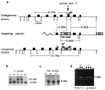
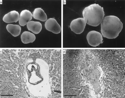
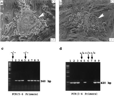
Targeted disruption of the mouse TOP3alpha gene encoding DNA topoisomerase IIIalpha was carried out to study the physiological functions of the mammalian type IA DNA topoisomerase. Whereas heterozygous top3alpha+/- mutant mice were found to resemble phenotypically their TOP3alpha+/+ litermates, no viable top3alpha-/- homozygotes were found among over 100 progeny of top3alpha+/- intercrosses. Examination of embryos dissected from decidual swellings and in vitro culturing of blastocysts from top3alpha+/- intercrosses showed that implantation of top3alpha-/- embryos and the induction of decidualization could occur, but viability of these embryos was severely compromised at an early stage of development. The requirement of mouse DNA topoisomerase IIIalpha during early embryogenesis is discussed in terms of its plausible role in chromosome replication and its interaction with the RecQ/SGS1 family of DNA helicases, whose members include the Bloom's syndrome and the Werner's syndrome gene products.


Figures

Figure 1
Figure 1
Targeted disruption of mouse TOP3α gene. (a) Schematics of the region of mouse TOP3α containing the active site tyrosine (Top), the targeting vector (Middle), and the Δtop3α allele after gene disruption (Bottom). Filled boxes represent exons. K, S, E, and A denote KpnI, SspI, EcoRI, and ApaI restriction sites, respectively, and boxes denoted neo and TK represent the neomycin and herpes simplex virus thymidine kinase markers used. The locations of the probes used in blot hybridization (5′ probe and neo probe) and primers used in PCR (arrows numbered 1–6) also are indicated. The nucleotide sequences of primers 1–4 are, respectively, 5′-CGCGGAAAAGCTCTATACACAA-3′, 5′-TGTAGCGCCAAGTGCCAGCGGGG-3′, 5′-CGTGTTCCGGCTGTCAGCGCA-3′, 5′-ATCGCCATGGGTCACGACGAGAT-3′. Primers 5 and 6 are the same as the YPRT and the FSES primer described in Materials and Methods. (b and c) Genotyping of tail biopsies by blot hybridization. (b) KpnI digests were fractionated by agarose gel electrophoresis, and blot hybridization was carried out by using the 5′ probe. (c) EcoRI digests and the neo probe were used. The sizes of the fragments detected are in agreement with those expected from the drawings in a. (d) Genotyping of tail biopsies by PCR, using the pair of primers 1 and 2. The presence of the 1.6-kb product signified the presence of at least one copy of the mutant allele. The leftmost lane contained the “1-Kb ladder” size markers (GIBCO/BRL).
Figure 2
Figure 2
(a and b) Decidual swellings from intercrosses of top3α+/− mice 8.5 dpc (a) and 10.5 dpc (b). (c and d) Histological sections of 7.5-dpc deciduae from intercrosses of top3α+/− mice. Whereas these decidual swellings showed a uniform external appearance, two distinct types were revealed by sectioning: the majority contained embryos of normal appearance, such as the one shown in c, and the remaining were devoid of embryonic material, such as the one shown in d. Embryos of normal appearance were invariably found by genotyping to possess at least one copy of unaltered TOP3α. The bars correspond to a length of 100 μm.
Figure 3
Figure 3
Culturing of a top3α+/− (a) and a top3α−/− (b) blastocyst on mouse embryonic fibroblast feeder layer cells. Embryos were collected 3.5 dpc and cultured for 4 days in vitro and photographed. The heterozygote showed normal trophetoderm outgrowth and inner cell mass proliferation, and the homozygote showed poor trophoblast outgrowth and a rather small inner cell mass (indicated by an arrowhead in b). The bars correspond to a length of 100 μm. (c and d) Genotyping of nine embryos cultured in vitro, using the primers specified in Fig. 1 (a). The leftmost lane of each gel slab contained the 1-Kb ladder size markers (GIBCO/BRL). The embryos shown in a and b corresponded to samples 9 and 5, respectively.

References

    1. Ellis N A, Groden J, Ye T Z, Straughen J, Lennon D J, Ciocci S, Proytcheva M, German J. Cell. 1995;83:655–666. - PubMed
    1. Yu C-E, Oshima J, Fu Y H, Wijsman E M, Hisama F, Alisch R, Mathews S, Nakura J, Miki T, Ouais S, Martin G M, Mulligan J, Schellenberg G D. Science. 1996;272:258–262. - PubMed
    1. Fritz E, Elsea S H, Patel P I, Meyn M S. Proc Natl Acad Sci USA. 1997;94:4538–4542. - PMC - PubMed
    1. Wang J C. Annu Rev Biochem. 1996;65:635–692. - PubMed
    1. Gangloff S, McDonald J P, Bendixen C, Arthur L, Rothstein R. Mol Cell Biol. 1994;14:8391–8398. - PMC - PubMed

Publication types

Substances

LinkOut - more resources