. 1998 Feb 3;95(3):1097-101.
doi: 10.1073/pnas.95.3.1097.

Shifts in the life history of parasitic wasps correlate with pronounced alterations in early development

Affiliations

Shifts in the life history of parasitic wasps correlate with pronounced alterations in early development

M Grbić et al. Proc Natl Acad Sci U S A. .

Abstract

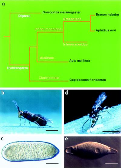
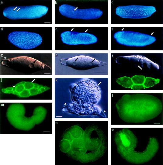
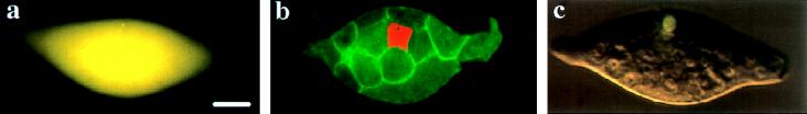
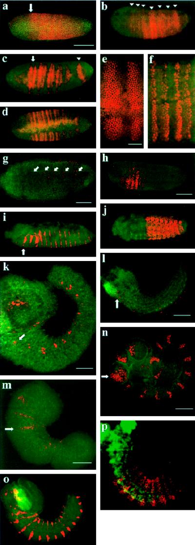
Developmental processes have been traditionally viewed to be invariant within higher taxa. However, examples are known whereby closely related species exhibit alterations in early embryogenesis yet appear very similar as adults. Such developmental changes are thought to occur in response to shifts in life history. In insects, the regulation of embryonic development has been intensively studied in model species like Drosophila melanogaster. Previous comparative studies suggest that the developmental processes documented in Drosophila well describe embryogenesis of advanced, holometabolous, insects generally. There have been few attempts, however, to take into account how life history has influenced early development of insects or to characterize early development of species with life histories fundamentally different from flies. Here we compared early development of two species from the same family of parasitic wasps that exhibit very different life histories. Bracon hebetor is an ectoparasite that lays large, yolky eggs on the integument of its host that develop much like the free-living honeybee and Drosophila. In contrast, Aphidius ervi is an endoparasite that lays small and apparently yolk-free eggs that develop in the hemocoel of the host. This wasp exhibits a radically different mode of early development at both the cellular and molecular level from B. hebetor. The developmental changes in A. ervi reflect functional adaptations for its derived life history and argue that departures from the fly paradigm may occur commonly among insects whose eggs develop under conditions different from typical terrestrial species.


Figures

Figure 1
Figure 1
Phylogeny and life history of Bracon hebetor, Aphidius ervi, and selected other hymenopterans (–17). (a) Both B. hebetor and A. ervi are in the family Braconidae (superfamily Ichneumonoidea). Almost all ichneumonoids develop as either ecto- or endoparasites of other arthropods. The sister group to the Ichneumonoidea is the Aculeata that includes the honeybee, Apis mellifera. The other parasitic wasp in the phylogeny, Copidosoma floridanum, is the only other endoparasite whose embryonic development has been studied at the cellular and molecular level (20, 34). This wasp is in the family Encyrtidae (superfamily Chalcidoidea) and is more distantly related to A. ervi than B. hebetor and A. mellifera. Despite this, its eggs are devoid of yolk and early development proceeds in a cellularized environment like A. ervi. Although not completely resolved, the sister group to the Hymenoptera is thought to be the Mecopteroidea that includes the order Diptera and the model species Drosophila melanogaster. (b) B. hebetor: this wasp is a 3 mm long ectoparasite that lays its eggs on the integument of moth larvae such as Plodia interpunctella. (c) A close-up view of a B. hebetor egg. Like eggs of other terrestrial flies and wasps (10, 11), B. hebetor eggs are large (0.5 mm), yolk-rich, surrounded by a thick chorion, and elongated along their anterior-posterior axis. After hatching, B. hebetor larvae develop by rasping a hole through the host’s integument and feeding on its tissues. (d) A. ervi: this wasp is a 1–2 mm long endoparasite that lays a single egg into the haemocoel of its aphid host, Acyrthosiphon pisum. (e) A close up view of an A. ervi egg. The egg is small (0.05 mm), yolkless, and is surrounded by a thin chorion. The A. ervi larva feeds inside the host and emerges as an adult by chewing a hole through the cuticle of the host. Scale bars = 1 mm (b, d); 100 μm (c); 15 μm (e).
Figure 2
Figure 2
Embryogenesis of B. hebetor and A. ervi. Confocal, fluorescent, and Nomarski images of embryonic development. (a) After oviposition the B. hebetor egg has a clear polarity corresponding to the dorsal-ventral and anterior-posterior embryonic axes. Embryonic nuclei (arrows) divide without cytokinesis. (b, c) During the first few syncytial cleavages nuclei remain in the yolk (arrow). (d) After the tenth cleavage nuclei migrate to the periphery of the egg where they undergo two additional division cycles in the syncytium before finally forming a cellular blastoderm (39). (e) The embryo then undergoes germband extension (anterior and posterior limits of the embryo marked by the arrows in e and f). (f) This is followed by germband retraction and segmentation. (g) After oviposition, the A. ervi egg is lemon-shaped and does not exhibit any axial polarity (nucleus marked by an arrow(s) in g and h and chorion by arrowhead). (h) The first nuclear division proceeds in a syncytium, without cytoplasmic cleavage. (i) The second cleavage results in formation of four nuclei that become separated by cell membranes (Upper, a single focal plane with two blastomeres, the cell membrane is marked by an arrow; Lower, the same stage embryo, phalloidin staining demarcates the cell cortex underlying the cell membranes in all four blastomeres). (j) The embryo undergoes cleavage to form large (which form the future extraembryonic membrane, arrow) and small blastomeres (which form the embryo proper, arrowhead). (k) The extraembryonic membrane surrounds the embryonic cells (arrow) and the embryo ruptures from the chorion (arrowhead). (l–o) The embryonic primordium remains surrounded by the extraembryonic membrane and initiates morphogenesis in the host’s haemocoel. (l) Embryonic primordium. (m) The embryo undergoes germband extension by posterior growth. (n) Fully extended germband stage whereby the embryo assumes a coiled shape. (o) This is followed by condensation and segmentation of the embryo. (l–o) The extraembryonic membrane was removed. Bars = 80 μm (a–f and m–o); 7 μm (g–k); 50 μm (l).
Figure 3
Figure 3
Fluorescent and confocal images of A. ervi blastomere injections. (a) Fluorescent image of an embryo injected at the four-cell stage. The tracer dye spread throughout the embryo (yellow) (n = 28). (b) Confocal image of a blastomere injected at the 16-cell stage (n = 36). Injected cell (red), cortical actin (green). (c) Fluorescent image of an embryonic cell injected at the putative 64-cell stage (n = 42). The dye remains confined to the injected cell (yellow). Bar = 10 μm (a–c).
Figure 4
Figure 4
Protein expression patterns of Eve, En, and Ubx/Abd-A in B. hebetor and A. ervi embryos. Confocal images of embryos stained with the anti-Eve antibody mAb2B813 (21), anti-En antibody EN4F11 (22), and anti-Ubx/Abd-A antibody mAbFP6.87 (23) (red) and counterstained with phalloidin (green). (ag) Eve expression in B. hebetor. (a) Eve expression in the syncytial blastoderm (arrow demarcates anterior boundary). (b) Eve expression resolved into a pair-rule pattern in the syncytial blastoderm (arrowheads). (c) Individual pair-rule stripes begin to split into two stripes in a brief anterior to posterior progression (arrow marks split of the first pair-rule stripe and an arrowhead last, undivided, stripe). (d) Complete split of pair-rule stripes results in segmental iteration of the stripes (ventral up). (e, f) High magnification of pair-rule and segmental Eve expression. (e) Pair-rule stripes are eight nuclei wide. (f) Antigen fades in the central nuclei and continues to be expressed in two rows of peripheral nuclei. (g) Eve expression in neurons in a bilateral pattern along the ventral midline (arrows, ventral up). (h, i) En expression pattern in B. hebetor. (h) Formation of En stripes in a brief anteroposterior progression. (i) Mature En pattern (arrow marks mandibular stripe in the posterior segment compartment). (j) Ubx/Abd-A expression in the posterior thorax and abdomen of B. hebetor. (k–l) Eve expression in A. ervi. Eve protein is not expressed prior to germband condensation. (k) Expression of Eve in the dorsolateral mesoderm (arrow marks the most anterior group of dorsal cells expressing Eve). (l) Expression of Eve in neurons along the ventral midline (arrow marks anterior neuroblasts expressing Eve). (m, n) Formation of En stripes in A. ervi. (m) Initial expression of En in gnathal segments (arrow marks labial stripe). (n) En stripes formed sequentially as the germband extends (arrow marks labial stripe). (o) Mature En pattern demarcating the posterior segmental compartment. (p) Mature pattern of Ubx/Abd-A from the posterior thorax to the penultimate abdominal segment. In all panels, except d and g, anterior is on the left and dorsal is up. Scale bars: a–d, 110 μm; e and f, 16 μm; g, 60 μm; h–j, 80 μm; k, 60 μm; l, 100 μm; m, 80 μm; n–p, 100 μm.

References

    1. von Baer K E. Entwicklungsgeschichte der Thiere: Beobachtung und Reflexion. Konigsberg, Germany: Born-trager; 1828.
    1. Gould S J. Ontogeny and Phylogeny. Cambridge, MA: Belknap; 1977.
    1. Wray G A. Science. 1995;267:115–116. - PubMed
    1. del Pino E M, Ellinson R P. Nature (London) 1983;306:589–591.
    1. Scott L B, Lennarz W J, Raff R A, Wray G A. Dev Biol. 1990;138:188–193. - PubMed

Publication types

LinkOut - more resources