. 1998 Feb 3;95(3):1120-5.
doi: 10.1073/pnas.95.3.1120.

Structure-function analysis of integrase interactor 1/hSNF5L1 reveals differential properties of two repeat motifs present in the highly conserved region

Affiliations

Structure-function analysis of integrase interactor 1/hSNF5L1 reveals differential properties of two repeat motifs present in the highly conserved region

A Morozov et al. Proc Natl Acad Sci U S A. .

Abstract

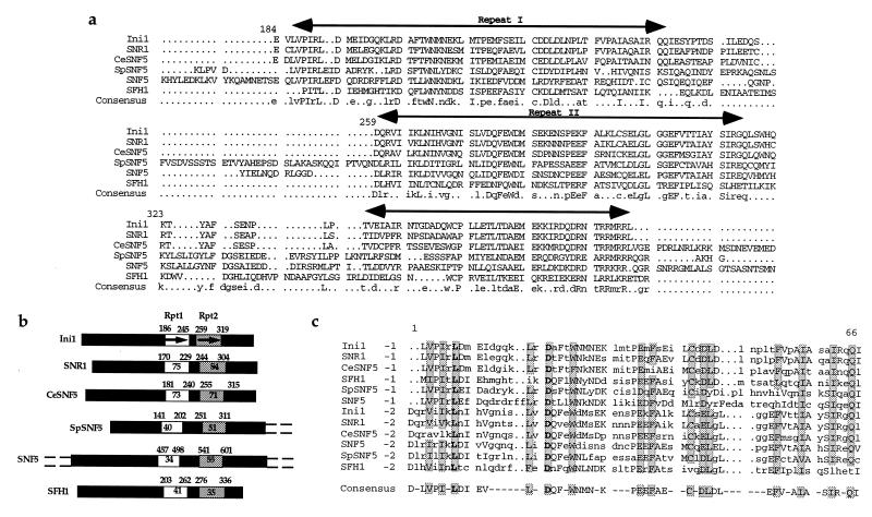
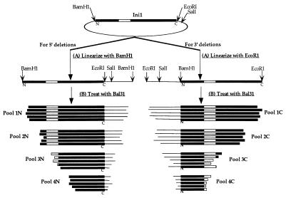
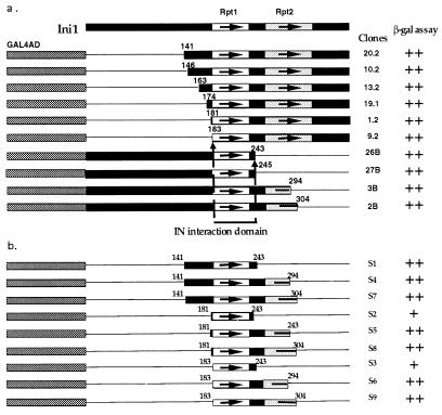
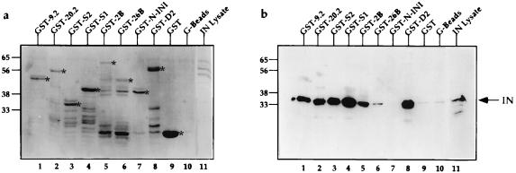
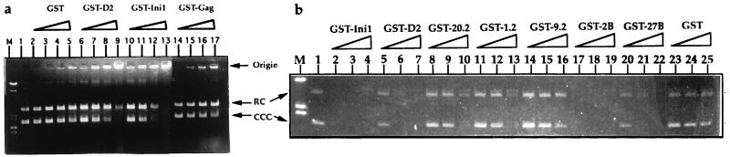
Retroviral integrase (IN) catalyzes the integration of retroviral cDNA into host chromosome. Ini1 (integrase interactor 1) is a host protein that specifically binds and stimulates in vitro joining activity of HIV-1 IN. Ini1 has homology to yeast transcription factor SNF5 and is a component of the analogous mammalian SWI/SNF complex that can remodel chromatin. Little is known about the function of Ini1 in mammalian cells. To gain insight into the functional domains of Ini1, and to understand the details of protein-protein interactions of IN and Ini1, a structure-function analysis of Ini1 was initiated. By means of the yeast two-hybrid system, the minimal IN binding domain of Ini1 was characterized. One of the two repeat motifs present in the highly conserved regions of Ini1 was found necessary and sufficient to bind to IN in yeast as well as in vitro. Because IN binds to only one of the two repeat motifs in this conserved region of Ini1, it appears that the IN-Ini1 interaction is very specific and functionally significant. Characterization of DNA-binding properties of Ini1 revealed that Ini1 can bind to plasmid DNA, binding more readily to supercoiled DNA than to the relaxed circular DNA. The minimal domain for DNA binding was localized to a region upstream of repeat 1. The DNA binding activity of Ini1 is not required for its ability to interact with IN. The finding that the two repeat motifs of Ini1 display differential binding to HIV-1 IN and that this discrete component of mammalian SWI/SNF complex binds to DNA will help understand the role of Ini1 in HIV-1 integration and in cellular process.


Figures

Figure 1
Figure 1
Alignment of proteins/ORFs with homology to Ini1 protein. (a) Alignment of highly conserved regions of six protein/ORFs, Drosophila protein SNR1, C. elegans ORF R07E5.3 (CeSNF5 protein), S. pombe ORF encoding C2F7.08C protein (SpSNF5), and the two yeast proteins, SNF5 and SFH1. The alignment was performed by using the program multialign. The three highly conserved regions are indicated by horizontal arrows. Consensus sequence is indicated at the bottom. The first amino acid residue of Ini1 in each lane is numbered. (b) Graphical representation of six Ini1 homologues displaying the two repeat motifs. The name of the protein is indicated on the left. The two repeats are indicated by thick arrows, and the highly conserved regions that contain the two repeats are indicated by boxes. Empty box, repeat 1 (Rpt1) and shaded box, repeat 2 (Rpt2). The numbers at the beginnings and ends of the boxes represent the position of the amino acid residue at each edge of the region. Percentage identity of primary sequence between Ini1 and a given protein within the particular repeat is indicated inside the boxed area. The N-terminal and C-terminal glutanine- and proline-rich regions of SNF5 and SpSNF5 are indicated by dashed lines. (c) Alignment of the sequence of two repeat motifs with each other and their consensus sequence. The repeats, 1 and 2 of six Ini1-related proteins, were aligned by using pileup program in GCG and displayed by using the pretty program. Consensus sequences of the repeat motif, determined by using the plurality of 6 of 12 identical or conserved residues, is indicated at the bottom. The invariant residues are indicated by bold letter. Highly conserved and invariant residues are indicated by shading.
Figure 2
Figure 2
Schematic representation of the strategy for generating 5′ and 3′ deletion pools of INI1 cDNA. The plasmid carrying the INI1 cDNA was first linearized with either BamHI or EcoRI and subsequently treated with Bal31 to generate different pools of DNA fragments carrying increasing extent of deletions (pools 1N-4N and 1C-4C). The deleted pools of cDNA fragments subsequently were cloned into pGADNot for expressing GAL4AC fusion proteins in yeast.
Figure 3
Figure 3
Interaction of HIV-1 IN with Ini1 truncations in the two-hybrid system. (a) Various truncations of Ini1 that retained the ability to interact with IN in the two-hybrid system. (b) Repeat 1 of Ini1 is sufficient for the interaction with IN. The top bar represents the full-length Ini1. The two highly conserved regions containing the two repeats are represented by open and shadowed boxes with arrows. Ini1 deletions obtained in the two-hybrid analysis are indicated as fusion to GAL4AD below the top bar. The thin lines represent the region of the protein deleted. The numbers on the right of deletions represent the clone number. The amino acid residue at the junction of both N- and C-terminal deletions are represented for each clone. The two shortest clones (9.2, and 26B) define the N- and C-terminal borders of IN-interaction domain. Relative strengths of interaction of Ini1 mutants with IN is indicated by ++, strong blue, and +, light blue.
Figure 4
Figure 4
In vitro binding assay of GST-Ini1 truncations to HIV-1 IN. (a) Coomassie-stained PAGE of bound proteins. (b) Western analysis using monoclonal anti-IN antibodies. Lanes are identical in a and b. Numbers on the left represent the position of molecular weight markers in kDa. GST-fusion proteins used for binding experiment is indicated on top of each lane. G beads, glutathione agarose beads not bound to any proteins. IN lysate in lane 11 represents the bacterial lysate expressing IN, before binding experiment. The bands of the expected size length GST or GST-fusion proteins are indicated by ∗ on the right side of each band.
Figure 5
Figure 5
DNA binding activity of Ini1. Ethidium bromide-stained gel of electrophoresis of plasmid DNA incubated with GST or GST-fusion proteins. (a) DNA binding was carried out by incubating 200 ng of plasmid pBluescript with increasing concentrations of GST or GST-fusion proteins in a 20-μl reaction. After the incubation, the plasmid DNAs were separated on agarose gels and stained with ethidium bromide. Lane M, lambda HindIII markers. Lane 1, pBluescript SK plasmid DNA. Plasmid DNA incubated with 5, 10, 20, and 40 pMols of GST (lanes 2–5); 1.4, 2.8, 5.6, and 11.2 pMols of GST-D2 (lanes 6–9); 0.4, 0.8, 1.6, and 3.2 pMols of GST-Ini1 (lanes 10–13); or GST-gag (lanes 14–17). Origin, wells in the agarose gel indicating the retention of protein–DNA complexes. RC, relaxed circle DNA. CCC, covalently closed circular DNA. (b) Deletion analysis to determine the DNA binding domain of Ini1. DNA binding assay was carried out as before with uniformly increasing concentrations (1.6, 3.2, and 6.4 pMols respectively) of all the GST and GST-fusion proteins and using 100 ng of pBluescript DNA. Lane M, lambda HindIII marker. Lane 1, pBluescript SK DNA. Lanes 2–25, plasmid DNA incubated with increasing concentrations of GST-Ini1 (lanes 2–4) GST-D2 (lanes 5–7), GST-20.2 (lanes 8–10), GST-1.2 (lanes 11–13), GST-9.2 (lanes 14–16), GST-2B (lanes 17–19), GST-27B (lanes 20–22), or GST (lanes 23–25) proteins as indicated on top of the gel. RC, relaxed circular DNA. CCC. covalently closed circular DNA.

References

    1. Goff S P. Annu Rev Genet. 1992;26:527–544. - PubMed
    1. Kulkosky J, Skalka A M. Pharmacol Ther. 1994;61:185–203. - PubMed
    1. Lieber M. Curr Biol. 1996;6:134–136. - PubMed
    1. Skalka A M. In: Integrative Recombination of Retroviral DNA. Kucherlapati R, Smith G R, editors. Washington, D.C.: Am. Soc. Microbiol. Publ.; 1989. pp. 701–724.
    1. Skalka A M. Gene. 1993;135:175–182. - PubMed

Publication types