

Marrow stromal cells as a source of progenitor cells for nonhematopoietic tissues in transgenic mice with a phenotype of osteogenesis imperfecta
- PMID: 9448299
- PMCID: PMC18700
- DOI: 10.1073/pnas.95.3.1142
Marrow stromal cells as a source of progenitor cells for nonhematopoietic tissues in transgenic mice with a phenotype of osteogenesis imperfecta
Abstract
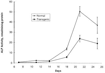
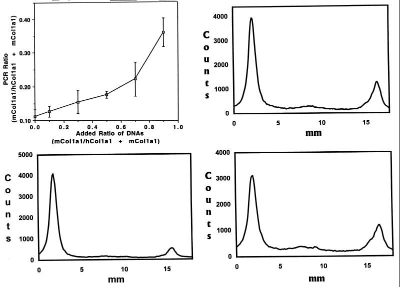
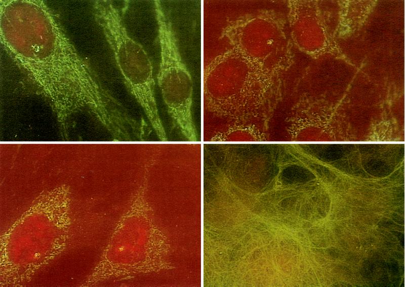
Marrow stromal cells from wild-type mice were infused into transgenic mice that had a phenotype of fragile bones resembling osteogenesis imperfecta because they expressed a human minigene for type I collagen. In mice that were irradiated with potentially lethal levels (700 cGy) or sublethal levels (350 cGy), DNA from the donor marrow stromal cells was detected consistently in marrow, bone, cartilage, and lung either 1 or 2.5 mo after the infusions. The DNA also was detected but less frequently in the spleen, brain, and skin. There was a small but statistically significant increase in both collagen content and mineral content of bone 1 mo after the infusion. Similar results were obtained with infusion of relatively large amounts of wild-type whole marrow cells into the transgenic mice. In experiments in which male marrow stromal cells were infused into a female osteogenesis imperfecta-transgenic mouse, fluorescense in situ hybridization assays for the Y chromosome indicated that, after 2.5 mo, donor male cells accounted for 4-19% of the fibroblasts or fibroblast-like cells obtained in primary cultures of the lung, calvaria, cartilage, long bone, tail, and skin. In a parallel experiment in which whole marrow cells from a male mouse were infused into a female immunodeficient rag-2 mouse, donor male cells accounted for 4-6% of the fibroblasts or fibroblast-like cells in primary cultures. The results support previous suggestions that marrow stromal cells or related cells in marrow serve as a source for continual renewal of cells in a number of nonhematopoietic tissues.
Figures
References
-
- Friedenstein A J, Gorskaja U, Kulagina N N. Exp Hematol (Charlottesville, Va) 1976;4:267–274. - PubMed
-
- Piersma A H, Ploemacher R E, Brockbank K G M. Br J Haematol. 1983;54:285–290. - PubMed
-
- Mardon H J, Bee J, von der Mark K, Owen M E. Cell Tissue Res. 1987;250:157–165. - PubMed
-
- Owen M E, Friedenstein A J. Cell and Molecular Biology of Vertebrate Hard Tissues: Ciba Foundation Symposium, 136. New York: Wiley; 1988. pp. 42–60. - PubMed
-
- Beresford J N, Bennett J H, Devlin C, Leboy P S, Owen M E. J Cell Sci. 1992;102:341–351. - PubMed
MeSH terms
Substances
LinkOut - more resources
Full Text Sources
Other Literature Sources
Medical
