. 1998 Feb 3;95(3):1224-9.
doi: 10.1073/pnas.95.3.1224.

PCR analysis of tissue samples from the 1979 Sverdlovsk anthrax victims: the presence of multiple Bacillus anthracis strains in different victims

Affiliations

PCR analysis of tissue samples from the 1979 Sverdlovsk anthrax victims: the presence of multiple Bacillus anthracis strains in different victims

P J Jackson et al. Proc Natl Acad Sci U S A. .

Abstract

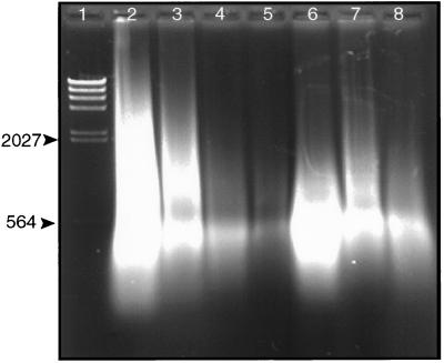
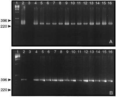
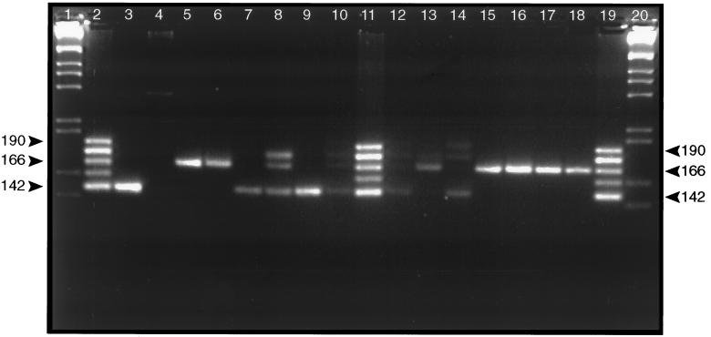
An outbreak of human anthrax occurred in Sverdlovsk, Union of Soviet Socialists Republic (now Ekaterinburg, Russia) in April 1979. Officials attributed this to consumption of contaminated meat, but Western governments believed it resulted from inhalation of spores accidentally released from a nearby military research facility. Tissue samples from 11 victims were obtained and methods of efficiently extracting high-quality total DNA from these samples were developed. Extracted DNA was analyzed by using PCR to determine whether it contained Bacillus anthracis-specific sequences. Double PCR using "nested primers" increased sensitivity of the assay significantly. Tissue samples from 11 persons who died during the epidemic were examined. Results demonstrated that the entire complement of B. anthracis toxin and capsular antigen genes required for pathogenicity were present in tissues from each of these victims. Tissue from a vaccination site contained primarily nucleic acids from a live vaccine, although traces of genes from the infecting organisms were also present. PCR analysis using primers that detect the vrrA gene variable region on the B. anthracis chromosome demonstrated that at least four of the five known strain categories defined by this region were present in the tissue samples. Only one category is found in a single B. anthracis strain.


Figures

Figure 1
Figure 1
Agarose gel electrophoresis of DNA extracted from formalin-fixed paraffin-embedded Sverdlovsk victim tissue samples. Total DNA extracted from tissues was analyzed by electrophoresis through a 1% agarose gel. Electrophoresis was for 1.5 hr at 85 V. DNA was visualized under UV after staining with ethidium bromide. Lanes: l, λ DNA digested with HindIII; 2–8, DNA extracted from 7.RA93.15.15 spleen, 31.RA93.39.3 spleen, 27.RA93.30.3 spleen, 37.RA93.35.4 vaccination site, 37.RA93.35.4 spleen, 37.RA93.35.6 lung, and 26.RA93.043 spleen, respectively. Numbers on the left side of the gel refer to the size of DNA markers in bp.
Figure 2
Figure 2
Agarose gel electrophoresis of PCR amplicons after amplification of capA and lef gene-specific DNA fragments from tissue DNA. Total DNA extracted from tissues was used as template in PCR containing capA-specific (A) or lef-specific (B) primers to amplify a portion of these genes. Products of the first amplification were subsequently used in a second set of reactions containing “nested” PCR primers to further amplify a portion of the targeted DNA fragment. Samples were analyzed by electrophoresis through 3% (wt/vol) agarose gels. Gels were stained with ethidium bromide and DNA was visualized under UV. Lanes: 1, 1 kb DNA ladder marker (Life Technologies); 2–16 (both A and B), results of second PCRs containing B. anthracis (strain Vollum) control DNA, uninfected human DNA control, 7.RA93.15.15 spleen DNA, 31.RA93.39.3 spleen DNA, 26.RA93.043 spleen DNA, 40.RA93.40.5 spleen DNA, 27.RA93.30.3 spleen DNA, 37.RA93.35.4 vaccination site DNA, 37.RA93.35.4 spleen DNA, 37.RA93.35.6 lung DNA, 3.RA93.1.1 meninges DNA, 25.RA93.031 meninges DNA, 1.RA93.42.1 meninges DNA, 33.RA93.20.5 meninges DNA, and 21.RA93.38.4 lymph node DNA. The numbers to the left refer to the size (bp) of marker DNA fragments. The triplet bands in the capA control reaction (A, lane 2) result from incomplete removal of the first primer set prior to running the second “nested PCR.” This results in amplicons of 397, 350, 349, and 342 bp.
Figure 3
Figure 3
VrrA VNTR amplicons produced by PCR of DNA extracted from 11 different Sverdlovsk anthrax victims. PCR amplicons were analyzed by electrophoresis through a 4.5% (wt/vol) Metaphor agarose gel. Electrophoresis was for 5 hr at 70 V. Gels were stained with ethidium bromide, and DNA was visualized under UV. Lanes: 1 and 20, 1 kb DNA ladder marker; 2, 11, and 19, VNTR ladder containing the five different VNTR categories found in B. anthracis (6); 3, results of PCR containing B. anthracis (strain Vollum) control DNA; 4, PCR containing uninfected human control DNA; 5–10, results of PCR containing 7.RA93.15.15 spleen DNA, 31.RA93.39.3 spleen DNA, 26.RA93.043 spleen DNA, 40.RA93.40.5 spleen DNA, 27.RA93.30.3 spleen DNA, and 37.RA93.35.4 vaccination site DNA, respectively; 12–18, results of PCR containing 37.RA93.35.4 spleen DNA, 37.RA93.35.6 lung DNA, 3.RA93.1.1 meninges DNA, 25.RA93.031 meninges DNA, 1.RA93.42.1 meninges DNA, 33.RA93.20.5 meninges DNA, and 21.RA93.38.4 lymph node DNA. Labels on the sides refer to the size of B. anthracis VNTR categories 2, 4, and 6 (142, 166, and 190 bp, respectively) (6).
Figure 4
Figure 4
Agarose gel electrophoresis of the B. anthracis vrrA VNTR amplicons in DNA extracted from formalin-fixed paraffin-embedded primate tissues infected with inhalational anthrax. DNA was extracted from primate tissue samples (8) and used as template in PCR containing primers that amplify the B. anthracis VNTR region. Amplification was as described for the results presented in Fig. 3 using nested primers. Lanes: 1 and 8, 1 kb DNA ladder marker; 2, VNTR ladder containing the five different VNTR categories found in B. anthracis (6); 3, results of amplification using tissue from an uninfected primate; 4–7, results of PCR amplification using template DNA from spleen, lung, liver, and brain of a single primate infected with a single strain of B. anthracis (8). Numbers on the left refer to the size of B. anthracis VNTR categories 2, 4, and 6 (6).

References

    1. Bezdenezhnykh I S, Nikiforov V N. Zh Mikrobiol Epidemiol Immunobiol. 1980;5:111–113. - PubMed
    1. Meselson M, Guillemin J, Hugh-Jones M, Langmuir A, Popova I, Shelokov A, Yampolskaya O. Science. 1994;266:1202–1208. - PubMed
    1. Abramova F A, Grinberg L M, Yampolskaya O V, Walker D H. Proc Natl Acad Sci USA. 1993;90:2291–2294. - PMC - PubMed
    1. Green B D, Battisti L, Koehler T M, Thorne C B, Ivins B E. Infect Immun. 1985;49:291–297. - PMC - PubMed
    1. Uchida I, Hashimoto K, Terakado N. J Gen Microbiol. 1986;132:557–559. - PubMed

Publication types