. 1998 Feb 3;95(3):1307-12.
doi: 10.1073/pnas.95.3.1307.

Activation of the atrial KACh channel by the betagamma subunits of G proteins or intracellular Na+ ions depends on the presence of phosphatidylinositol phosphates

Affiliations

Activation of the atrial KACh channel by the betagamma subunits of G proteins or intracellular Na+ ions depends on the presence of phosphatidylinositol phosphates

J L Sui et al. Proc Natl Acad Sci U S A. .

Abstract

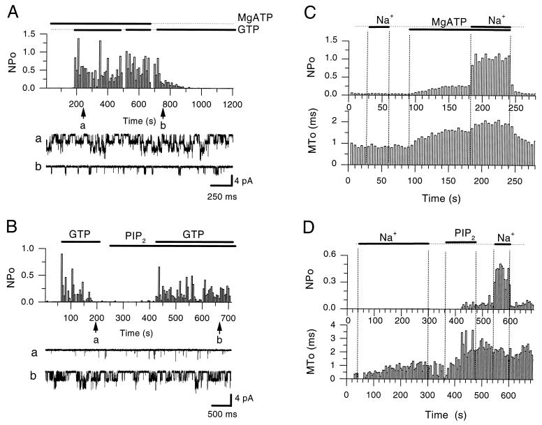
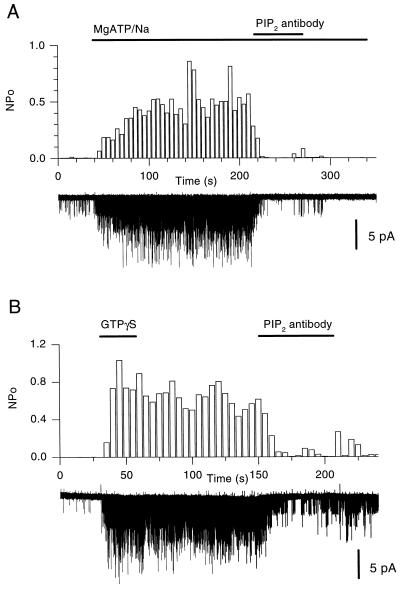
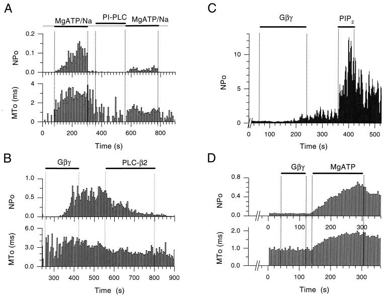
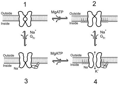
The betagamma subunits of GTP-binding proteins (Gbetagamma) activate the muscarinic K+ channel (KACh) in heart by direct binding to both of its component subunits. KACh channels can also be gated by internal Na+ ions. Both activation mechanisms show dependence on hydrolysis of intracellular ATP. We report that phosphatidylinositol 4,5-bisphosphate (PIP2) mimics the ATP effects and that depletion or block of PIP2 retards the stimulatory effects of Gbetagamma subunits or Na+ ions on channel activity, effects that can be reversed by restoring PIP2. Thus, regulation of KACh channel activity may be crucially dependent on PIP2 and phosphatidylinositol signaling. These striking functional results are in agreement with in vitro biochemical studies on the PIP2 requirement for Gbetagamma stimulation of G protein receptor kinase activity, thus implicating phosphatidylinositol phospholipids as a potential control point for Gbetagamma-mediated signal transduction.


Figures

Figure 1
Figure 1
PIP2 mimics the MgATP dependence of the rundown of G protein-mediated activation as well as channel sensitization to gating by internal Na+ ions. (A) NPo plot of KACh channel activity in an inside-out patch from a chicken embryonic atrial myocyte, showing that in the presence of ATP (2 mM, sodium salt, in the presence of 0.6 mM Mg2+ in the bath solution), GTP (100 μM) produced sustained activity (10 μM acetylcholine was present in the pipette). Withdrawal of ATP caused a gradual rundown of the GTP-induced activity. As can be seen at the beginning of the record ATP perfusion alone did not activate the channel. Application of each nucleotide is indicated by the bars. Representative single-channel currents are shown before and after the rundown of the activity, as indicated at time points a and b during the course of the experiment. (B) Plot similar to that in A, showing that PIP2 restored the ability of GTP to stimulate channel activity and mimicked ATP in its ability to prevent rundown of the GTP-induced channel activity. Acetylcholine at 5 μM was present in the pipette solution. Perfusion of the inside-out patch with GTP (100 μM) and PIP2 (5 μM) in the bath solution is indicated by bars. The arrows labeled a and b show time points during which rundown and restored single channel activity is shown, respectively. PIP2, like ATP, did not stimulate channel activity when perfused alone. (C) NPo and MTo plots of KACh channel activity in inside-out patches from Xenopus oocyte coinjected with GIRK1/GIRK4 cRNAs. In the absence of MgATP, the channels did not respond to Na+ stimulation (20 mM). MgATP (5 mM) modified channels to a longer-lived open state but produced little activation. After this effect, channels responded to Na+ by increasing NPo with no further significant change in MTo. (D) Experiment similar to that in A from another patch showing that PIP2 (1 μM) produced similar effects on MTo and sensitization to gating by intracellular Na+ (20 mM). PIP2 application, unlike ATP, had long lasting effects and, therefore, it was not necessary to apply it with Na+ ions for maximal effects.
Figure 2
Figure 2
PIP2-Ab blocks the MgATP/Na or GTP[γS] activation of KACh channels. (A) Single-channel records and analyzed activity (NPo) plots at the same compressed time scale, from an inside-out patch obtained from a Xenopus oocyte expressing KACh (GIRK1/GIRK4). KACh activation with MgATP/Na (5/20 mM) was blocked by PIP2-antibody (titer, 1:1 or 1:500 dilution from manufacturer’s stock) rendering further MgATP/Na perfusion ineffective. (B) Plot similar to that in A showing GTP[γS]-mediated persistent activation (10 μM) and PIP2-Ab (titer, 1:2 or 1:250 dilution from manufacturer’s stock) block from an inside-out patch obtained from an atrial cell (5 μM acetylcholine was present in the pipette solution).
Figure 3
Figure 3
Depletion of PIP2 by PLCs blocks MgATP and Gβγ stimulation of KACh channel activity and restoration of PIP2 reverses these effects. (A) Single-channel NPo and MTo plots as a function of time in the experiment were obtained from an inside-out patch of a Xenopus oocyte expressing GIRK1/GIRK4. The KACh channels were first activated by application of MgATP/Na (5/20 mM). Both NPo and MTo were increased by MgATP/Na. Subsequent treatment of the inside-out patch with phosphatidylinositol-specific PLC (1 unit/ml) for approximately 2.5 min, significantly reduced activation by MgATP/Na, causing a concomitant decrease in MTo. (B) Single-channel NPo and MTo plots, obtained from an inside-out patch of a Xenopus oocyte expressing GIRK1/GIRK4. Soon after excision, Gβγ application (20 nM) caused persistent channel activation. Subsequent treatment of the patch with PLC-β2 (5 μg/ml) inhibited activity with a concomitant decrease in MTo. (C) NPo plot as a function of time in the experiment from an inside-out patch of Xenopus oocyte expressing GIRK1/GIRK4. The patch was exposed to PLC-β2 (5 μg/ml) for a period greater than 5 min, before Gβγ (20 nM) application. PLC-β2 treatment greatly retarded Gβγ effectiveness. Subsequent perfusion with PIP2 (5 μM) revealed high channel activity, thus rescuing the Gβγ action. The number of active KAch channels in the membrane was greater than 5, thus precluding analysis of MTo [see Sui et al. (13)]. (D) Single-channel NPo and MTo plots, obtained from an inside-out patch of Xenopus oocyte expressing GIRK1/GIRK4. Perfusion of the patch with ATP-free solutions for several minutes rendered Gβγ (20 nM) ineffective. Subsequent addition of MgATP (5 mM) to the membrane revealed high channel activity, thus rescuing the Gβγ action, which was presumably still bound but ineffective.
Figure 4
Figure 4
Two-gate model of KACh activation. State 1. In the absence of MgATP and Na+ ions or Gβγ subunits both channel gates are closed. State 2. In the presence of MgATP, inositol phospholipids are phosphorylated by lipid kinases to produce phosphoinositides. Direct interaction of phosphoinositides with the channel protein results in opening of the bottom gate. However, K+ ion permeation is limited or absent, because the top gate is closed. State 3. In the presence of the channel gating molecules (Na+ ions or Gβγ complex) but in the absence of MgATP, the top gate is open but K+ ion permeation is limited or absent, because the bottom gate is closed. State 4. In the presence of intact phosphoinositides and gating molecules (Na+ ions or Gβγ subunits), both gates are open resulting in K+ ion permeation.

References

    1. Sui, J.-L., Chan, K., Langan, M.-N., Vivaudou, M. & Logothetis, D. E. in Advances in Second Messenger and Phosphoprotein Research, eds. Armstrong, D. & Rossie, S., in press. - PubMed
    1. Soejima M, Noma A. Pflügers Arch. 1984;400:424–431. - PubMed
    1. Logothetis D E, Kurachi Y, Galper J, Neer E J, Clapham D E. Nature (London) 1987;325:321–326. - PubMed
    1. Krapivinsky G, Gordon E A, Wickman K, Velimirovic B, Krapivinsky L, Clapham D E. Nature (London) 1995;374:135–141. - PubMed
    1. Reuveny E, Slesinger P A, Inglese J, Morales J M, Iniguez-Lluhi J A, Lefkowitz R J, Bourne H R, Jan Y N, Jan L Y. Nature (London) 1994;370:143–146. - PubMed

Publication types

MeSH terms

Substances

LinkOut - more resources