. 1998 Feb 3;95(3):1325-30.
doi: 10.1073/pnas.95.3.1325.

Homology and functional similarity of an hrp-linked pathogenicity locus, dspEF, of Erwinia amylovora and the avirulence locus avrE of Pseudomonas syringae pathovar tomato

Affiliations

Homology and functional similarity of an hrp-linked pathogenicity locus, dspEF, of Erwinia amylovora and the avirulence locus avrE of Pseudomonas syringae pathovar tomato

A J Bogdanove et al. Proc Natl Acad Sci U S A. .

Abstract

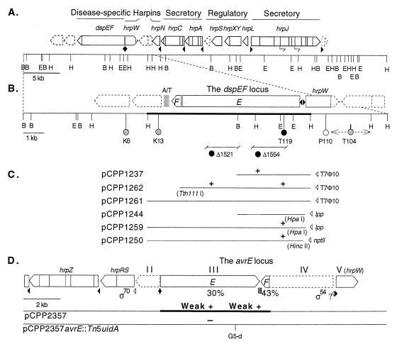
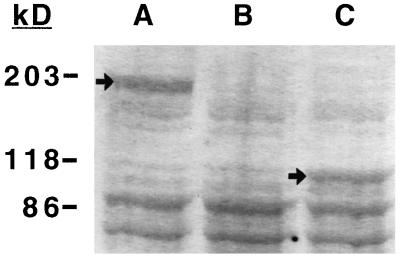
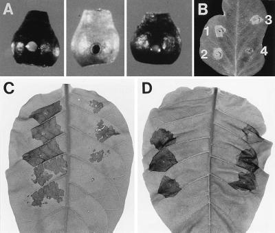
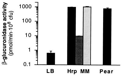
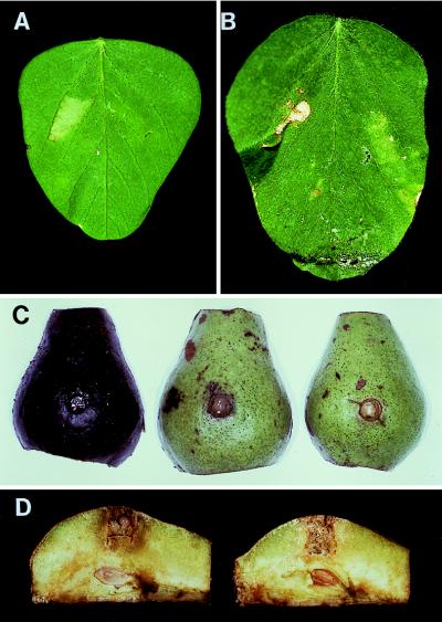
The "disease-specific" (dsp) region next to the hrp gene cluster of Erwinia amylovora is required for pathogenicity but not for elicitation of the hypersensitive reaction. A 6.6-kb apparent operon, dspEF, was found responsible for this phenotype. The operon contains genes dspE and dspF and is positively regulated by hrpL. A BLAST search revealed similarity in the dspE gene to a partial sequence of the avrE locus of Pseudomonas syringae pathovar tomato. The entire avrE locus was sequenced. Homologs of dspE and dspF were found in juxtaposed operons and were designated avrE and avrF. Introduced on a plasmid, the dspEF locus rendered P. syringae pv. glycinea race 4 avirulent on soybean. An E. amylovora dspE mutant, however, elicited a hypersensitive reaction in soybean. The avrE locus in trans restored pathogenicity to dspE strains of E. amylovora, although restored strains were low in virulence. DspE and AvrE are large (198 kDa and 195 kDa) and hydrophilic. DspF and AvrF are small (16 kDa and 14 kDa) and acidic with predicted amphipathic alpha helices in their C termini; they resemble chaperones for virulence factors secreted by type III secretion systems of animal pathogens.


Figures

Figure 1
Figure 1
The dspEF locus of E. amylovora: mutagenesis, complementation and heterologous expression constructs, and homology with and restoration of mutants by the avrE locus of P. syringae. Dashed boxes are uncharacterized ORFs; a solid triangle indicates an hrp box, and an open triangle indicates another promoter. Thick lines delineate the DNA for which sequence was accessioned. (A) The dsp/hrp gene cluster of E. amylovora in pCPP430. Operon names and types of proteins encoded are indicated at the top. B, BamHI; E, EcoRI; H, HindIII. Half-arrows indicate internal promoters without similarity to the hrp box consensus. (B) The region downstream of hrpN containing the dspEF locus. Circles mark deletion mutations and representative transposon insertions: black, nonpathogenic and HR+ or HR-reduced (dsp); gray, reduced virulence and HR; white, wild type. T104 lies within the area marked by the dashed double arrow. K, Tn5miniKm; P, Tn5phoA; T, Tn10tetr; Δ, deletion mutation. The gray box is A/T-rich DNA. (C) Clones and subclones of the dspEF locus. Plasmid designations are indicated at the left, and vector-borne promoters are indicated at the right. Restriction sites used for subcloning not shown above are shown in parentheses. A “+” aligned with a circle representing a mutation in B indicates that the subclone complements that mutation. (D) The avrE locus (transcription units III and IV) of P. syringae pv. tomato DC3000 in pCPP2357. Percent amino acid identity of the predicted proteins AvrE and AvrF to DspE and DspF, respectively, are indicated. Solid rectangles are transcriptional terminators (inverted repeats). Ability to restore mutations depicted in B are indicated, aligned as for complementation data in C.
Figure 2
Figure 2
Expression of the full-length and the N-terminal half of DspE in recombinant E. coli DH5α. Lysates of cells carrying either pCPP1259, containing the entire dspEF locus (lane A); pCPP50, the cloning vector (lane B); or pCPP1244, containing only the 5′ half of the dspE gene (lane C), were subjected to SDS/PAGE (7.5% acrylamide) followed by Coomassie staining. Bands corresponding to DspE (lane A) and the N-terminal half of DspE (lane C) are marked by arrows. Migration of molecular mass markers is indicated on the left.
Figure 3
Figure 3
The role of the dspE gene in pathogenicity and HR elicitation. (A) Immature pear fruit 4 days after inoculation with (left to right) strains Ea321, Ea321dspEΔ1554, or Ea321dspEΔ1554 harboring the 5′ half of dspE on pCPP1237. (B) Norchief soybean leaf 24 hr after infiltration with 5 × 10⁸ cfu/ml suspensions of (1) Ea321, (2) Ea321dspEΔ1554, (3) Ea321hrpN∷Tn5 (ref. 4), and (4) Ea321hrpL∷Tn5 (ref. 1). (C) Tobacco leaf 48 hr after infiltration with parallel dilution series of suspensions of strains Ea321 (Left) and Ea321dspEΔ1554 (Right). The concentrations infiltrated (top to bottom) are 1 × 10¹⁰, 1 × 10⁹, 5 × 10⁸, 1 × 10⁸, and 5 × 10⁷ cfu/ml. (D) As for C, except the more virulent strain, Ea273, and corresponding mutant Ea273dspEΔ1554 were used, and concentrations ranged from 5 × 10⁹ to 5 × 10⁵ cfu/ml in log increments.
Figure 4
Figure 4
Expression of a promoterless GUS construct fused to dspE in E. amylovora Ea273. Ea273 and Ea273dspEuidA (a merodiploid containing both a wild-type dspE and a truncated dspE fused to the uidA gene; solid bars) were grown in LB or Hrp MM, or inoculated to immature pear fruit. Ea273dspEuidAhrpL∷Tn5 (darkly shaded bar) and Ea273hrcV∷Tn5uidA (lightly shaded bar) were also grown in hrp MM. Values shown represent means of triplicate samples normalized for bacterial cell concentration. Standard deviations are represented by lines extending from each bar. The mean values for three samples of Ea273 in each assay were subtracted from, and standard deviations added to, the corresponding values obtained for the other strains.
Figure 5
Figure 5
Transgeneric avirulence function of the dspEF locus and restoration of dspE mutants with the avrE locus. Norchief soybean leaves were either (A) infiltrated with 1 × 10⁸ cfu/ml suspensions of P. syringae pv. glycinea race 4 carrying pCPP1250 (containing the dspEF locus) (Left) or pML122 (the cloning vector) and photographed after 24 hr at room temperature (Right), or (B) infiltrated with 8 × 10⁵ cfu/ml suspensions of the same strains and photographed after 7 days at 22°C and high relative humidity. Tissue collapse is apparent on both leaves where the strain carrying pCPP1250 was infiltrated. On the leaf incubated for 7 days, chlorosis extending beyond the infiltrated area, typical of disease, is apparent on the half infiltrated with the strain carrying the vector only. The dark section on the side of the leaf infiltrated with the strain carrying pCPP1250 is a shadow caused by a buckle in the leaf. Pear halves are shown (C) 10 days after inoculation with (left to right) Ea273, Ea273dspEΔ1521(pCPP2357, containing the avrE locus), or Ea273dspEΔ1521(pCPP2357avrE∷Tn5uidA), and (B) cross-sectioned through the well 10 days after inoculation with Ea321dspEΔ1521(pCPP2357) (Left) and Ea321dspEΔ1521(pCPP2357avrE∷Tn5uidA) (Right). Although greatly reduced relative to wild type, water soaking and necrosis are apparent around and ooze can be seen within the wells of fruit inoculated with dspE strains carrying the intact avrE locus. Fruit inoculated with dspE strains carrying a disrupted clone of avrE is symptomless and shows no ooze.

References

    1. Wei Z-M, Beer S V. J Bacteriol. 1995;177:6201–6210. - PMC - PubMed
    1. Bogdanove A J, Wei Z-M, Zhao L, Beer S V. J Bacteriol. 1996;178:1720–1730. - PMC - PubMed
    1. Kim J-H, Wei Z-M, Beer S V. J Bacteriol. 1997;179:1690–1697. - PMC - PubMed
    1. Wei Z M, Laby R J, Zumoff C H, Bauer D W, He S Y, Collmer A, Beer S V. Science. 1992;257:85–88. - PubMed
    1. Kim J F. Ph.D. dissertation. Ithaca, NY: Cornell University; 1997.

Publication types

Substances

Associated data