

Review
doi: 10.1128/AAC.42.1.1.
Kinship and diversification of bacterial penicillin-binding proteins and beta-lactamases
Affiliations
- PMID: 9449253
- PMCID: PMC105448
- DOI: 10.1128/AAC.42.1.1
Item in Clipboard
Review
Kinship and diversification of bacterial penicillin-binding proteins and beta-lactamases
Antimicrob Agents Chemother.
1998 Jan.
No abstract available
Figures
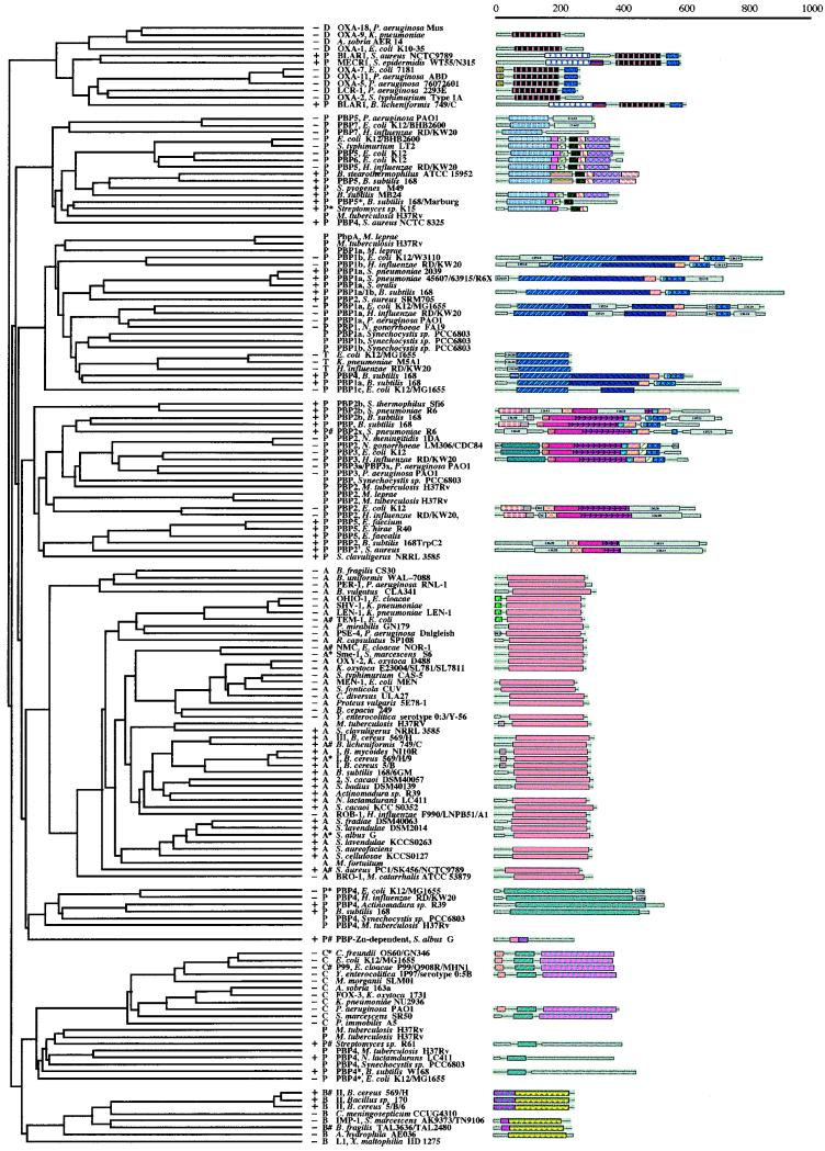
Multiple-sequence alignment of PBPs and β-lactamases made by the use of the program PileUp from the WISCONSIN package. The first column on the left indicates whether the organism is gram positive (+), gram negative (−), or unspecified. The second column states if a given entry is a penicillin-binding protein (P); a class A (A), a class B (B), a class C (C), or a class D (D) β-lactamase; or a monofunctional transglycosylase (T). The pound sign indicates a protein for which a crystal structure is available. The asterisk denotes a protein for which a crystal structure was published but for which the coordinates are not available. The next column indicates the source for the given entry. The last column indicates the domain structures for the proteins from the ProDom library, when they are available. The color and the pattern codes for the domains are arbitrary and were obtained directly from the ProDom library (47a); their utility in this figure is for ready and immediate visualization of homologous domains in different proteins. The scale at the top indicates the lengths of the proteins (in numbers of amino acids).
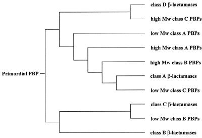
Simplified schematic for the multiple-sequence alignment shown in Fig. 1. Mw, molecular weight.
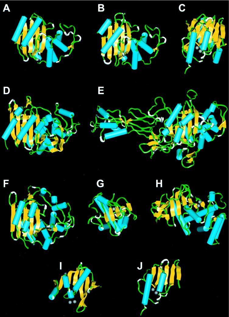
Backbone ribbon presentations for the class A TEM-1 β-lactamase from E. coli (A), the class A β-lactamase from B. licheniformis 749/C (B), the class B β-lactamase from B. fragilis (C), the class C β-lactamase from E. cloacae P99 (D), transpeptidase PBP 2x from S. pneumoniae R6 (E), the dd -peptidase–transpeptidase from Streptomyces sp. strain R61 (F) zinc-dependent dd -peptidase from S. albus G (G), thermolysin from B. thermoproteolyticus (H), the N-terminal half of the class B β-lactamase from B. fragilis (I), and the C-terminal half of the class B β-lactamase from B. fragilis (J). The structures in panels E, F, and G are PBPs. The helices are shown in cyan, the β-strands are in yellow, and the zinc ion is in gray spheres.
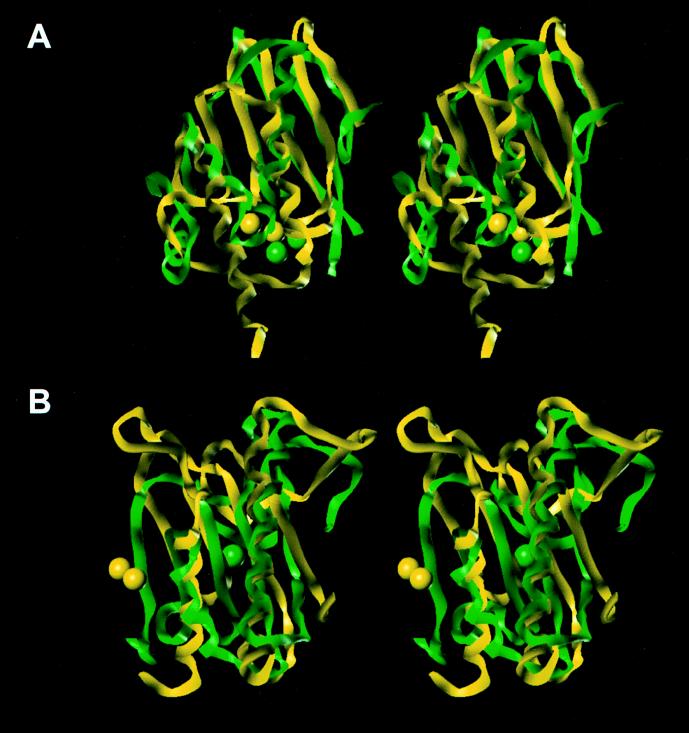
Stereoviews of the three-dimensional folds for the superimposed N-terminal (green) and C-terminal (yellow) subdomains of the class B β-lactamase from B. fragilis (A) and of the Leu-91 to Ile-123 stretch of dd -peptidase (a PBP) from S. albus G (green) with the Asn-33 to Leu-155 stretch of thermolysin from B. thermoproteolyticus (yellow) (B). The spheres represent the zinc ions.
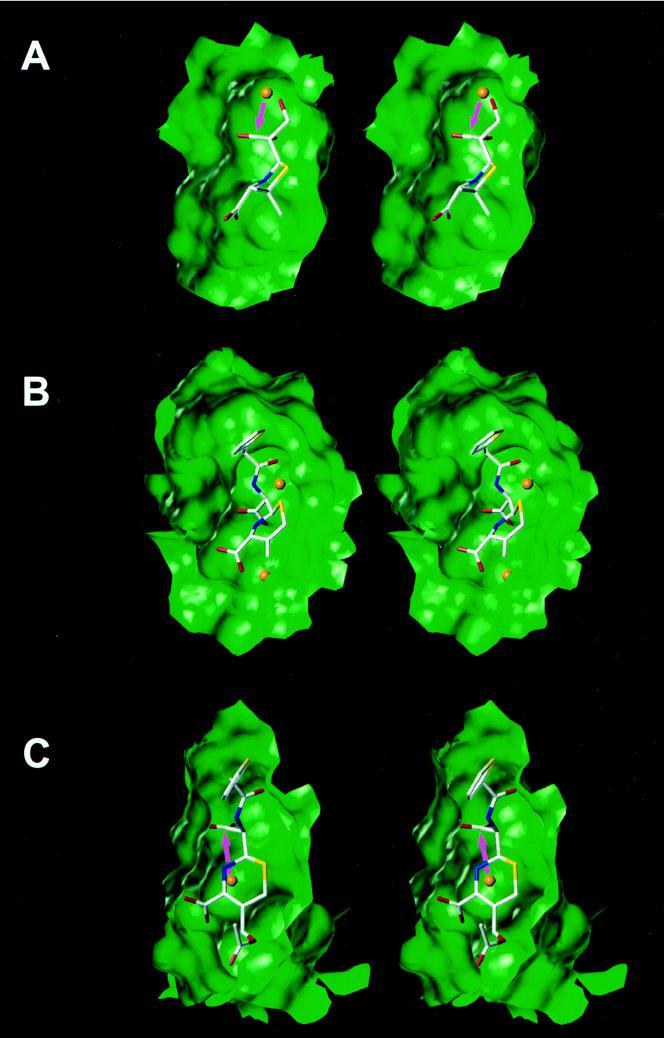
Views of the crystal structure of the acyl enzyme intermediate for 6α-(hydroxymethyl)penicillanate in the class A TEM-1 β-lactamase (A), the crystal structure of the dd -peptidase–transpeptidase from Streptomyces sp. strain R61 modified by cephalothin (the C3 substituent is eliminated because of the longevity of the species) (B), and the energy-minimized structure of the acyl enzyme intermediate for cephalothin in the active site of the class C β-lactamase from E. cloacae P99 (the C3 substituent is not eliminated because of the fleeting existence of the species) (C). The active-site cavities are shown as Connolly water-accessible surfaces. Water molecules are represented as orange spheres.
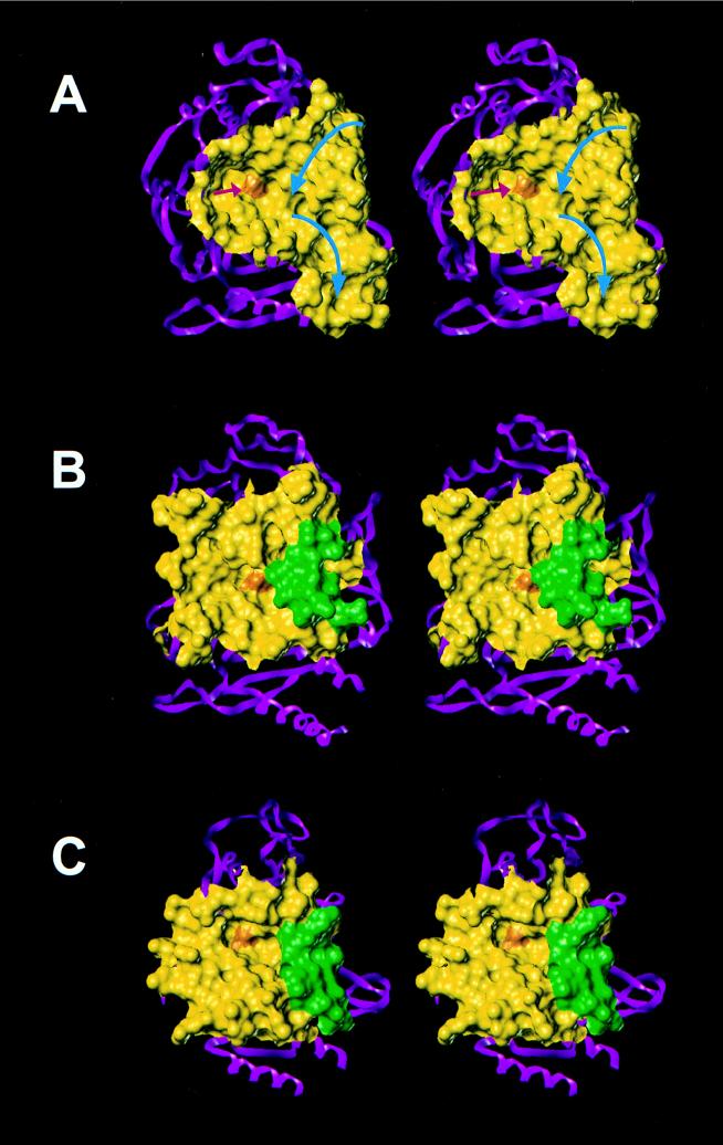
Active sites, shown as Connolly water-accessible surfaces, of the bifunctional dd -peptidase–transpeptidase from Streptomyces sp. strain R61 (A), the class C β-lactamase from E. cloacae P99 (B), and the class A TEM-1 β-lactamase from E. coli (C). The red and blue arrows indicate the grooves where the first and second peptidoglycan strands, respectively, bind to the active site. The first peptidoglycan strand (red arrow) would approach the active-site serine, represented by the orange surface, essentially orthogonally to the plain of the figure in the depicted perspective. The green areas of surfaces B and C are contributed by residues Glu-285 to Ile-296 in class C β-lactamases and Asp-214 to Ala-224 residues in class A β-lactamases. The yellow areas constitute the remainder of the active-site regions. The rest of each protein is depicted in the ribbon presentation in magenta.
References
-
- Adachi H, Ohta T, Matsuzawa H. Site-directed mutants, at position 166, of RTEM-1 β-lactamase that form a stable acyl-enzyme intermediate with penicillin. J Biol Chem. 1991;266:3186–3191. - PubMed
-
- Bulychev A, Massova I, Miyashita K, Mobashery S. Evolution of the versatile β-lactam hydrolase activity: from biosynthetic enzymes to drug resistance factors. J Am Chem Soc. 1997;119:7619–7625.
-
- Bush K, Jacoby G. Nomenclature of TEM β-lactamases. J Antimicrob Chemother. 1997;39:1–3. - PubMed
Publication types
MeSH terms
Substances
LinkOut - more resources
Full Text Sources
Molecular Biology Databases
