. 1998 Feb 2;187(3):329-39.
doi: 10.1084/jem.187.3.329.

Adhesion of activated platelets to endothelial cells: evidence for a GPIIbIIIa-dependent bridging mechanism and novel roles for endothelial intercellular adhesion molecule 1 (ICAM-1), alphavbeta3 integrin, and GPIbalpha

Affiliations

Adhesion of activated platelets to endothelial cells: evidence for a GPIIbIIIa-dependent bridging mechanism and novel roles for endothelial intercellular adhesion molecule 1 (ICAM-1), alphavbeta3 integrin, and GPIbalpha

T Bombeli et al. J Exp Med. .

Abstract

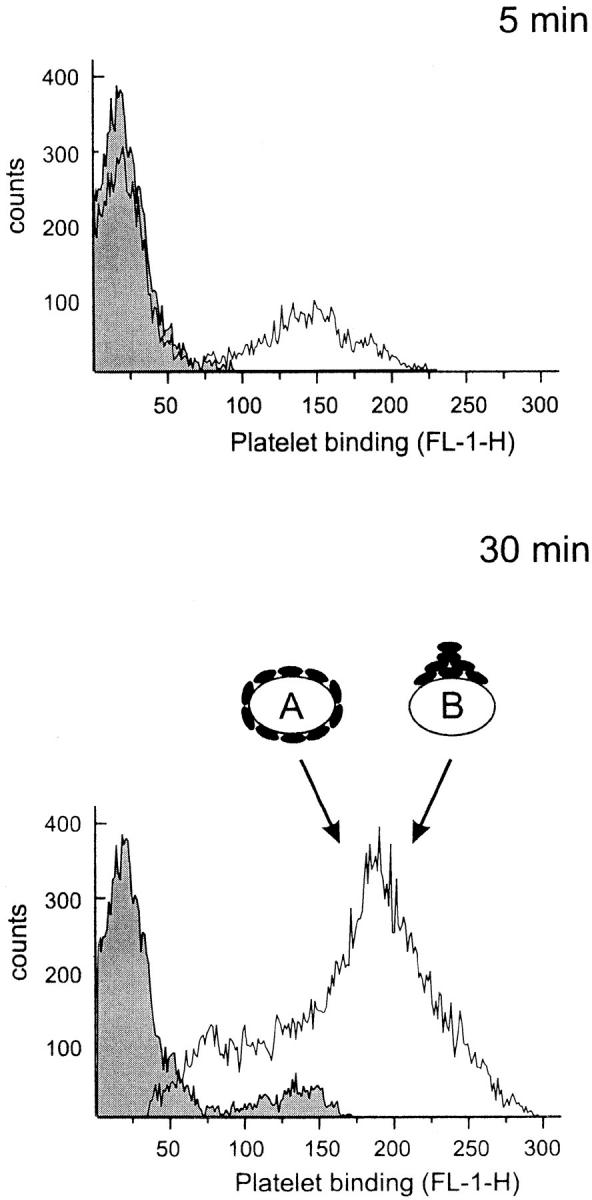
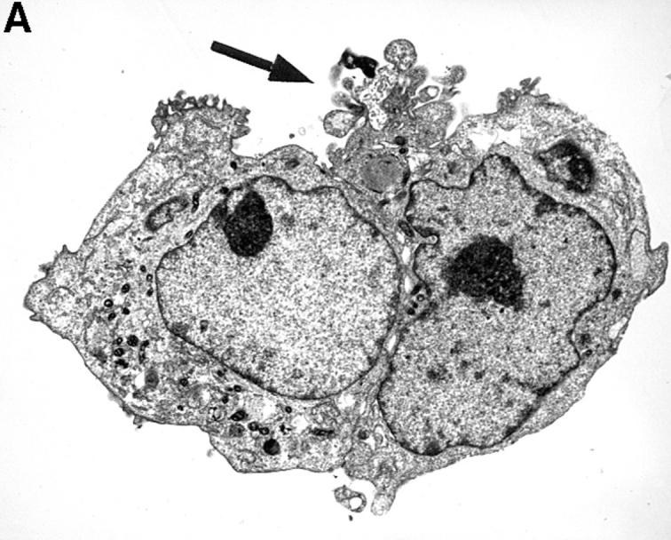
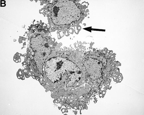
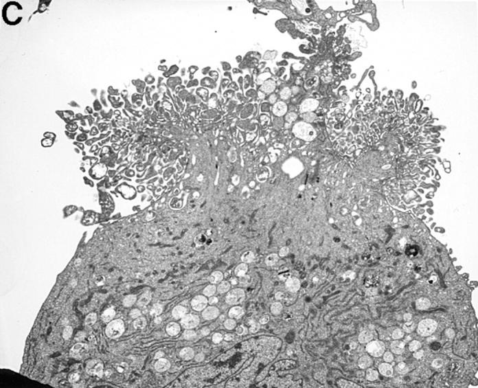
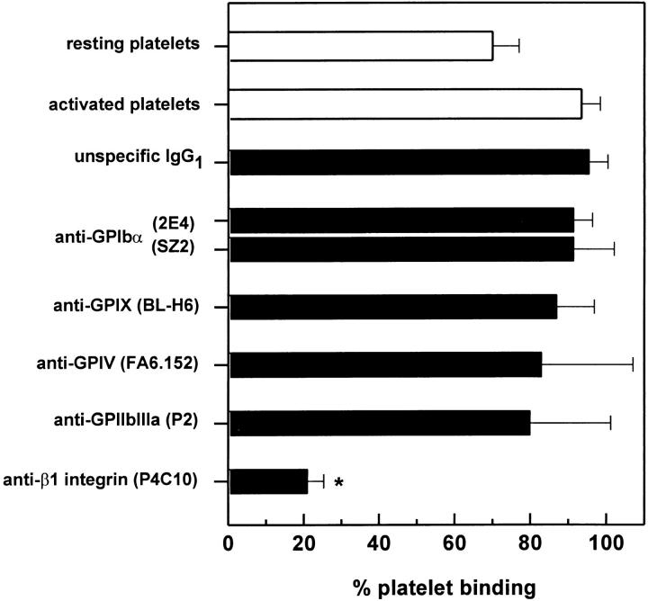
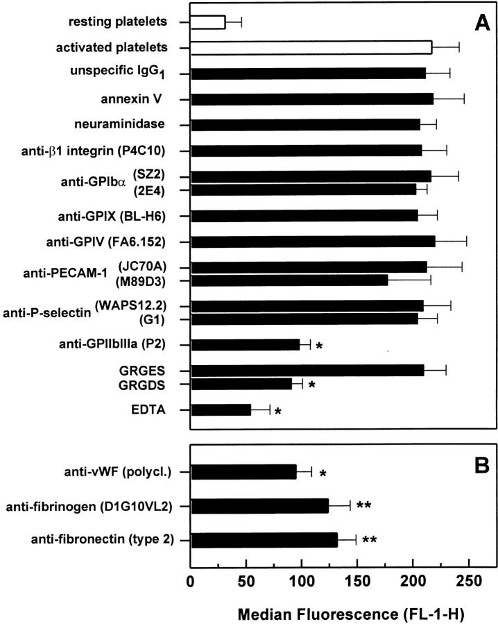
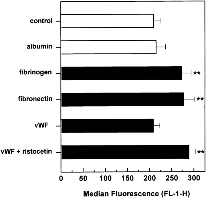
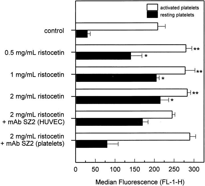
Although it has been reported that activated platelets can adhere to intact endothelium, the receptors involved have not been fully characterized. Also, it is not clear whether activated platelets bind primarily to matrix proteins at sites of endothelial cell denudation or directly to endothelial cells. Thus, this study was designed to further clarify the mechanisms of activated platelet adhesion to endothelium. Unstimulated human umbilical vein endothelial cell (HUVEC) monolayers were incubated with washed, stained, and thrombin-activated human platelets. To exclude matrix involvement, HUVEC were harvested mechanically and platelet binding was measured by flow cytometry. Before the adhesion assay, platelets or HUVEC were treated with different receptor antagonists. Whereas blockade of platelet beta1 integrins, GPIbalpha, GPIV, P-selectin, and platelet-endothelial cell adhesion molecule (PECAM)-1 did not reduce platelet adhesion to HUVEC, blockade of platelet GPIIbIIIa by antibodies or Arg-Gly-Asp (RGD) peptides markedly decreased adhesion. Moreover, when platelets were treated with blocking antibodies to GPIIbIIIa-binding adhesive proteins, including fibrinogen and fibronectin, and von Willebrand factor (vWF), platelet binding was also reduced markedly. Addition of fibrinogen, fibronectin, or vWF further increased platelet adhesion, indicating that both endogenous platelet-exposed and exogenous adhesive proteins can participate in the binding process. Evaluation of the HUVEC receptors revealed predominant involvement of intercellular adhesion molecule (ICAM)-1 and alphavbeta3 integrin. Blockade of these two receptors by antibodies decreased platelet binding significantly. Also, there was evidence that a component of platelet adhesion was mediated by endothelial GPIbalpha. Blockade of beta1 integrins, E-selectin, P-selectin, PECAM-1, vascular cell adhesion molecule (VCAM)-1 and different matrix proteins on HUVEC did not affect platelet adhesion. In conclusion, we show that activated platelet binding to HUVEC monolayers is mediated by a GPIIbIIIa-dependent bridging mechanism involving platelet-bound adhesive proteins and the endothelial cell receptors ICAM-1, alphavbeta3 integrin, and, to a lesser extent, GPIbalpha.


Figures

Figure 1
Figure 1
Time course of platelet binding to HUVEC monolayers. Washed and calcein-loaded resting or thrombin-activated platelets were allowed to adhere to a HUVEC monolayer for 5 or 30 min. HUVEC were then harvested and assayed by flow cytometry to determine platelet binding that is expressed as Platelet binding (FL-1-H) of the entire HUVEC population. The shaded and open curves represent HUVEC incubated with resting and activated platelets, respectively. When estimated by an arbitrary gate, HUVEC bound resting and activated platelets by ∼10 and 65% after 30 min, respectively. As depicted by the symbols A and B, the increased fluorescence of HUVEC incubated with activated platelets may be due to either numerous single platelets (symbol A) or only one large aggregate (symbol B). Using flow cytometry, it is not possible to distinguish between these two mechanisms. A representative experiment of three performed is shown.
Figure 2
Figure 2
TEM of HUVEC monolayers incubated with platelets. A illustrates two adherent HUVEC that were incubated with resting platelets. On the entire circumference of the cell surfaces, there is only one area (black arrow) exhibiting adherent platelets (A). However, when incubated with activated platelets HUVEC were covered completely with single platelets (black arrow, B). Occasionally, large platelet aggregates were found (C). The magnification is 1:5,000–6,000.
Figure 2
Figure 2
TEM of HUVEC monolayers incubated with platelets. A illustrates two adherent HUVEC that were incubated with resting platelets. On the entire circumference of the cell surfaces, there is only one area (black arrow) exhibiting adherent platelets (A). However, when incubated with activated platelets HUVEC were covered completely with single platelets (black arrow, B). Occasionally, large platelet aggregates were found (C). The magnification is 1:5,000–6,000.
Figure 3
Figure 3
Platelet adhesion to HUVEC monolayers after blockade of HUVEC receptors. Thrombin-activated platelets were allowed to adhere to a HUVEC monolayer that was treated with either antagonists of different endothelial cell adhesion molecules (A) or antibodies to adhesive proteins (B). Platelet binding was then determined by flow cytometry as described above. The following concentrations were used: 5 μM annexin V, 0.5 U/ml neuraminidase, 50 μg/ml GRGES and GRGDS peptides, 1 mM EDTA, 40 μg/ml mAb, and a dilution of 1:250 for polyclonal antibodies. Results are expressed as the mean ± SD of the median fluorescence (FL-1-H) of at least four experiments. *P <0.01 and **P <0.05 by Student's t test, compared with activated platelet adhesion to HUVEC treated with the isotype-specific control mAb.
Figure 4
Figure 4
Platelet adhesion to HUVEC matrix after blockade of platelet receptors. Thrombin-activated platelets treated with antagonists to different platelet receptors were allowed to adhere to HUVEC matrix for 30 min. Platelet binding was then determined by a fluorescence plate reader as described above. The following concentrations were used: 40 μg/ml for mAb and a dilution of 1:250 for polyclonal antibodies. Results are expressed as the mean ± SD of the percentage of adherent platelets of at least three experiments. *P <0.01 by Student's t test, compared with adhesion of activated platelets treated with the isotype-specific control mAb.
Figure 5
Figure 5
Platelet adhesion to HUVEC monolayers after blockade of platelet receptors. Thrombin-activated platelets treated with either antagonists to different platelet receptors (A) or antibodies to adhesive proteins (B) were allowed to adhere to a HUVEC monolayer for 30 min. Platelet binding was determined by flow cytometry as described above. The following concentrations were used: 5 μM annexin V, 0.5 U/ml neuraminidase, 50 μg/ml GRGES and GRGDS peptides, 1 mM EDTA, 40 μg/ml mAb, and a dilution of 1:250 for polyclonal antibodies. Results are expressed as the mean ± SD of the median fluorescence (FL-1-H) of at least four experiments. *P <0.01 and P <0.05 by Student's t test, compared with adhesion of activated platelets treated with the isotype-specific control mAb.
Figure 6
Figure 6
Platelet adhesion to HUVEC monolayers after addition of adhesive proteins. Thrombin-activated platelets were allowed to adhere to HUVEC monolayers for 30 min in the presence of different adhesive proteins. Platelet binding was then determined by flow cytometry as described above. The following concentrations were used: 4 mg/ml albumin, 75 μg/ ml fibrinogen, 15 μg/ml fibronectin, and 0.25 μg/ml vWF, which corresponded to a plasma concentration of 10, 2.5, 5, and 2.5%, respectively. Ristocetin was added together with vWF at a final concentration of 1 mg/ml. Results are expressed as the mean ± SD of the median fluorescence (FL-1-H) of at least four experiments. **P <0.05 by Student's t test, compared with adhesion without proteins.
Figure 7
Figure 7
Adhesion of resting platelets to HUVEC monolayers in the presence of zinc or manganese. Resting platelets were allowed to adhere to HUVEC monolayers for 30 min in the presence of 0.5 mM zinc (open bars) or 2 mM manganese (filled bars) with or without the addition of 375 μg/ml fibrinogen. Both zinc and manganese activate platelets without inducing secretion. Platelet binding was then determined by flow cytometry as described above. Adhesion was also measured in the presence of 40 μg/ml anti-GPIIbIIIa mAb P2 and antifibrinogen mAb D1G10VL2. Results are expressed as the mean ± SD of the median fluorescence (FL-1-H) of at least three experiments. *P <0.01 by Student's t test, compared with adhesion with fibrinogen.
Figure 8
Figure 8
Platelet adhesion to HUVEC monolayers after addition of vWF and ristocetin. Resting (filled bars) and thrombin-activated platelets (open bars) were allowed to adhere to HUVEC monolayers for 30 min in the presence of 1 μg/ml vWF and increasing concentrations of ristocetin. Platelet binding was then determined by flow cytometry as described above. To test involvement of endothelial and platelet GPIbα, HUVEC and platelets were pretreated separately with 40 μg/ml anti–human GPIbα mAb, clone SZ2. Results are expressed as the mean ± SD of the median fluorescence (FL-1-H) of at least four experiments. *P <0.01 and **P <0.05 by Student's t test, compared with the corresponding controls without vWF and ristocetin.
Figure 9
Figure 9
Proposed model of adhesion of activated platelets to HUVEC. Upon activation, platelet GPIIbIIIa receptors bind different adhesive proteins, such as fibrinogen, vWF, and fibronectin. These proteins are either present in plasma or secreted by the platelets themselves. Dependent upon the bound adhesive protein, adhesion of platelets to endothelial cells is mediated by different endothelial counter receptors. Whereas fibronectin binds only to αvβ3-integrin, fibrinogen binds to ICAM-1 or αvβ3-integrin and vWF binds primarily to αvβ3-integrin or possibly also to GPIbα.

Similar articles

Cited by

References

    1. Abrams C, Shattil SJ. Immunological detection of activated platelets in clinical disorders. Thromb Haemostasis. 1991;65:467–473. - PubMed
    1. Czervionke RL, Hoak JC, Fry GL. Effect of aspirin on thrombin-induced adherence of platelets to cultured cells from the blood vessel wall. J Clin Invest. 1978;62:847–856. - PMC - PubMed
    1. Fry GL, Czervionke RL, Hoak JC, Smith JB, Haycraft DL. Platelet adherence to cultured vascular cells: influence of prostacyclin (PGI2) Blood. 1980;55:271–275. - PubMed
    1. Kaplan JE, Moon DG, Weston LK, Minnear FL, Del Vecchio PJ, Shepard JM, Fenton JW. Platelets adhere to thrombin-treated endothelial cells in vitro. Am J Physiol. 1989;257:H423–H433. - PubMed
    1. Tloti MA, Moon DG, Weston LK, Kaplan JE. Effect of 13-hydroxy octadeca-9,11-dienoic acid (13-HODE) on thrombin induced platelet adherence to endothelial cells in vitro. Thromb Res. 1991;62:305–317. - PubMed

Publication types

MeSH terms