. 1998 Feb 2;187(3):389-402.
doi: 10.1084/jem.187.3.389.

Interleukin 7 transgenic mice develop chronic colitis with decreased interleukin 7 protein accumulation in the colonic mucosa

Affiliations

Interleukin 7 transgenic mice develop chronic colitis with decreased interleukin 7 protein accumulation in the colonic mucosa

M Watanabe et al. J Exp Med. .

Abstract

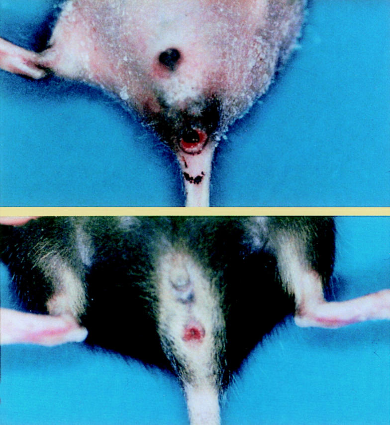
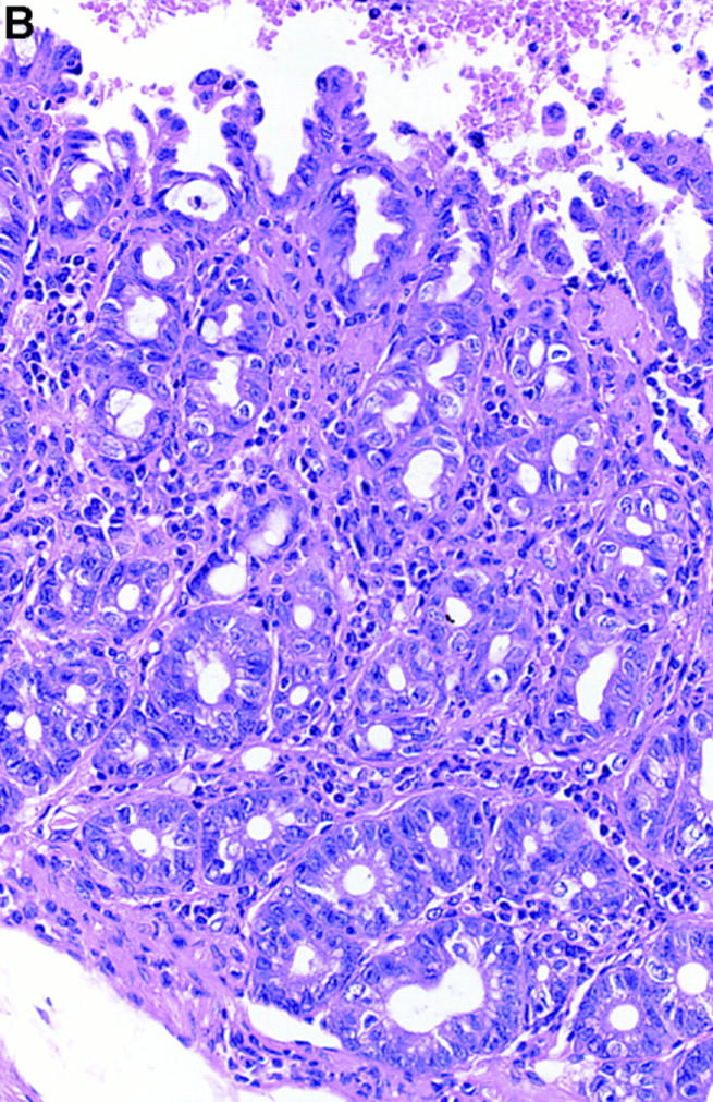
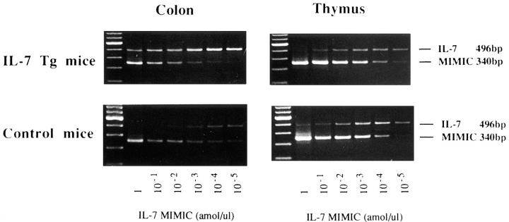
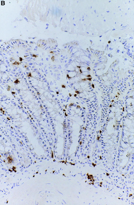
We have demonstrated that intestinal epithelial cells produce interleukin 7 (IL-7), and IL-7 serves as a potent regulatory factor for proliferation of intestinal mucosal lymphocytes expressing functional IL-7 receptor. To clarify the mechanism by which locally produced IL-7 regulates the mucosal lymphocytes, we investigated IL-7 transgenic mice. Here we report that transgenic mice expressing murine IL-7 cDNA driver by the SRalpha promoter developed chronic colitis in concert with the expression of SRalpha/IL-7 transgene in the colonic mucosa. IL-7 transgenic but not littermate mice developed chronic colitis at 4-12 wk of age, with histopathological similarity to ulcerative colitis in humans. Southern blot hybridization and competitive PCR demonstrated that the expression of IL-7 messenger RNA was increased in the colonic mucosal lymphocytes but not in the colonic epithelial cells. IL-7 protein accumulation was decreased in the goblet cell-depleted colonic epithelium in the transgenic mice. Immunohistochemical and cytokine production analysis showed that lymphoid infiltrates in the lamina propria were dominated by T helper cell type 1 CD4+ T cells. Flow cytometric analysis demonstrated that CD4+ intraepithelial T cells were increased, but T cell receptor gamma/delta T cells and CD8alpha/alpha cells were not increased in the area of chronic inflammation. Increased IL-7 receptor expression in mucosal lymphocytes was demonstrated in the transgenic mice. These findings suggest that chronic inflammation in the colonic mucosa may be mediated by dysregulation of colonic epithelial cell-derived IL-7, and this murine model of chronic colitis may contribute to the understanding of the pathogenesis of human inflammatory bowel disease.


Figures

Figure 1
Figure 1
Transgenic mice carrying murine IL-7 cDNA by the SRα promoter developed acute colitis with infiltrating neutrophils and lymphocytes at 1–3 wk of age (A and B). At that time, the SRα/IL-7 transgene and IL-7 mRNA expression in the colonic mucosa was revealed (C). IL-7 protein was also significantly expressed in the inflamed colonic mucosa of IL-7 transgenic mice (D). Infiltrating T cells in the acute colitis lesion were mainly CD4+ T cells (E) and T cell receptor γ/δ T cells (F).
Figure 1
Figure 1
Transgenic mice carrying murine IL-7 cDNA by the SRα promoter developed acute colitis with infiltrating neutrophils and lymphocytes at 1–3 wk of age (A and B). At that time, the SRα/IL-7 transgene and IL-7 mRNA expression in the colonic mucosa was revealed (C). IL-7 protein was also significantly expressed in the inflamed colonic mucosa of IL-7 transgenic mice (D). Infiltrating T cells in the acute colitis lesion were mainly CD4+ T cells (E) and T cell receptor γ/δ T cells (F).
Figure 2
Figure 2
A line 34 transgenic mice carrying murine IL-7 cDNA by the SRα promoter developed rectal prolapse at 6 wk (A) and 10 wk (B). The IL-7 transgenic line 34 mice frequently showed rectal prolapse and remittent intestinal bleeding at 6–10 wk of age, suggesting the development of chronic colitis.
Figure 3
Figure 3
Histopathological examination (hematoxylin-eosin staining) of the colonic tissues revealed the development of chronic colitis in the IL-7 transgenic mice. Erosions and neutrophil infiltration were observed in the anal ring (original magnification: 100, A). The inflammatory cell infiltration and goblet cell depletion was most prominent in the rectum (original magnification: 200, B), but observed diffusely in the entire colon (original magnification: 100, C; original magnification: 200, D). Crypt abscess (original magnification: 200, E), paneth cell metaplasia (original magnification: 200, F), and infiltration of eosinophils (original magnification: 200, G) was observed in the lesions. These mimicked some histopathological characteristics of ulcerative colitis in humans.
Figure 3
Figure 3
Histopathological examination (hematoxylin-eosin staining) of the colonic tissues revealed the development of chronic colitis in the IL-7 transgenic mice. Erosions and neutrophil infiltration were observed in the anal ring (original magnification: 100, A). The inflammatory cell infiltration and goblet cell depletion was most prominent in the rectum (original magnification: 200, B), but observed diffusely in the entire colon (original magnification: 100, C; original magnification: 200, D). Crypt abscess (original magnification: 200, E), paneth cell metaplasia (original magnification: 200, F), and infiltration of eosinophils (original magnification: 200, G) was observed in the lesions. These mimicked some histopathological characteristics of ulcerative colitis in humans.
Figure 4
Figure 4
The expression of SRα/IL-7 transgene in the colonic mucosa. In the IL-7 transgenic mice, 278-bp PCR amplified products of mRNA from the SRα/IL-7 transgene were expressed in the colonic mucosa. The SRα/IL-7 transgene expression in the colonic mucosa is parallel with development of colitis in the IL-7 transgenic mice. SRα/IL-7 transgene was expressed in the colonic mucosa of transgenic mice with colitis at both 4 and 8 wk, but not expressed in the colonic mucosa of mice without colitis at 4 and 8 wk (A). In the colitis lesion of transgenic mice, SRα/IL-7 transgene was expressed in the colonic epithelial cells (EC), IELs, and LPLs (B). In contrast, the transgene was constitutively expressed in the thymus, spleen, kidney, brain, lung, and skin, but not in the liver and muscle as previously reported (data not shown).
Figure 4
Figure 4
The expression of SRα/IL-7 transgene in the colonic mucosa. In the IL-7 transgenic mice, 278-bp PCR amplified products of mRNA from the SRα/IL-7 transgene were expressed in the colonic mucosa. The SRα/IL-7 transgene expression in the colonic mucosa is parallel with development of colitis in the IL-7 transgenic mice. SRα/IL-7 transgene was expressed in the colonic mucosa of transgenic mice with colitis at both 4 and 8 wk, but not expressed in the colonic mucosa of mice without colitis at 4 and 8 wk (A). In the colitis lesion of transgenic mice, SRα/IL-7 transgene was expressed in the colonic epithelial cells (EC), IELs, and LPLs (B). In contrast, the transgene was constitutively expressed in the thymus, spleen, kidney, brain, lung, and skin, but not in the liver and muscle as previously reported (data not shown).
Figure 5
Figure 5
The expression of IL-7 mRNA in the colonic mucosa. RT-PCR analysis demonstrated IL-7 mRNA expression in normal murine colonic tissues (A). The specificity of amplified bands was validated by their predicted size (496-bp). To ensure the correct predicted fragments are present, we digested the amplified IL-7 PCR product by restriction enzyme EcoRI. 496-bp PCR products from murine colonic mucosa were digested into two predicted fragments with 263 and 233 bp. Markers represented 100-bp DNA ladder. Southern blot analysis confirmed the expression of IL-7 mRNA in normal colonic mucosa (B). PCR products were blotted onto nylon membrane and hybridized with IL-7 gene-specific cDNA oligonucleotide probe labeled with digoxigenin. IL-7 mRNA expression was detected in the colon, and a detectable expression of IL-7 was in small intestinal tissues and stomach tissues. Markers represented the 100-bp DNA ladder. For internal standard, amplified bands for G3PDH were used as the housekeeping gene.
Figure 5
Figure 5
The expression of IL-7 mRNA in the colonic mucosa. RT-PCR analysis demonstrated IL-7 mRNA expression in normal murine colonic tissues (A). The specificity of amplified bands was validated by their predicted size (496-bp). To ensure the correct predicted fragments are present, we digested the amplified IL-7 PCR product by restriction enzyme EcoRI. 496-bp PCR products from murine colonic mucosa were digested into two predicted fragments with 263 and 233 bp. Markers represented 100-bp DNA ladder. Southern blot analysis confirmed the expression of IL-7 mRNA in normal colonic mucosa (B). PCR products were blotted onto nylon membrane and hybridized with IL-7 gene-specific cDNA oligonucleotide probe labeled with digoxigenin. IL-7 mRNA expression was detected in the colon, and a detectable expression of IL-7 was in small intestinal tissues and stomach tissues. Markers represented the 100-bp DNA ladder. For internal standard, amplified bands for G3PDH were used as the housekeeping gene.
Figure 6
Figure 6
Quantity of IL-7 mRNA expression in the colon and thymus tissues in the IL-7 transgenic mice. Competitive PCR demonstrated that 100-fold increase in IL-7 mRNA transcripts was detected in the inflamed colonic mucosa of the IL-7 transgenic mice (IL-7 Tg), as compared to those in wild-type mice (Control). On the contrary, no difference of the IL-7 mRNA expression was observed in the thymus tissues between control mice and IL-7 transgenic mice.
Figure 7
Figure 7
Analysis of the differential expression of IL-7 mRNA in the colonic mucosa. PCR analysis demonstrated that colonic IELs and LPLs isolated from the inflamed colonic tissues of IL-7 transgenic mice expressed a significant amount of IL-7 mRNA (A). However, IELs and LPLs from the colonic tissues of littermate mice showed no expression of IL-7 mRNA. Interestingly, competitive PCR analysis demonstrated that IL-7 mRNA expression in the colonic epithelial cells was not changed between control mice and the transgenic mice (B).
Figure 7
Figure 7
Analysis of the differential expression of IL-7 mRNA in the colonic mucosa. PCR analysis demonstrated that colonic IELs and LPLs isolated from the inflamed colonic tissues of IL-7 transgenic mice expressed a significant amount of IL-7 mRNA (A). However, IELs and LPLs from the colonic tissues of littermate mice showed no expression of IL-7 mRNA. Interestingly, competitive PCR analysis demonstrated that IL-7 mRNA expression in the colonic epithelial cells was not changed between control mice and the transgenic mice (B).
Figure 8
Figure 8
Immunohistochemical analysis using rabbit anti–murine IL-7 Ab confirmed IL-7 protein expression in the human intestinal epithelial cells and epithelial goblet cells. The reactivity of anti–murine IL-7 IgG Ab but not control rabbit IgG is confined to the colonic epithelial cells and epithelial goblet cells in the normal murine intestinal mucosal tissues (A). Note that biotin-conjugated goat anti–rabbit Ab alone did not show the reactivity. IL-7 protein expression was not changed in the colonic mucosa of IL-7 transgenic mice at 4–6 wk of age (data not shown). However, IL-7 protein expression seemed to be decreased in the goblet-depleted colonic epithelial cells in the inflamed colonic mucosa of IL-7 transgenic mice at 8–10 wk of age (B).
Figure 8
Figure 8
Immunohistochemical analysis using rabbit anti–murine IL-7 Ab confirmed IL-7 protein expression in the human intestinal epithelial cells and epithelial goblet cells. The reactivity of anti–murine IL-7 IgG Ab but not control rabbit IgG is confined to the colonic epithelial cells and epithelial goblet cells in the normal murine intestinal mucosal tissues (A). Note that biotin-conjugated goat anti–rabbit Ab alone did not show the reactivity. IL-7 protein expression was not changed in the colonic mucosa of IL-7 transgenic mice at 4–6 wk of age (data not shown). However, IL-7 protein expression seemed to be decreased in the goblet-depleted colonic epithelial cells in the inflamed colonic mucosa of IL-7 transgenic mice at 8–10 wk of age (B).
Figure 9
Figure 9
The phenotype of the infiltrating mucosal lymphocytes at the colitis lesion was analyzed by immunohistochemistry. Sections of colonic tissues from IL-7 transgenic mice were stained with anti-CD4 (A) and anti-CD8 (B) antibodies. Lymphoid infiltrates in the lamina propria at the colitis lesion were dominated by T cells bearing CD4+. Both TCR-α/β T cells and γ/δ T cells infiltrated in the lamina propria of colonic mucosa (data not shown).
Figure 9
Figure 9
The phenotype of the infiltrating mucosal lymphocytes at the colitis lesion was analyzed by immunohistochemistry. Sections of colonic tissues from IL-7 transgenic mice were stained with anti-CD4 (A) and anti-CD8 (B) antibodies. Lymphoid infiltrates in the lamina propria at the colitis lesion were dominated by T cells bearing CD4+. Both TCR-α/β T cells and γ/δ T cells infiltrated in the lamina propria of colonic mucosa (data not shown).
Figure 10
Figure 10
The phenotype of the infiltrating intraepithelial lymphocytes in the colitis lesion was analyzed by flow cytometric analysis. Flow cytometric analysis of isolated cells from IEL demonstrated that T cell receptor γ/δ IELs were not increased in the IL-7 transgenic mice (IL-7 Tg) as compared to those in control littermate mice (Control), although γ/δ T cells were the main components of mononuclear cells infiltrating in the dermatitis lesion of IL-7 transgenic mice as previously reported (17). CD4+ T cells, but not CD8+ T cells were significantly increased in the colitis lesions. Moreover, infiltrating CD8+ IELs in the IL-7 transgenic mice expressed mainly α/β heterodimers, indicating that these cells were thymus dependent. A minimum of 5,000 cells was analyzed for each histogram.
Figure 11
Figure 11
Cytokine protein production by isolated cells from the chronic inflamed colonic mucosa of IL-7 transgenic mice (shaded bars) and from normal mucosa of normal littermate mice (white bars). Isolated cells were cultured and culture supernatants were analyzed for concentration of Th1 (IL-2, A; IFN-γ, B) and Th2 (IL-4, C) cytokines by specific ELISA. Unstimulated lamina propria CD4+ T cells from IL-7 transgenic mice and littermate mice produced marginal amounts of both IL-2 and IFN-γ. Isolated lamina propria CD4+ T cells from the inflamed colonic mucosa of IL-7 transgenic mice produced significantly higher levels of both IL-2 and IFN-γ than lamina propria CD4+ T cells from littermate mice after stimulation with anti-CD3 and anti-CD28 mAbs. In contrast to Th1 type cytokine production, secretion of IL-4 by unstimulated lamina propria CD4+ T cells from IL-7 transgenic mice was identical to lamina propria CD4+ T cells from control littermate mice, and in anti-CD3 and anti-CD28 mAb-stimulated lamina propria CD4+ T cells from IL-7 transgenic mice, the average secretion of IL-4 was reduced compared with littermate mice. Data represent mean ± standard error from three independent experiments done in triplicate. *P <0.05, statistically significant as compared with control littermate mice by Mann-Whitney U test.
Figure 12
Figure 12
Southern blot analysis demonstrated that IL-7 receptor (IL-7R) mRNA was not readily detectable in the colonic mucosa from normal littermate mice. However, IL-7 receptor expression was strongly detected in the inflamed colonic mucosa of transgenic mice. The specificity of the amplified bands for IL-7 receptor mRNA was validated by their predicted size (600 bp). Markers represented 100-bp DNA ladder. For internal standard, amplified bands for G3PDH were used. There was no difference in the expression of IL-7 receptor in the thymus between IL-7 transgenic mice and normal littermate mice (data not shown).

Similar articles

Cited by

References

    1. Namen AE, Lupton S, Hjerrild K, Wignall J, Mochizuki DY, Schmierer AE, Mosley B, March CJ, Urdal DL, Gillis S, et al. Stimulation of B-cell progenitors by cloned murine interleukin-7. Nature. 1988;333:571–573. - PubMed
    1. Goodwin RG, Lupton S, Schmierer A, Hjerrild KJ, Jerzy R, Clevenger W, Gillis S, Cosman D, Namen AE. Human interleukin 7: molecular cloning and growth factor activity on human and murine B-lineage cells. Proc Natl Acad Sci USA. 1989;86:302–306. - PMC - PubMed
    1. Morrissey PJ, Goodwin RG, Nordan RP, Anderson D, Grabstein KH, Cosman D, Sims J, Lupton S, Acres B, Reed SG. Recombinant interleukin 7, pre–B cell growth factor, has costimulatory activity on purified mature T cells. J Exp Med. 1989;169:707–716. - PMC - PubMed
    1. Chazen GD, Pereira GMB, LeGros G, Gillis S, Shevach EM. Interleukin 7 is a T-cell growth factor. Proc Natl Acad Sci USA. 1989;86:5923–5927. - PMC - PubMed
    1. Welch PA, Namen AE, Goodwin RG, Armitage R, Cooper MD. Human IL-7: a novel T cell growth factor. J Immunol. 1989;143:3562–3567. - PubMed

Publication types

MeSH terms