. 1998 Jan;116(1):357-67.
doi: 10.1104/pp.116.1.357.

Two isoforms of NADPH:cytochrome P450 reductase in Arabidopsis thaliana. Gene structure, heterologous expression in insect cells, and differential regulation

Affiliations

Two isoforms of NADPH:cytochrome P450 reductase in Arabidopsis thaliana. Gene structure, heterologous expression in insect cells, and differential regulation

M Mizutani et al. Plant Physiol. 1998 Jan.

Abstract

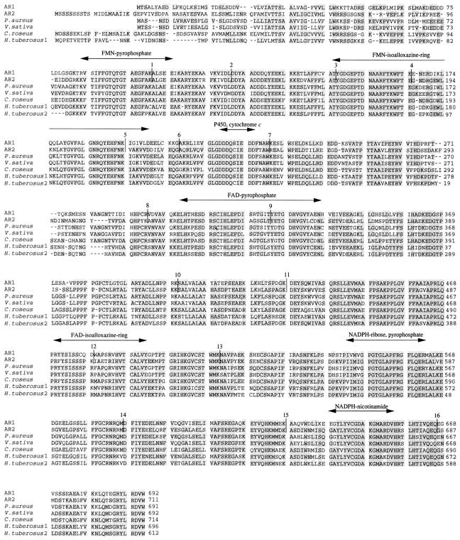
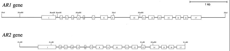
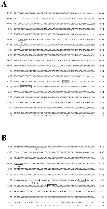
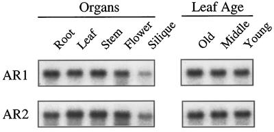
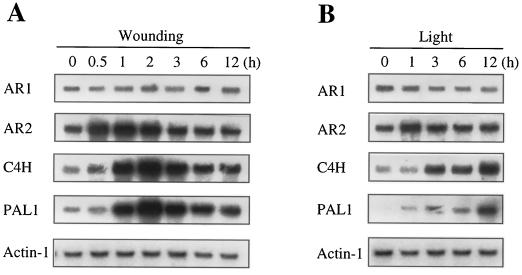
We have investigated two NADPH-cytochrome (Cyt) P450 reductase isoforms encoded by separate genes (AR1 and AR2) in Arabidopsis thaliana. We isolated AR1 and AR2 cDNAs using a mung bean (Phaseolus aureus L.) NADPH-Cyt P450 reductase cDNA as a probe. The recombinant AR1 and AR2 proteins produced using a baculovirus expression system showed similar Km values for Cyt c and NADPH, respectively. In the reconstitution system with a recombinant cinnamate 4-hydroxylase (CYP73A5), the recombinant AR1 and AR2 proteins gave the same level of cinnamate 4-hydroxylase activity (about 70 nmol min-1 nmol-1 P450). The AR2 gene expression was transiently induced by 4- and 3-fold within 1 h of wounding and light treatments, respectively, and the induction time course preceded those of CYP73A5 and a phenylalanine ammonia-lyase (PAL1) gene. On the contrary, the AR1 expression level did not change during the treatments. Analysis of the AR1 and AR2 gene structure revealed that only the AR2 promoter contained three putative sequence motifs (boxes P, A, and L), which are involved in the coordinated expression of CYP73A5 and other phenylpropanoid pathway genes. These results suggest the possibility that AR2 transcription may be functionally linked to the induced levels of phenylpropanoid pathway enzymes.


Figures

Figure 1
Figure 1
Amino acid alignment of plant P450 reductases. The deduced amino acid sequences of AR1 and AR2 are aligned with those of P450 reductases from P. aureus, V. sativa, C. roseus, and H. tuberosus by the program Clustal W (Thompson et al., 1994). Identical amino acid residues are shadowed, and dashes were inserted to maximize the sequence homology. The putative functional regions involved in the interaction with FMN, FAD, NADPH, P450, and Cyt c (Porter and Kasper, 1986; Nisimoto, 1986; Yabusaki et al., 1988) are indicated above the sequences by an arrow. Intron positions are shown by vertical lines with the number of the position from the N terminus.
Figure 2
Figure 2
Heterologous expression of recombinant AR1 and AR2 proteins in insect cells. SDS-PAGE was performed using 10% polyacrylamide slab gel, and proteins were visualized with Coomassie brilliant blue R-250. Lane 1, Solubilized fraction of microsomes of mock-infected Sf21 cells (10 μg of protein); lane 2, solubilized fraction of microsomes of Sf21 cells (10 μg of protein) infected with the recombinant virus containing AR1 cDNA; lane 3, solubilized fraction of microsomes of Sf21 cells (10 μg of protein) infected with the recombinant virus containing AR2 cDNA; lane 4, purified AR1 protein (200 μg of protein); and lane 5, purified AR2 protein (0.2 μg of protein). The migration of size standard is shown to the left of the gel.
Figure 3
Figure 3
Absolute absorption spectra of the purified recombinant AR1 and AR2 proteins. A, Purified recombinant AR1 protein; B, purified recombinant AR2. The one-electron reduced semiquinone forms were prepared by adding 1 mm NADPH to a final concentration of 25 μm, and the spectra were recorded after incubating for 10 min at 25°C. A few grains of sodium dithionite were added to completely reduce the reductases. [——], Oxidized form; [… . .], semiquinone form; and [– – – –], completely reduced form.
Figure 4
Figure 4
Southern-blot analysis of the P450 reductase genes. A. thaliana Columbia genomic DNA (1 μg) was digested with the restriction enzymes X (XbaI), H (HindIII), and E (EcoRI). The digested DNA was separated on 0.7% agarose gel, blotted onto a nylon membrane, and hybridized with a 32P-labeled probe. A, Full-length AR1 cDNA was used as a probe at high stringency; B, AR1 cDNA fragment obtained by digestion with BamHI and HindIII was used as a probe at low stringency. The weak-hybridization bands, which was ascribed to the AR2 gene, are indicated by arrows. C, Full-length AR2 cDNA was used as a probe under high stringency. The migration of size marker is shown to the right of the blots.
Figure 5
Figure 5
Gene organization of the AR1 and AR2 genes. Exons are indicated by boxes 1 to 17, and introns are indicated by solid lines.
Figure 6
Figure 6
The nucleotide sequences of the TATA-proximal regions of the AR1 and AR2 genes. A, AR1 promoter region; B, AR2 promoter region. The translation initiation codon ATG is located at +1. A putative TATA box and a CAAT box are boxed. Putative cis-acting elements homologous to sequence motifs for boxes P, A, and L (Logemann at al., 1995; Mizutani et al., 1997) are underlined. The deduced amino acid sequences of the coding regions are shown below the nucleotide sequences.
Figure 7
Figure 7
Tissue-specific expression of the AR1 and AR2 genes in Arabidopsis. A, Total RNA was isolated from the roots and leaves of 3-week-old plants, from inflorescence stems and flowers of 4-week-old plants, and from the siliques of 5-week-old plants. B, Total RNA was isolated from leaves of 3-week-old plants with 12 leaves. The first and second leaves represent the older (Old) leaves. The middle-aged leaves (Middle) were collected from the 4th and 5th positions, and the younger (Young) leaves were collected from the 9th and 10th positions, counted from the bottom. Plants were grown under continuous light. Five micrograms of total RNA was separated on formaldehyde agarose gels, transferred to nylon membranes, and hybridized to the indicated probes.
Figure 8
Figure 8
Effect of wounding and light treatment on expression levels of the AR1 and AR2 genes. A, Leaves were harvested from 3-week-old plants grown under continuous light. The harvested samples were cut into 2-mm-wide strips and incubated for 9 h under continuous light in a Petri dish containing GM. Total RNA was isolated at the times indicated after wounding and analyzed by RNA gel blotting (5 μg per lane) using the probes indicated. B, Two-week-old plants grown under continuous light were placed in the dark for 2 d and returned to the light condition. Total RNA was isolated from the leaves at the times indicated after the onset of the light period (0–9 h) and analyzed by RNA gel blotting (5 μg per lane) using the probes indicated.

Similar articles

Cited by

References

    1. Ausubel FM, Brent R, Kingston RE, Moore DD, Siedman JG, Smith JA, Struhl K (1987) Current Protocols in Molecular Biology. John Wiley, New York
    1. Bell-Lelong DA, Cusumano JC, Meyer K, Chapple C. Cinnamate 4-hydroxylase expression in Arabidopsis. Regulation in response to development and the environment. Plant Physiol. 1997;113:729–738. - PMC - PubMed
    1. Benveniste I, Lesot A, Hasenfratz MP, Durst F. Immunocharacterization of NADPH-cytochrome P450 reductase from Jerusalem artichoke and other higher plants. Biochem J. 1989;259:847–853. - PMC - PubMed
    1. Benveniste I, Lesot A, Hasenfratz MP, Kochs G, Durst F. Multiple forms of NADPH-cytochrome P450 reductase in higher plants. Biochem Biophys Res Commun. 1991;177:105–112. - PubMed
    1. Bolwell GP, Bozak K, Zimmerlin A. Plant cytochrome P450. Phytochemistry. 1994;37:1491–1506. - PubMed

MeSH terms