. 1998 Feb 1;12(3):290-303.
doi: 10.1101/gad.12.3.290.

Regulation of dorsal somitic cell fates: BMPs and Noggin control the timing and pattern of myogenic regulator expression

Affiliations

Regulation of dorsal somitic cell fates: BMPs and Noggin control the timing and pattern of myogenic regulator expression

R Reshef et al. Genes Dev. .

Abstract

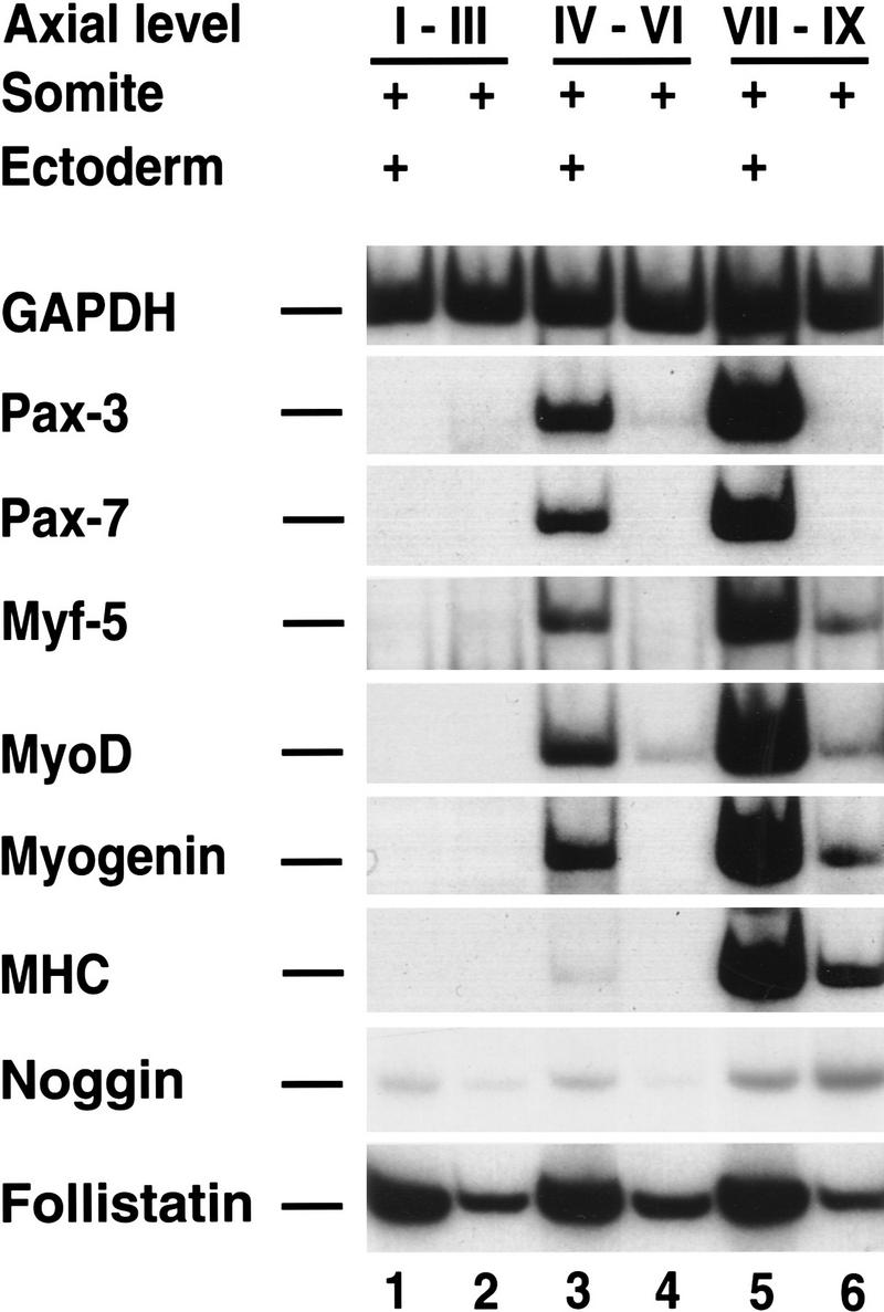
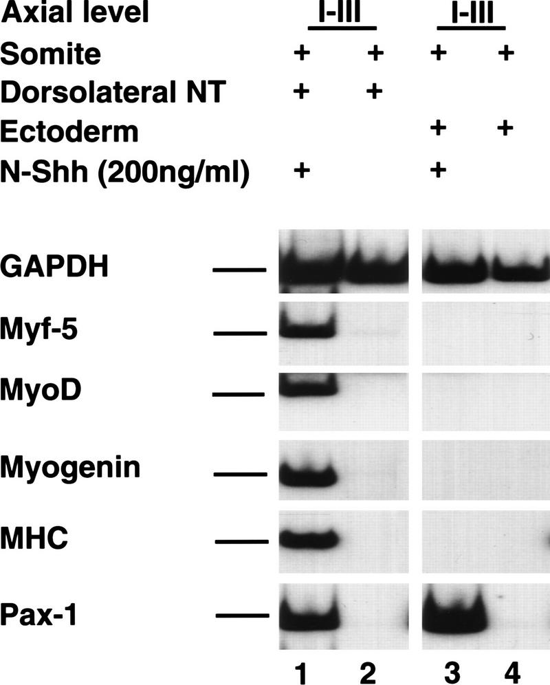
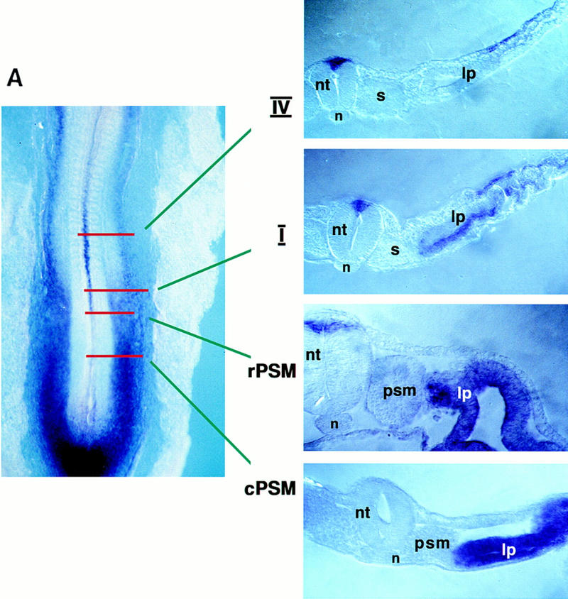
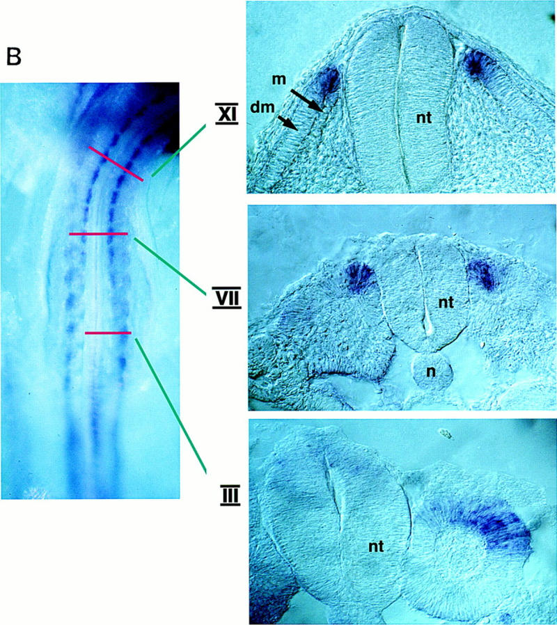
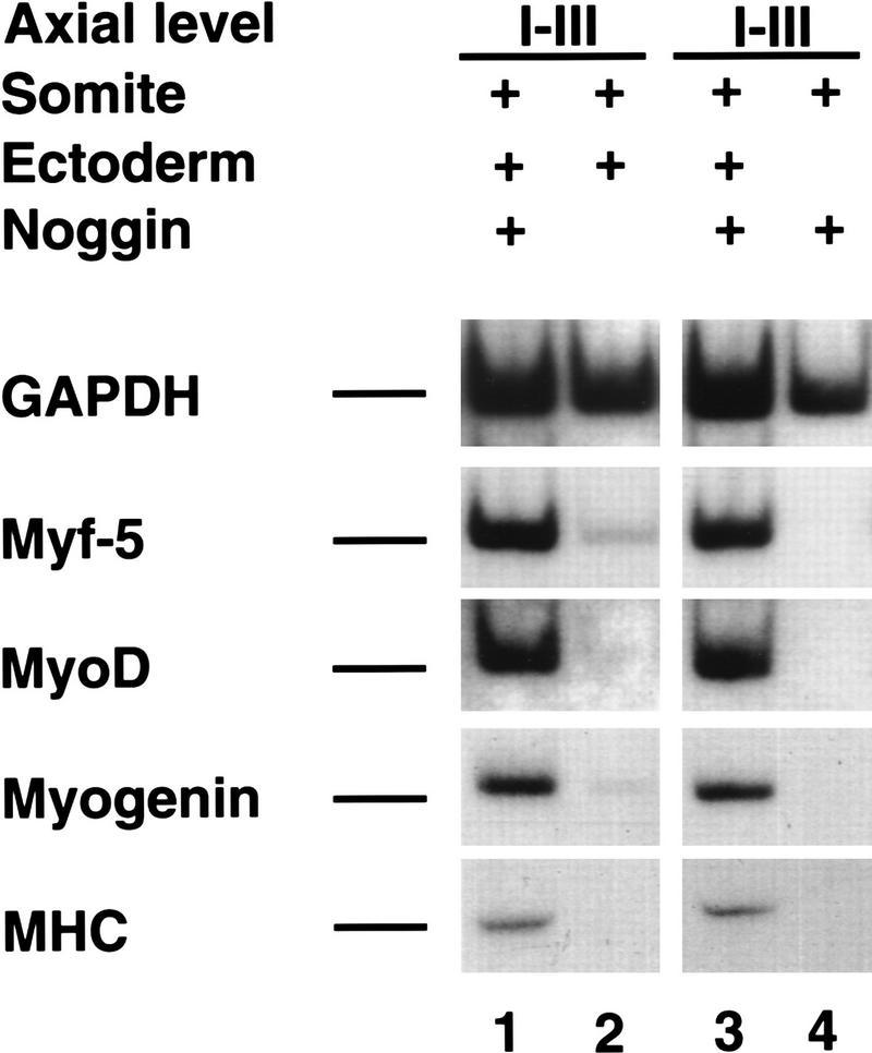
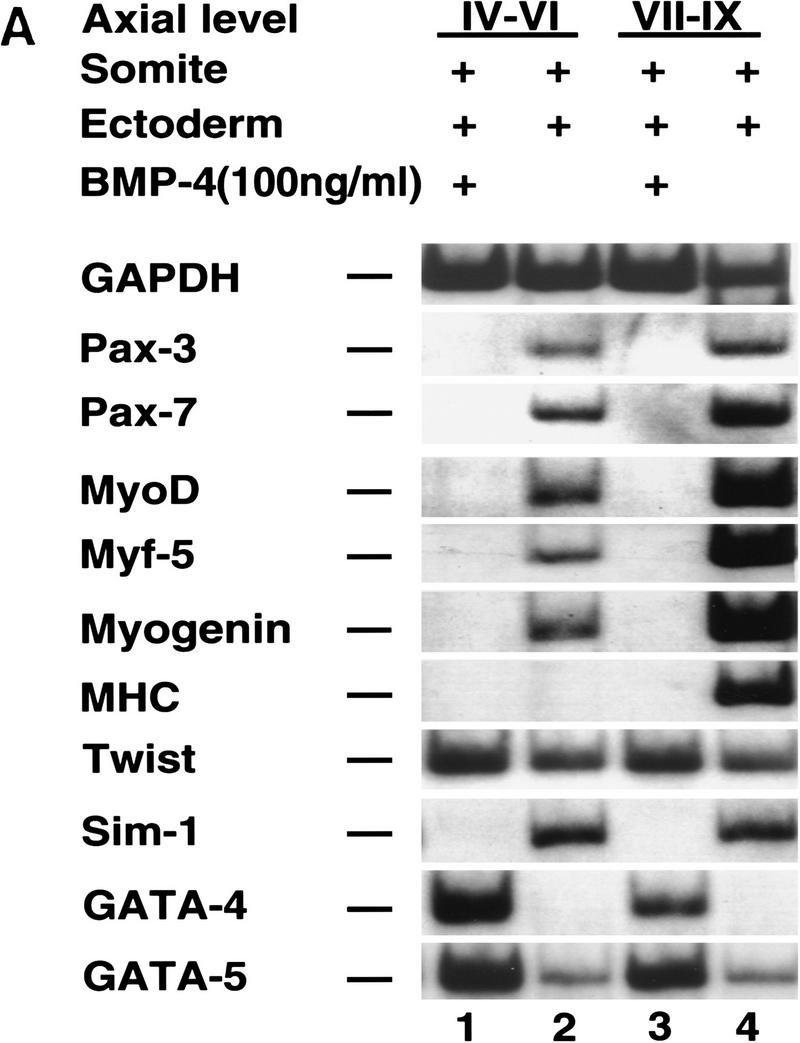
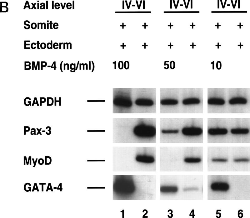
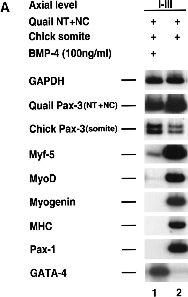
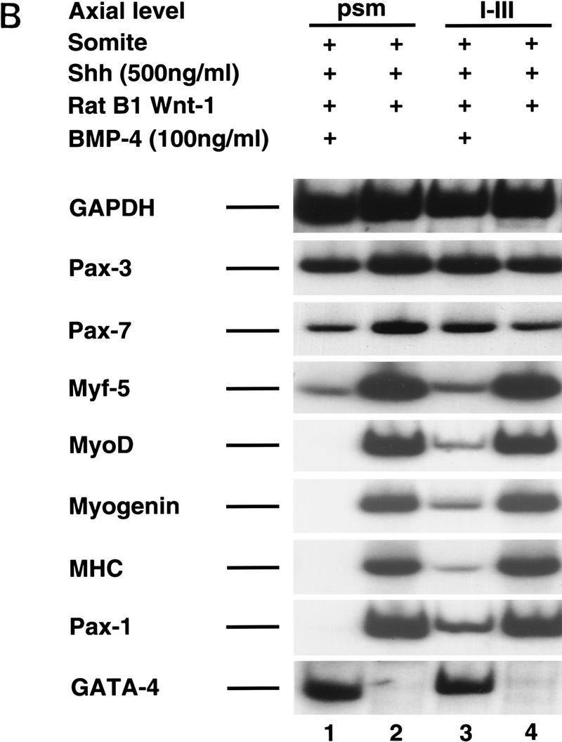
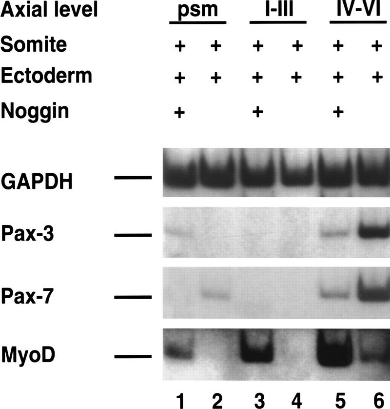
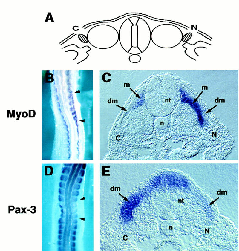
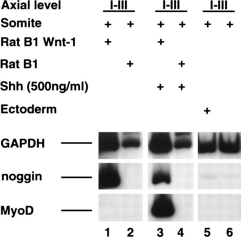
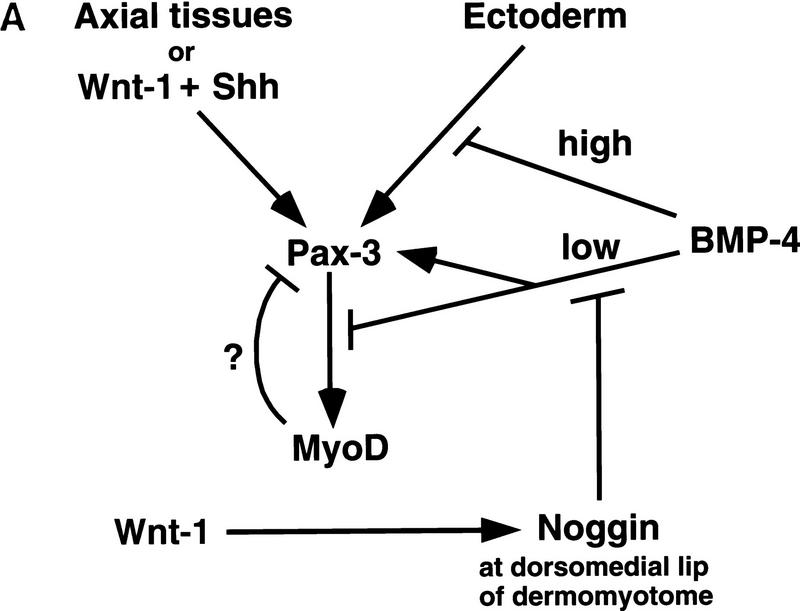
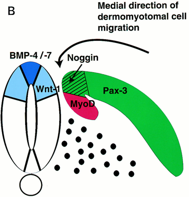
Previous work has indicated that signals from the neural tube, notochord, and surface ectoderm promote somitic myogenesis. Here, we show that somitic myogenesis is under negative regulation as well; BMP signaling serves to inhibit the activation of MyoD and Myf5 in Pax3-expressing cells. Furthermore, we show that the BMP antagonist Noggin is expressed within the dorsomedial lip of the dermomyotome, where Pax3-expressing cells first initiate the expression of MyoD and Myf5 to give rise to myotomal cells in the medial somite. Consistent with the expression of Noggin in dorsomedial dermomyotomal cells that lie adjacent to the dorsal neural tube, we have found that coculture of somites with fibroblasts programmed to secrete Wnt1, which is expressed in dorsal neural tube, can induce somitic Noggin expression. Ectopic expression of Noggin lateral to the somite dramatically expands MyoD expression into the lateral regions of the somite, represses Pax3 expression in this tissue, and induces formation of a lateral myotome. Together, our findings indicate that the timing and location of myogenesis within the somite is controlled by relative levels of BMP activity and localized expression of a BMP antagonist.


Figures

Figure 1
Figure 1
Signals from the surface ectoderm can activate somitic myogenesis in rostral but not in caudal paraxial mesoderm. Paraxial mesoderm isolated from differing axial levels of a stage 10 chick embryo was cultured for 72 hr either in the absence (lanes 2,4,6) or in the presence (lanes 1,3,5) of overlying ectoderm. Gene expression was analyzed by RT–PCR. Similar results were obtained in at least 10 independent experiments.
Figure 2
Figure 2
Signals from dorsolateral neural tube, but not from surface ectoderm induce myogenesis in caudal somites in the presence of Shh. Somites I–III isolated from a stage 10 chick embryo were cultured with either the adjacent dorsolateral neural tube (lacking the floor plate; lanes 1,2) or with the overlying ectoderm (lanes 3,4) in the presence (lanes 1,3) or absence (lanes 2,4) of 200 ng/ml of the amino-terminal fragment of Shh (N-Shh). Gene expression was analyzed by RT–PCR. Similar results were obtained in at least seven independent experiments.
Figure 3
Figure 3
In situ hybridization analysis of BMP4 (A) and Noggin (B) gene expression. (A) A whole mount in situ hybridization of a stage 11 chick embryo displaying the expression of BMP4 transcripts. Transverse sections show the expression of BMP4 at the indicated axial levels in tissues adjacent to the paraxial mesoderm: lateral plate mesoderm at caudal levels and roof plate and dorsal ectoderm at more rostral levels. (B) A whole mount in situ hybridization of a stage 13 chick embryo displaying the expression of Noggin transcripts. Transverse sections show the expression of Noggin at the indicated axial levels. Lateral somitic expression of Noggin is observed in the psm and in newly formed epithelial somites, dorsomedial expression of Noggin can be detected rostral to somite III. (dm) Dermomyotome; (lp) lateral plate; (m) myotome; (n) notochord; (nt) neural tube; (psm) presegmented mesoderm; (s) somite; (cPSM) caudal presegmented mesoderm; (rPSM) rostral presegmented mesoderm.
Figure 3
Figure 3
In situ hybridization analysis of BMP4 (A) and Noggin (B) gene expression. (A) A whole mount in situ hybridization of a stage 11 chick embryo displaying the expression of BMP4 transcripts. Transverse sections show the expression of BMP4 at the indicated axial levels in tissues adjacent to the paraxial mesoderm: lateral plate mesoderm at caudal levels and roof plate and dorsal ectoderm at more rostral levels. (B) A whole mount in situ hybridization of a stage 13 chick embryo displaying the expression of Noggin transcripts. Transverse sections show the expression of Noggin at the indicated axial levels. Lateral somitic expression of Noggin is observed in the psm and in newly formed epithelial somites, dorsomedial expression of Noggin can be detected rostral to somite III. (dm) Dermomyotome; (lp) lateral plate; (m) myotome; (n) notochord; (nt) neural tube; (psm) presegmented mesoderm; (s) somite; (cPSM) caudal presegmented mesoderm; (rPSM) rostral presegmented mesoderm.
Figure 4
Figure 4
Alleviation of BMP signaling by Noggin induces myogenesis in caudal somites cultured with ectoderm. Somites I–III isolated from a stage 10 chick embryo were cultured with overlying ectoderm (lanes 1–3) or cultured alone (lane 4) either in the presence of medium condition by Noggin secreting CHO cells (lanes 1,3,4) or medium conditioned by control CHO cells (lane 2). Gene expression was analyzed by RT–PCR. Similar results were obtained in at least eight independent experiments.
Figure 5
Figure 5
Varying BMP4 concentrations differentially affect the induction of Pax3 and MyoD in paraxial mesoderm by signals from the surface ectoderm. (A) Somites from the indicated axial level were isolated from a stage 10 chick embryo and cultured with overlying ectoderm in either the presence (lanes 1,3) or absence (lanes 2,4) of 100 ng/ml BMP4. Similar results were obtained in at least 10 independent experiments. (B) Somites IV–VI were isolated from a stage 10 chick embryo and cultured with overlying ectoderm in either the absence (lanes 2,4,6) or presence (lanes 1,3,5) of increasing concentrations of BMP4 as indicated. Gene expression was analyzed by RT–PCR. Similar results were obtained in at least four independent experiments.
Figure 5
Figure 5
Varying BMP4 concentrations differentially affect the induction of Pax3 and MyoD in paraxial mesoderm by signals from the surface ectoderm. (A) Somites from the indicated axial level were isolated from a stage 10 chick embryo and cultured with overlying ectoderm in either the presence (lanes 1,3) or absence (lanes 2,4) of 100 ng/ml BMP4. Similar results were obtained in at least 10 independent experiments. (B) Somites IV–VI were isolated from a stage 10 chick embryo and cultured with overlying ectoderm in either the absence (lanes 2,4,6) or presence (lanes 1,3,5) of increasing concentrations of BMP4 as indicated. Gene expression was analyzed by RT–PCR. Similar results were obtained in at least four independent experiments.
Figure 6
Figure 6
BMP4 blocks the muscle promoting activities of either the axial tissues or Wnt1 plus Shh downstream of Pax3 expression in paraxial mesoderm. (A) BMP4 blocks somitic myogenesis but not the induction of Pax3 or Pax7 by the axial tissues. Somites I–III were isolated from a chick embryo and recombined with neural tube and notochord isolated from the same axial level of a quail embryo and either cultured in the presence (lane 1) or absence (lane 2) of 100 ng/ml of BMP4. Gene expression was analyzed by RT–PCR. Chick and quail Pax3 PCR products were distinguished by a restriction enzyme polymorphism. Similar results were obtained in at least three independent experiments. (B) BMP4 blocks somitic myogenesis but not the induction of Pax3 or Pax7 by the combination of Wnt1 and Shh signals. Paraxial mesoderm from the indicated axial level was cultured in the presence of RatB1a cells programmed to express Wnt1 (RatB1a–Wnt1) plus 500 ng/ml of N-Shh in either the presence (lanes 1,3) or absence (lanes 2,4) of 100 ng/ml of BMP4. Gene expression was analyzed by RT–PCR. Similar results were obtained in at least four independent experiments.
Figure 6
Figure 6
BMP4 blocks the muscle promoting activities of either the axial tissues or Wnt1 plus Shh downstream of Pax3 expression in paraxial mesoderm. (A) BMP4 blocks somitic myogenesis but not the induction of Pax3 or Pax7 by the axial tissues. Somites I–III were isolated from a chick embryo and recombined with neural tube and notochord isolated from the same axial level of a quail embryo and either cultured in the presence (lane 1) or absence (lane 2) of 100 ng/ml of BMP4. Gene expression was analyzed by RT–PCR. Chick and quail Pax3 PCR products were distinguished by a restriction enzyme polymorphism. Similar results were obtained in at least three independent experiments. (B) BMP4 blocks somitic myogenesis but not the induction of Pax3 or Pax7 by the combination of Wnt1 and Shh signals. Paraxial mesoderm from the indicated axial level was cultured in the presence of RatB1a cells programmed to express Wnt1 (RatB1a–Wnt1) plus 500 ng/ml of N-Shh in either the presence (lanes 1,3) or absence (lanes 2,4) of 100 ng/ml of BMP4. Gene expression was analyzed by RT–PCR. Similar results were obtained in at least four independent experiments.
Figure 7
Figure 7
BMP signals attenuate MyoD induction by surface ectoderm at varying axial levels. Paraxial mesoderm was isolated from the indicated axial level and cultured with the overlying ectoderm in the presence of medium condition by Noggin-secreting CHO cells (lanes 1,3,5) or medium conditioned by control CHO cells (lanes 2,4,6). Gene expression was analyzed by RT–PCR. Similar results were obtained in at least three independent experiments.
Figure 8
Figure 8
Alleviation of BMP signals by Noggin expands MyoD expression within the dermomyotome. Implantation of Noggin-expressing COS cells between the paraxial and lateral plate mesoderm leads to the expansion of MyoD expression in the lateral regions of the dermomyotome and down-regulation of Pax3 expression in this domain. COS cells transfected with either a Noggin expression vehicle or a control vehicle were implanted between the presegmental plate and lateral plate mesoderm of a stage 10 chick embryo. Noggin (N)-transfected cells were implanted on the right side and control (C)-transfected COS cells were implanted on the left side of the operated embryo (position of COS cell pellets diagrammed in A). The embryo was allowed to develop in ovo for 15 hr until the implanted COS cells were approximately adjacent to somite X. Whole mount in situ hybridization was performed for MyoD (B,C) and Pax3 (D,E). (dm) Dermomyotome; (m) myotome; (n) notochord; (nt) neural tube. Similar results were obtained in at least six independent experiments.
Figure 9
Figure 9
Wnt1 signals are sufficient to induce somitic Noggin expression. Somites I–III from a stage 10 chick embryo were cultured either in the presence of RatB1a cells programmed to express Wnt1 (RatB1a–Wnt1; lanes 1,3) or the parental RatB1a cells (lanes 2,4), in either the absence (lanes 1,2) or presence (lanes 3,4) of 500 ng/ml of N-Shh. In addition, somites I–III were cultured with either overlying ectoderm (lane 5) or alone (lane 6). Gene expression was analyzed by RT–PCR. Similar results were obtained in at least three independent experiments.
Figure 10
Figure 10
(A) Diagram of regulatory networks controlling somitic expression of Pax3 and MyoD. Combinatorial signals from the dorsolateral neural tube and floor plate/notochord or signals from the surface ectoderm activate Pax3 expression and, subsequently, Pax3 induces MyoD. We speculate that high levels of BMP signals in lateral plate mesodermal tissue serve to block the induction of Pax3 in this tissue by signals from the surface ectoderm. In contrast, in somitic tissue, lower levels of BMP signaling are both necessary to maintain Pax3 expression in response to signals from either the axial tissues or the surface ectoderm and to block MyoD expression within the dermomyotome. Potential sources of these BMP signals are the epidermal ectoderm that overlies the invaginating paraxial mesoderm and expresses BMP4 and BMP7 (Liem et al. 1995; Schultheiss et al. 1997); the lateral plate mesoderm that expresses BMP4 (Pourquié et al. 1996); the dorsal neural tube that expresses BMP2, BMP7, and Dorsalin (Liem et al. 1995); the mesonephric duct that expresses BMP2 (R. Reshef, M. Maroto, and A.B. Lassar, data not shown); and the dermomyotome of mature somites that expresses BMP4 (A. McMahon, pers. comm.) and BMP2 in the dorsomedial lip of this tissue (R. Reshef, M. Maroto, and A.B. Lassar, data not shown). Our data suggests that Wnt signals sent from the dorsal neural tube induce Noggin expression at the dorsomedial lip of the dermomyotome, and that Noggin acts in combination with other BMP antagonists expressed within the somite (i.e., Follistatin) to relieve a BMP-mediated inhibition of MyoD expression and thereby initiates MyoD expression and myotome formation. For simplicity, Myf5 is not shown in this diagram; however, our data indicate that BMP signals similarly repress the expression of Myf5 by signals from either the axial tissues or the surface ectoderm. (B) Diagram of the domains of dermomyotomal gene expression. The somitic expression domains of Pax3, Noggin, and MyoD are indicated within the dermomyotome, the dorsomedial lip of the dermomyotome, and the myotome, respectively. Also diagrammed is the expression domain of BMP4, BMP7, and Wnt1 in the neural tube.
Figure 10
Figure 10
(A) Diagram of regulatory networks controlling somitic expression of Pax3 and MyoD. Combinatorial signals from the dorsolateral neural tube and floor plate/notochord or signals from the surface ectoderm activate Pax3 expression and, subsequently, Pax3 induces MyoD. We speculate that high levels of BMP signals in lateral plate mesodermal tissue serve to block the induction of Pax3 in this tissue by signals from the surface ectoderm. In contrast, in somitic tissue, lower levels of BMP signaling are both necessary to maintain Pax3 expression in response to signals from either the axial tissues or the surface ectoderm and to block MyoD expression within the dermomyotome. Potential sources of these BMP signals are the epidermal ectoderm that overlies the invaginating paraxial mesoderm and expresses BMP4 and BMP7 (Liem et al. 1995; Schultheiss et al. 1997); the lateral plate mesoderm that expresses BMP4 (Pourquié et al. 1996); the dorsal neural tube that expresses BMP2, BMP7, and Dorsalin (Liem et al. 1995); the mesonephric duct that expresses BMP2 (R. Reshef, M. Maroto, and A.B. Lassar, data not shown); and the dermomyotome of mature somites that expresses BMP4 (A. McMahon, pers. comm.) and BMP2 in the dorsomedial lip of this tissue (R. Reshef, M. Maroto, and A.B. Lassar, data not shown). Our data suggests that Wnt signals sent from the dorsal neural tube induce Noggin expression at the dorsomedial lip of the dermomyotome, and that Noggin acts in combination with other BMP antagonists expressed within the somite (i.e., Follistatin) to relieve a BMP-mediated inhibition of MyoD expression and thereby initiates MyoD expression and myotome formation. For simplicity, Myf5 is not shown in this diagram; however, our data indicate that BMP signals similarly repress the expression of Myf5 by signals from either the axial tissues or the surface ectoderm. (B) Diagram of the domains of dermomyotomal gene expression. The somitic expression domains of Pax3, Noggin, and MyoD are indicated within the dermomyotome, the dorsomedial lip of the dermomyotome, and the myotome, respectively. Also diagrammed is the expression domain of BMP4, BMP7, and Wnt1 in the neural tube.

Similar articles

Cited by

References

    1. Amthor H, Connolly D, Patel K, Brand-Saberi B, Wilkinson DG, Cooke J, Christ B. The expression and regulation of follistatin and a follistatin-like gene during avian somite compartmentalization and myogenesis. Dev Biol. 1996;178:343–362. - PubMed
    1. Avery G, Chow M, Holtzer H. An experimental analysis of the development of the spinal column. J Exp Zool. 1956;132:409–425.
    1. Barth J, Ivarie R. Polyvinyl alcohol enhances detection of low abundance transcripts in early stage quail embryos in a nonradioactive whole mount in situ hybridization technique. Biotechniques. 1994;17:324–327. - PubMed
    1. Basler K, Edlund T, Jessel T, Yamada T. Control of cell pattern in the neural tube: Regulation of cell differentiation by dorsalin-1, a novel TGF-β family member. Cell. 1993;73:687–702. - PubMed
    1. Buckingham M. Making muscle in mammals. Trends Genet. 1992;8:144–149. - PubMed

Publication types

MeSH terms

Substances

LinkOut - more resources