. 1998 Feb 1;12(3):304-15.
doi: 10.1101/gad.12.3.304.

Inscuteable and numb mediate asymmetric muscle progenitor cell divisions during Drosophila myogenesis

Affiliations

Inscuteable and numb mediate asymmetric muscle progenitor cell divisions during Drosophila myogenesis

A Carmena et al. Genes Dev. .

Erratum in

  • Genes Dev 1998 Apr 15;12(8):1241

Abstract

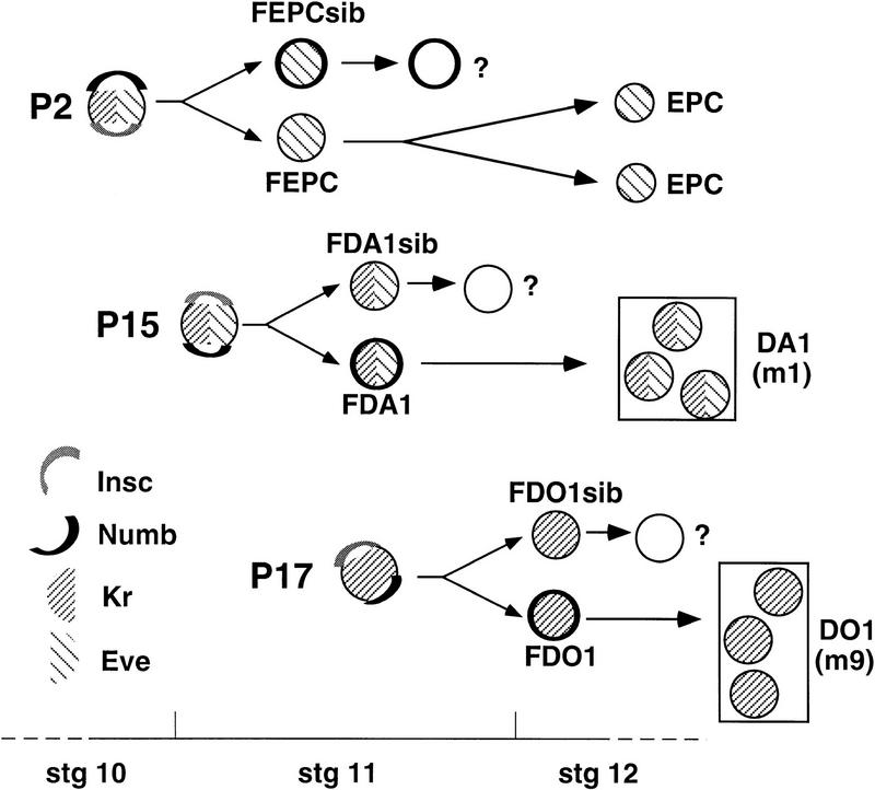
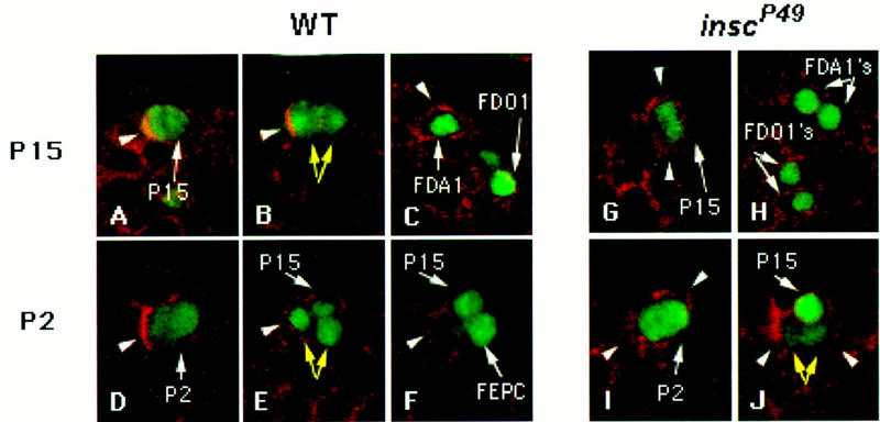
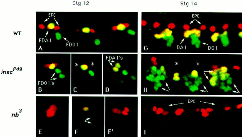
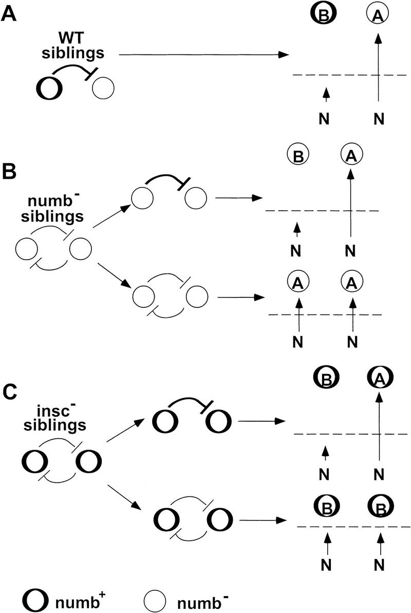
Each larval hemisegment comprises approximately 30 uniquely specified somatic muscles. These derive from muscle founders that arise as distinct sibling pairs from the division of muscle progenitor cells. We have analyzed the progenitor cell divisions of three mesodermal lineages that generate muscle (and pericardial cell) founders. Our results show that Inscuteable and Numb proteins are localized as cortical crescents on opposite sides of dividing progenitor cells. Asymmetric segregation of Numb into one of the sibling myoblasts depends on inscuteable and is essential for the specification of distinct sibling cell fates. Loss of numb or inscuteable results in opposite cell fate transformations-both prevent sibling myoblasts from adopting distinct identities, resulting in duplicated or deleted mesodermal structures. Our results indicate that the muscle progenitor cell divisions are intrinsically asymmetric; moreover, the involvement of both inscuteable and numb/N suggests that the specification of the distinct cell fates of sibling myoblasts requires intrinsic and extrinsic cues.


Figures

Figure 1
Figure 1
Three progenitors and their mesodermal derivatives. P2, P15, and P17 can be identified using anti-Kr and/or anti-Eve. Each localizes Insc (gray) and Nb (black) as crescents on opposite sides of the cell cortex just before division and divides to produce two sibling cells only one of which inherits Nb. In the case of P2, it is the Nb progeny that retains Eve expression, becomes the founder of the Eve+ pericardial cells (FEPC), and divides to generate the EPC. Its Nb+ sibling (FEPCsib) initially expresses Eve but soon extinguishes marker expression and its fate is unknown. For P15 and P17, the Nb+ daughter cell retains marker expression and become founders (FDA1 and FDO1) for the muscle DA1 (=m1) and the muscle DO1 (=m9), respectively. Their Nb siblings (FDA1sib and FDO1sib) extinguish marker expression and their fates are unknown. The stages during development when these cells can be detected and the schematic representations used for Kr, Eve, Nb, and Insc are indicated. Our lineage model is based on a careful analysis of wild-type embryos at different stages with several markers.
Figure 2
Figure 2
Insc protein accumulates as cortical crescents in progenitors of the mesoderm. Embryos were stained for Insc (red) and either L’sc (A, C–E, green), Kr (B, green), or DNA (F, green). Anterior is left and dorsal is up in all panels. Insc crescents in muscle progenitors do not have a fixed orientation (see arrowheads in A showing Insc crescents in two different progenitors within a hemisegment) but localize to similar positions in a given type of progenitor (B, cf. arrowheads showing Insc crescents in P17 in two adjacent hemisegments). All three progenitors analyzed in this study, P2, P15, and P17, enlarged their nuclei and display Insc crescents shortly before division (arrows in C–E). (F) A metaphase progenitor stained for Insc (arrowhead) and DNA (yellow arrow) to show the location of the Insc crescent relative to (the deduced orientation of) the mitotic spindle.
Figure 3
Figure 3
Numb protein becomes polarized in muscle progenitors and segregates preferentially with one of the two progeny cells. Embryos were stained for Nb (red) and either Kr (A–D, green) or Insc (E, green). Anterior is left and dorsal is up in all panels. (A) A lateral view of two hemisegments of the mesoderm. Each hemisegment contains a domain of high Nb expression (white arrows) from which progenitors segregate. Some progenitors express Kr, and their nuclei enlarge as they enter mitosis (yellow arrow). Kr is also detected in pairs of postmitotic cells (yellow arrowheads), derived from recently divided progenitors. (B) P7 in hemisegments 1, 2, and 4 (from left to right) showing Nb crescents (white arrowheads) at the anterior half of each of these progenitors. In hemisegment 3, P7 has already divided and the two sibling progeny (yellow arrowheads) can be better seen at a deeper plane of focus (see inset); the progeny placed anteriorly has inherited Nb. (C) Polarization of Nb as a cortical crescent (arrowhead) in P17 as it begins to divide. (D) After division, the two progeny cells of P17 (arrows) still express Kr. However, the cell that has presumably inherited parental Nb (arrowhead) retains higher levels of Kr than its Nb sibling. (E) The Nb cortical crescent (arrowhead) is located opposite the Insc cortical crescent (arrow) in muscle progenitors.
Figure 4
Figure 4
The effects of various genotypes on the number of EPCs, DA1s, and DO1S. (A–D,G) Embryos were double stained for Eve (black nuclear stain) and MHC (stains all muscles) using horseradish peroxidase-conjugated secondary antibodies. (AB44) The P-lacZ insertion from which inscP49 and inscP72 excisions were generated, showed little muscle defects and was used to represent wild type. The EPCs express Eve from stage 12 onward, and there are usually two per hemisegment in wild type (A). Their numbers increase in nb embryos (B) and in the insc,nb double mutant (G). In insc embryos (C) and in embryos overexpressing Nb (D), a significant reduction of EPC is seen. Because Eve expression in DA1 diminishes after stage 15, embryos at stages 14–15 were used for scoring this muscle (see top panels for DA1s). Additional DA1s are seen in insc mutants and in embryos overexpressing Nb, whereas they are essentially eliminated in nb3 embryos. In the insc,nb double mutant, there is a complete loss of all dorsal muscles, DA1, DO1, DA2 (m2), and DO2 (m10), as seen by the absence of MHC-stained somatic muscles in the dorsal region at stages 15–16 (G). Muscle DO1 was identified by its position at stage 16. Its numbers were reduced in nb mutants (*). In contrast, additional dorsal oblique muscles were present in insc and in embryos overexpressing Nb (bottom panels). However, in such cases, we could not distinguish between DO1 and DO2. (E,F) Confocal images showing inscP49 embryos where Insc was expressed under the control of the hsp70 promoter. Embryos were stained with anti-β-galactosidase and either anti-MHC (E) or anti-Eve (F), and mutant embryos homozygous for both inscP49 and hsp70–insc were identified by the absence of β-galactosidase expression from lacZ-marked balancers. The hemisegments shown in E and F exhibit wild-type musculature; in F rescue of the EPC phenotype is seen in all but the middle hemisegment. For quantitation of the various phenotypes, see Table 1. For all panels, anterior is toward the left.
Figure 5
Figure 5
Segregation of Nb and progression of Kr expression in the descendants of progenitors in wild-type and insc mutant embryos. Wild-type (A–F) and inscP49 (G–J) embryos were stained for Nb (red) and either Kr (green) (A–C,G,H) or Eve (green) (D–F,I,J). (A) At early stage 11, a Nb crescent (arrowhead) is observed in P15 (arrow), which also expresses Kr. (B) As P15 divides to give two daughter cells (yellow arrows), Nb segregates with one of the progeny (arrowhead) that is larger than its sibling. (C) At early stage 12, Kr is only detected in the descendant from P15 that inherits Nb (FDA1, arrowhead); its sibling has already extinguished Kr expression. The progeny of P17 are also detected. Note that Kr expression is maintained in the Nb+ FDO1 but has already decayed considerably in its Nb sibling. (G) In early stage 11 inscP49 embryos, Nb (arrowheads) is not polarized and is distributed throughout the cortex of a dividing P15. (H) In an early stage 12 inscP49 mutant embryo, siblings derived from P15 and P17 both inherit Nb and maintain similar high levels of Kr expression, characteristics of FDA1 and FDO1, respectively. (D) A Nb crescent (arrowhead) in wild-type P2 that expresses Eve as it begins to divide at late stage 10. (E) After P2 divides at early stage 11, Nb (arrowhead) segregates with one of the two progeny cells (yellow arrows); at this time, P15, which also expresses Eve, has been singled out but has not yet enlarged. (F) Soon thereafter, Eve expression is extinguished from the Nb+ progeny of P2 (arrowhead). The Nb sibling (FEPC) is the precursor of the two EPCs. (I) In a inscP49 embryo, a dividing P2 does not exhibit Nb polarization (arrowheads). (J) Both P2 progeny cells (yellow arrows, just ventral to P15) appear to have inherited Nb (arrowheads), and Eve expression is decaying in both cells.
Figure 6
Figure 6
The fate of the progeny from P2, P15, and P17 progenitors in wild-type, inscP49, and numb3 embryos. Embryos were double stained for Eve (red) and Kr (green). Stage 12 (A–F′) and stage 14 (G–I) wild-type (A,G), inscP49 (B–D,H), and nb3 (E–F′, I) embryos are shown. (A) Three consecutive wild-type hemisegments at mid- to late-stage 12. At this stage, the two EPC (red) are already present. The Kr+ Eve+ FDA1 (yellow) and the Kr+ FDO1 (green) are also evident. (B–D) In inscP49 embryos, the incomplete expressivity of the mutant phenotype is evident in different hemisegments and is characterized by duplication of FDO1 (B), loss of the two EPC (C, *), and duplication of the FDA1 (D). (E–F′) The opposite phenotype is found in nb3 embryos: Two FEPCs are detected at early stage 12, which are enlarging to divide (E), and no putative FDA1 and FDO1 are detected that express Eve and/or Kr. At mid-stage 12, extra EPCs are detected (F′). (F,F′) Two different focal planes of the same mutant hemisegment at mid-stage 12. (F) The FDA1 (yellow cells) is losing Eve and Kr expression; likewise Kr expression is decaying in both siblings produced by division of P17 (arrows). (G) The characteristic pattern of EPC and precursors of DA1 and DO1 in a wild-type embryo at stage 14. (H) In an inscP49 embryo, loss of EPC (*) and duplication of the precursors of muscles DA1 (yellow syncytia, arrows) and DO1 (green syncytia, arrows) are evident. (I) The opposite phenotype is observed in a nb3 embryo: extra EPCs and the absence of DA1 and DO1 muscle precursors.
Figure 7
Figure 7
A model to explain the phenotypes of nb and insc loss of function. (A) In wild type (WT) sibling pairs, the presence of Nb in only one cell ensures a sufficiently large difference in N signaling between the two cells such that the feedback mechanism mediated by N/Dl always becomes irreversible (see text). As a consequence, the level of N signaling (represented by vertical arrows) is maximal in the Nb cell and minimal in the Nb+ cell. Because the level of N signaling exceeds the necessary threshold (broken line) in the Nb cell, it adopts an A-type fate; its Nb+ sibling adopts the B-type fate because its level of N signaling falls below the threshold required for an A-type fate. In this context, where B represents the primary fate, FDA1, FDO1, and FEPCsib cells all have B-type fate. (B) In nb siblings a sufficiently large difference in the level of N signaling is not guaranteed, as neither cell inherits functional Nb. Nevertheless, the N/Dl feedback mechanism can become irreversible some of the time (see text). When this happens (top alternative) a wild-type situation ensues. However, if a sufficiently large difference in N signaling never arises and the feedback mechanism is never resolved (bottom alternative), the default level of N signaling in both cells is sufficiently above the postulated threshold required to specify an A-type fate. (C) Both sibling cells in insc embryos are Nb+ due to the failure of mutant progenitors to asymmetrically localize and segregate Nb. Hence, the difference in the level of N signaling required to irreversibly resolve the feedback mechanism can only arise through stochastic variations (top alternative) and statistically this will occur only a proportion of the time. When this fails (bottom alternative), the default level of N signaling in the siblings (due to the presence of Nb in both cells) falls below the threshold required to specify an A-type fate and both cells adopt the default B-type fate.

Similar articles

Cited by

References

    1. Artavanis-Tsakonas A, Matsuno K, Fortini ME. Notch signaling. Science. 1995;268:225–232. - PubMed
    1. Baker R, Schubiger G. Ectoderm induces muscle-specific gene expression in Drosophila embryos. Development. 1995;121:1387–1398. - PubMed
    1. Bate M. The embryonic development of larval muscles in Drosophila. Development. 1990;110:791–804. - PubMed
    1. ————— . The mesoderm and its derivatives. In: Bate M, Martínez-Arias A, editors. The development of Drosophila melanogaster. Cold Spring Harbor, NY: Cold Spring Harbor Laboratory Press; 1993. pp. 1013–1090.
    1. Bate, M., E. Rushton, and M. Frasch. 1993. A dual requirement for neurogenic genes in Drosophila myogenesis. Development (Suppl.) pp. 149–161. - PubMed

Publication types

MeSH terms

LinkOut - more resources