. 1998 Feb;111(2):243-55.
doi: 10.1085/jgp.111.2.243.

Rabphilin-3A: a multifunctional regulator of synaptic vesicle traffic

Affiliations

Rabphilin-3A: a multifunctional regulator of synaptic vesicle traffic

M E Burns et al. J Gen Physiol. 1998 Feb.

Abstract

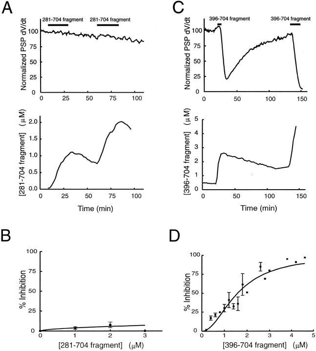
We have investigated the function of the synaptic vesicle protein Rabphilin-3A in neurotransmitter release at the squid giant synapse. Presynaptic microinjection of recombinant Rabphilin-3A reversibly inhibited the exocytotic release of neurotransmitter. Injection of fragments of Rabphilin-3A indicate that at least two distinct regions of the protein inhibit neurotransmitter release: the NH2-terminal region that binds Rab3A and is phosphorylated by protein kinases and the two C2 domains that interact with calcium, phospholipid, and beta-adducin. Each of the inhibitory fragments and the full-length protein had separate effects on presynaptic morphology, suggesting that individual domains were inhibiting a subset of the reactions in which the full-length protein participates. In addition to inhibiting exocytosis, constructs containing the NH2 terminus of Rabphilin-3A also perturbed the endocytotic pathway, as indicated by changes in the membrane areas of endosomes, coated vesicles, and the plasma membrane. These results indicate that Rabphilin-3A regulates synaptic vesicle traffic and appears to do so at distinct stages of both the exocytotic and endocytotic pathways.


Figures

Figure 4
Figure 4
Rabphilin-3A redistributes membranes among organelles of the synaptic vesicle cycle. The surface area of several organelles was measured in 20 sections taken from terminals injected with either full-length Rabphilin-3A (FL), the 1–280 fragment (N), the 396–704 fragment (C), or control solutions (Con). Although these treatments produced significant changes (*P < 0.05) in the membrane area of individual organelles, the total amount of organellar membrane (bottom right) was conserved.
Figure 1
Figure 1
Rabphilin-3A inhibits neurotransmitter release. Microinjection of Rabphilin-3A reversibly inhibited the postsynaptic potential PSP evoked by presynaptic action potentials (A). The time course of Rabphilin-3A inhibition of PSP slope (d  V/dt), normalized to the maximum value measured during the experiment (B), was correlated with the amount of protein that was microinjected. Microinjection of Rabphilin-3A during the time indicated by solid horizontal bars (top) was monitored by coinjection of 10 kD fluorescein dextran, which was used to estimate the concentration of injected protein (bottom; see methods). The extent of inhibition was a function of the intracellular concentration of Rabphilin-3A, measured at the time of peak inhibition and at 10 min before and after (C). These data were fit with the Hill equation; half-maximal inhibition required ∼500 nM Rabphilin-3A. The data points are the mean ± SEM of one to three injections to each concentration.
Figure 2
Figure 2
Rabphilin-3A injection alters presynaptic structure. Representative images of active zones from terminals injected with either buffer alone or the HA tag (Control), full-length Rabphilin-3A (Rabphilin-3A), the 1–280 (NH2-terminal) fragment, or the 396–704 (COOH-terminal) fragment. CV, coated vesicle; ISE, irregularly-shaped endosome; VE, vesicular endosome.
Figure 3
Figure 3
Differential actions of Rabphilin-3A fragments on the spatial distribution of synaptic vesicles. The mean number of synaptic vesicles (SVs) found in 50-nm spatial compartments within the active zone (AZ) was determined for presynaptic terminals injected with control solutions or Rabphilin-3A proteins (left). These data are also expressed as a percentage of the total number of vesicles at a given active zone (right). Comparison of terminals injected with buffer alone (n = 482 active zones) or full-length Rabphilin-3A (n = 321 active zones) revealed no change in the spatial distribution of synaptic vesicles despite full inhibition of transmitter release (A). The spatial distribution of vesicles at active zones from terminals injected with the 1–280 fragment (B; n = 423 active zones) is significantly different from that of controls injected with buffer alone or an inactive NH2-terminal fragment (n = 300 active zones). Likewise, the 396–704 fragment (C; n = 381 active zones) also differed from controls injected with the HA tag (n = 356 active zones). Both fragments reduced vesicle number in each spatial compartment (left) and caused a relative accumulation of vesicles 50–100 nm from the plasma membrane (right).
Figure 5
Figure 5
The NH2-terminal fragment of Rabphilin-3A inhibits transmitter release. Fragments encompassing the various domains of Rabphilin-3A were selected for microinjection (A). Microinjection of the 1–280 fragment inhibited neurotransmitter release in a manner correlated with the time and extent of injection (B). The degree of inhibition was a saturating function of the 1–280 fragment, with half-maximal inhibition occurring at 0.8 μM (C). The data points are the mean ± SEM of one to seven injections to each concentration.
Figure 6
Figure 6
The C2 domains of Rabphilin-3A regulate transmitter release. Microinjection of the 281–704 fragment of Rabphilin-3A has no effect on neurotransmitter release (A), even at concentrations of several micromolar (B; one to five injections to each concentration). However, microinjection of the 396–704 fragment did inhibit release reversibly (C). This inhibition was dose dependent (D ; one to seven injections to each concentration), with half-maximal inhibition requiring injection of ∼1.7 μM of this fragment.

References

    1. Augustine GJ, Burns ME, DeBello WM, Pettit DL, Schweizer FE. Exocytosis: proteins and perturbations. Annu Rev Pharmacol Toxicol. 1996;36:659–701. - PubMed
    1. Augustine GJ, Charlton MP, Horn R. Role of calcium-activated potassium channels in transmitter release and the squid giant synapse. J Physiol (Camb) 1988;398:149–164. - PMC - PubMed
    1. Augustine GJ, Charlton MP, Smith SJ. Calcium entry into voltage-clamped presynaptic terminals of squid. J Physiol (Camb) 1985;367:143–162. - PMC - PubMed
    1. Augustine GJ, Eckert R. Divalent cations differentially support transmitter release at the squid giant synapse. J Physiol (Camb) 1984;346:257–271. - PMC - PubMed
    1. Augustine GJ, Neher E. Calcium requirements for secretion in bovine chromaffin cells. J Physiol (Camb) 1992;450:247–271. - PMC - PubMed

Publication types

MeSH terms