. 1998 Feb;111(2):381-94.
doi: 10.1085/jgp.111.2.381.

Ligand-insensitive state of cardiac ATP-sensitive K+ channels. Basis for channel opening

Affiliations

Ligand-insensitive state of cardiac ATP-sensitive K+ channels. Basis for channel opening

A E Alekseev et al. J Gen Physiol. 1998 Feb.

Abstract

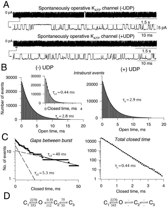
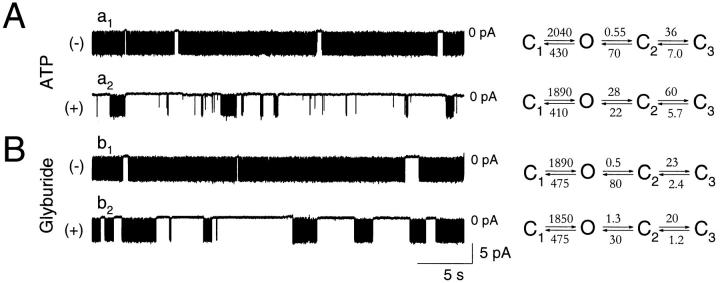
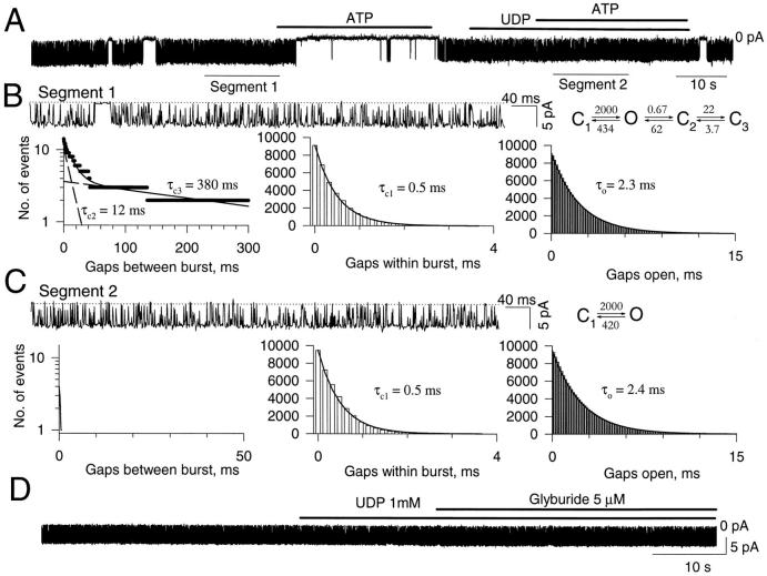
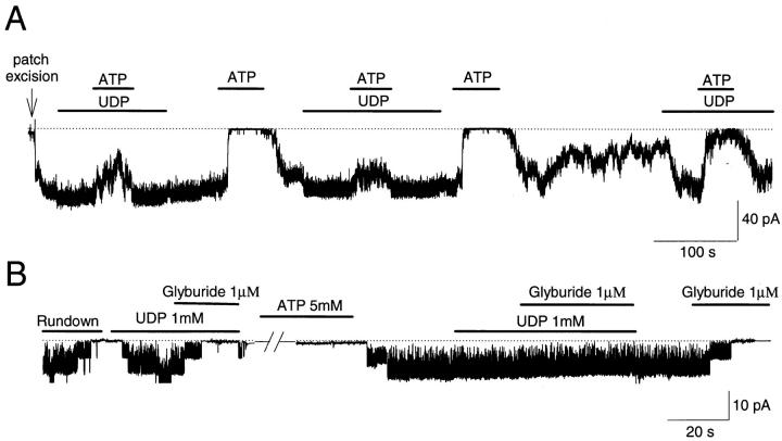
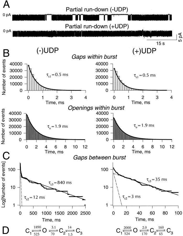
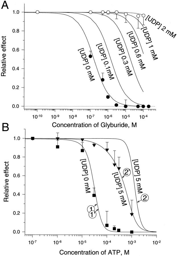
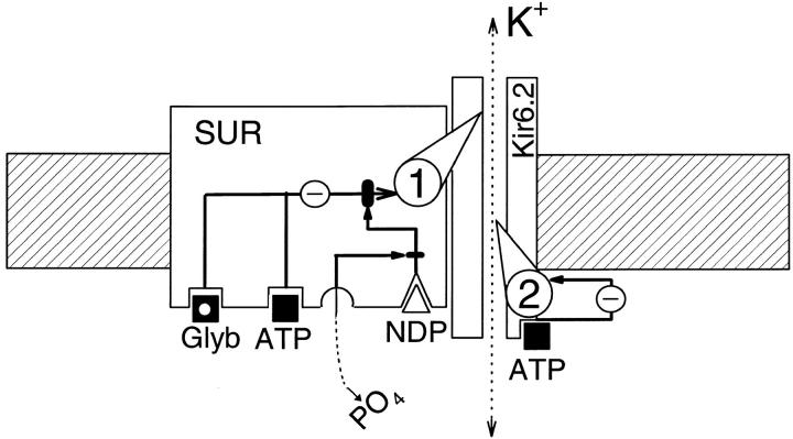
The mechanism by which ATP-sensitive K+ (KATP) channels open in the presence of inhibitory concentrations of ATP remains unknown. Herein, using a four-state kinetic model, we found that the nucleotide diphosphate UDP directed cardiac KATP channels to operate within intraburst transitions. These transitions are not targeted by ATP, nor the structurally unrelated sulfonylurea glyburide, which inhibit channel opening by acting on interburst transitions. Therefore, the channel remained insensitive to ATP and glyburide in the presence of UDP. "Rundown" of channel activity decreased the efficacy with which UDP could direct and maintain the channel to operate within intraburst transitions. Under this condition, the channel was sensitive to inhibition by ATP and glyburide despite the presence of UDP. This behavior of the KATP channel could be accounted for by an allosteric model of ligand-channel interaction. Thus, the response of cardiac KATP channels towards inhibitory ligands is determined by the relative lifetime the channel spends in a ligand-sensitive versus -insensitive state. Interconversion between these two conformational states represents a novel basis for KATP channel opening in the presence of inhibitory concentrations of ATP in a cardiac cell.


Figures

Figure 1
Figure 1
UDP induces loss of interburst transition. (A) Single KATP channel records (80 s in duration) in the absence (top) and presence (bottom) of 1 mM UDP. Records under both conditions are presented on compressed and extended time scales. Zero-current level is indicated by 0 pA in the compressed, and by a dotted line in the extended time record. (B, left) In the absence of UDP, open and closed (inset) time distributions of intraburst events were fitted by single exponents (solid lines) with characteristic times τo andτc,1. (right) Distribution of openings within a burst in the presence of 1 mM of UDP. (C, left) In the absence of UDP, distribution of gaps between bursts was fitted by the sum of two exponents (solid line) with characteristic times τc,2 andτc,3. Dashed lines correspond to individual exponents. (right) In the presence of UDP, there were no interburst events, and the distribution of closed times could be fitted by a single exponent with a characteristic time, τc, essentially identical to the mean closed time for intraburst events (τc,1) obtained in the absence of UDP. Kinetic scheme constructed based on calculated rates of transitions (in s−1) using Eq. 8 in the absence (left) and presence (right) of 1 mM UDP. Holding potential was −60 mV.
Figure 2
Figure 2
Inhibitory ligands act on KATP channels outside intraburst transition. Portions of original single channel records in the absence (a1 and b1) and presence (a2) of 100 μM ATP or 1 μM glyburide (b2). Corresponding kinetic schemes with calculated rates of transitions (in s−1, Eq. 8) are provided for each record in the absence and presence of ATP (A) and in the absence and presence of glyburide (B). Holding potential was −60 mV throughout.
Figure 3
Figure 3
Loss of interburst events induced by UDP associated with loss of ATP or glyburide sensitivity of KATP channels. (A) Single-channel record of KATP channel activity inhibited by ATP (200 μM) in the absence but not in the presence of UDP (1 mM). (B) Single-channel record of segment 1 (in A) presented on an extended time scale with distributions for gaps between burst (left), gaps within burst (middle), and open gaps (right). Corresponding kinetic scheme with rates of transitions (s−1, Eq. 8) is provided. (C) Single-channel record of segment 2 (in A), obtained in the presence of UDP (1 mM) and ATP (200 μM) and presented on an extended time scale with distributions for gaps between burst (left), gaps within burst (middle), and open gaps (right). In the presence of both UDP and ATP, essentially no gaps between bursts were visible, while no effect on intraburst kinetics was observed. A corresponding kinetic scheme, without interburst transitions and with calculated rates of intraburst transitions (s−1), is provided. (D) Single-channel record of KATP channel activity depicting UDP-induced antagonism of glyburide-mediated channel inhibition. Holding potential was −60 mV throughout.
Figure 4
Figure 4
Dual responsive behavior of KATP channels, in the presence of UDP, towards ATP and glyburide. (A) After patch excision, KATP channel activity was vigorous and sustained at maximal level by UDP (2 mM), under which condition ATP (300 μM) produced only partial channel inhibition. Removal of UDP was associated with channel inhibition by ATP. Such effect was reproducible. With time after patch excision, KATP channel activity was observed to rundown. Under partial rundown, UDP enhanced channel activity, but only partially antagonized ATP- induced channel inhibition. The dotted line corresponds to the zero-current level. (B) Conversion of rundown to spontaneous KATP channel activity by Mg-ATP switches on the UDP-induced antagonism of glyburide-dependent channel block. A 10-min long pretreatment of rundown KATP channels with 5 mM Mg-ATP restored spontaneous channel activity and with it the UDP-induced antagonism of glyburide-dependent channel block lost in rundown channels. In the absence of UDP, channel activity was readily inhibited by glyburide. The dotted line with original trace corresponds to the zero-current level. nP o values, corresponding to the trace record, were calculated over 1.02-s-long intervals. Holding potential was −60 mV throughout.
Figure 5
Figure 5
Rundown prevents UDP from locking KATP channels within intraburst transitions. (A) Single KATP channel records during partial rundown in the absence (top) and presence (bottom) of 1 mM UDP. Zero-current level indicated by 0 pA. (B) Intraburst kinetic properties. UDP (right) did not affect distributions of closed (top) and open (bottom) times within bursts of channel activity. Both distributions were well-fitted by single exponents, with τc,1 and τo representing characteristic closed and open times, respectively. (C) Distribution of gaps between bursts in the absence (left) and presence (right) of UDP. Under both conditions, distributions needed to be fitted by the sum of two exponents with characteristic times τc,2 andτc,3. Solid lines correspond to the sum of both exponents drawn by fitting, whereas dashed lines correspond to individual exponents. Holding potential was −60 mV. (D) Kinetic scheme constructed based on calculated rates of transitions (in s−1, Eq. 8) in the absence (left) and presence (right) of 1 mM UDP.
Figure 6
Figure 6
Use of an allosteric model to predict the effect of glyburide or ATP on KATP channel opening in the absence and presence of UDP. (A) Concentration dependence of glyburide-induced KATP channel inhibition in the absence (•) and presence (○) of 1 mM UDP. Data points are from five to nine patches. Relative effect of glyburide was calculated in each patch as a ratio between slopes of cumulative nP o measured in the presence over the value obtained in the absence of glyburide (see Brady et al., 1998). Solid curves were constructed using Eq. 13 at various concentrations of UDP (see text for values of parameters). (B) Concentration dependence of ATP-induced KATP channel inhibition in the absence (▪) and presence (▾) of 5 mM UDP. Data points are from 4 to 10 patches. Relative effect of ATP was calculated in each patch as a ratio between nP o values measured in the presence over the value obtained in the absence of ATP (see Terzic et al., 1994a ). Curves 1 and 1′ (at 0 mM UDP), 2 and 2′ (at 5 mM UDP) were constructed using Eqs. 13 and 14, respectively. See text for values of parameters.
Figure 7
Figure 7
Scheme of UDP-induced change in ATP- and glyburide-dependent inhibitory channel gating. This mechanistic model, which takes into account the proposed structure of the KATP channel (Inagaki et al., 1995, 1996, 1997; Clement et al., 1997; Tucker et al., 1997), as well as the kinetic and allosteric properties of channel behavior, suggests the existence of two inhibitory gating mechanisms of KATP channels labeled 1 and 2. The gating mechanism number 1 transduces inhibitory signals from glyburide (Glyb) and ATP-binding sites on the SUR channel subunit. This inhibitory gating can be intercepted after binding of a nucleotide-diphosphate (NDP) to the SUR subunit. Presumed dephosphorylation of the channel affects nucleotide diphosphate–dependent regulation of channel gating. The gating mechanism number 2 transduces inhibitory signals from the ATP-binding site on the Kir6.2 channel subunit, which appears to be insensitive to nucleotide-diphosphate regulation.

Similar articles

Cited by

References

    1. Aguilar-Bryan L, Nichols CG, Rajan AS, Parker C, Bryan J. Co-expression of sulfonylurea receptors and KATPchannels in hamster insulinoma tumor (HIT) cells. Evidence for direct association of the receptor with the channel. J Biol Chem. 1992;267:14934–14940. - PubMed
    1. Aguilar-Bryan L, Nichols CG, Wechsler SW, Clement JP, IV, Boyd AE, III, Gonzalez G, Herrera-Sosa H, Nguy K, Bryan J, Nelson DA. Cloning of the beta cell high-affinity sulfonylurea receptor: a regulator of insulin secretion. Science. 1995;268:423–426. - PubMed
    1. Alekseev AE, Gomez LA, Aleksandrova LA, Brady PA, Terzic A. Opening of cardiac sarcolemmal KATPchannels by dinitrophenol separate from metabolic inhibition. J Membr Biol. 1997a;157:203–214. - PubMed
    1. Alekseev AE, Kennedy ME, Navarro B, Terzic A. Burst kinetics of co-expressed Kir6.2/SUR1 clones: comparison of recombinant with native ATP-sensitive K+channel behavior. J Membr Biol. 1997b;159:161–168. - PubMed
    1. Alekseev AE, Jovanovic A, Lopez JR, Terzic A. Adenosine slows the rate of K+-induced membrane depolarization in ventricular cardiomyocytes: possible implication in hyperkalemic cardioplegia. J Mol Cell Cardiol. 1996a;28:1193–1202. - PubMed

Publication types