. 1998 Feb;9(2):237-47.
doi: 10.1091/mbc.9.2.237.

Evidence for four cytoplasmic dynein heavy chain isoforms in rat testis

Affiliations
Free PMC article

Evidence for four cytoplasmic dynein heavy chain isoforms in rat testis

P S Criswell et al. Mol Biol Cell. 1998 Feb.
Free PMC article

Abstract

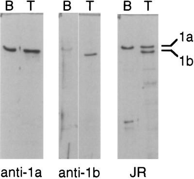
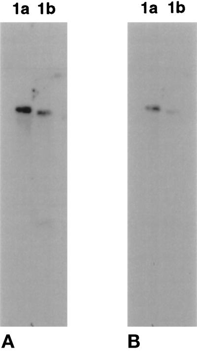
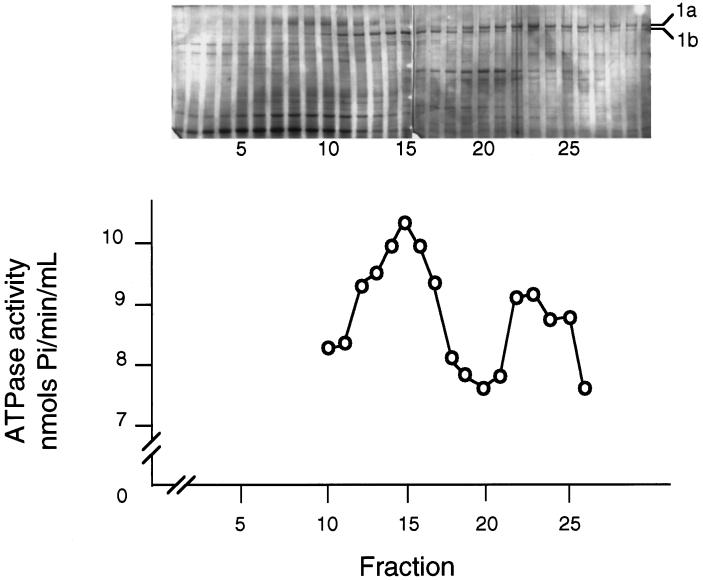
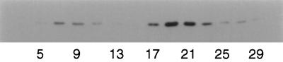
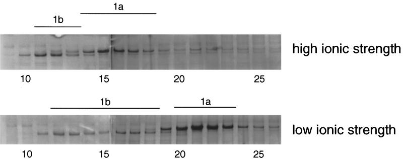
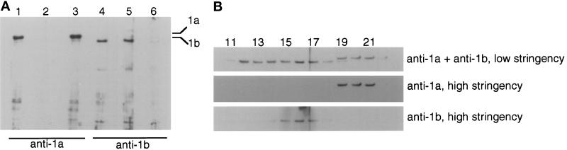
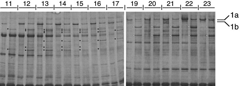
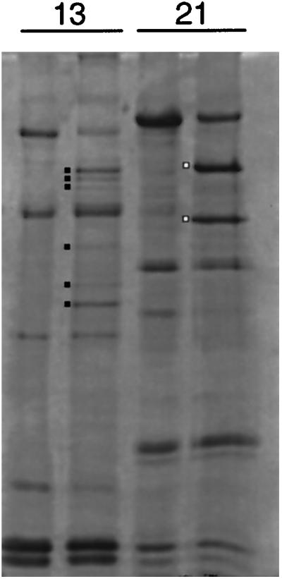
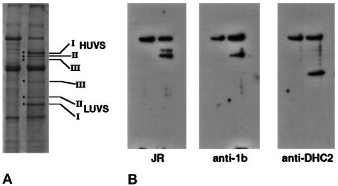
Recent studies have revealed the expression of multiple putative cytoplasmic dynein heavy chain (DHC) genes in several organisms, with each gene encoding a separate protein isoform. This finding is consistent with the hypothesis that different isoforms do different things, as is the case for the axonemal dyneins. Furthermore, the large number of tasks ascribed to cytoplasmic dynein suggests that there may be additional isoforms not yet identified. Two of the mammalian cytoplasmic dynein heavy chains are DHC1a and DHC1b. DHC1a is conventional cytoplasmic dynein and is found in all organisms examined. DHC1b is expressed in organisms that have multiple dyneins, and has been implicated in the intracellular trafficking of molecules in unciliated and ciliated cells. In the present study, we examined the DHC1b protein from rat testis. Testis cytoplasmic dynein contains a large amount of dynein heavy chain reactive with an antibody raised against a peptide sequence of rat DHC1b. The testis anti-DHC1b immunoreactive protein is slightly smaller than testis DHC1a, as assessed by SDS-PAGE. In Northern blots, the DHC1b mRNA is smaller than the DHC1a mRNA. In sucrose gradients made in low ionic strength, DHC1a sedimented at approximately 20S, and the anti-1b immunoreactive heavy chains sedimented in a broad band centered at approximately 14S. The V1-photolysis reaction of individual sucrose gradient fractions revealed three distinct patterns of photolysis, suggesting that there are at least three separate 1b-like heavy chain isoforms in testis. Using a high-stringency Western blotting protocol, the anti-1b antibody and the anti-DHC2 antibody recognized the same heavy chain and specifically bound to one of the three 1b-like heavy chains. We conclude that rat testis contains three 1b-like dynein heavy chains, and one of these is the product of the DHC1b/DHC2 gene previously identified.


Figures

Figure 1
Figure 1
Western blots of cytoplasmic dyneins from rat brain and testis. Three identical blots containing cytoplasmic dyneins isolated from rat brain (B) and rat testis (T) were resolved by 6.7% SDS-PAGE, transferred to nitrocellulose, and probed with antibodies to DHC1a (anti-1a) and DHC1b (anti-1b) and with the pan-reactive anticytoplasmic dynein antibody (JR). Blots were developed with horseradish peroxidase–conjugated secondary antibodies, followed by chemiluminescence and exposure to x-ray film. The anti-1b-immunoreactive protein from testis, but not brain, was smaller than DHC1a.
Figure 2
Figure 2
Northern blots of rat testis RNA. Twenty micrograms of testis poly(A)+ RNA were resolved on a 0.8% agarose gel under denaturing conditions. Samples were blotted onto membranes and hybridized overnight with 32P-labeled 300-bp cDNA probes derived from DHC1a and DHC1b. Identical strips were individually probed with one of the cDNAs, and the strips were exposed side by side to the x-ray film. (A) Autoradiogram after low-stringency wash (100 mM sodium phosphate, 50°C). (B) Autoradiogram of the same blots after high-stringency wash (50 mM sodium phosphate, 65°C). The DHC1b transcript is smaller and appears to be less abundant than the DHC1a mRNA.
Figure 3
Figure 3
Sucrose density centrifugation of testis cytoplasmic dyneins. The 0.5 M NaCl extract of testis microtubules was sedimented in a linear 5–20% sucrose gradient made in low ionic strength (see MATERIALS AND METHODS), and the gradient was fractionated into 30 equal-volume samples. In separate gradients run in parallel, catalase (11.3S) sedimented in fractions 11–12, and thyroglobulin (19.2S) sedimented in fractions 21–22. Top, silver-stained 6.7% SDS-PAGE of 20 μl of each gradient fraction. The samples were electrophoresed in two identical minigels and are joined together. The positions of DHC1a and anti-1b-immunoreactive protein are indicated. The top of the gradient (fraction 1) is to the left. Bottom, Mg-ATPase activity of each fraction from the same gradient shown in the top panel.
Figure 4
Figure 4
Western blot of dynein intermediate chain IC74 in the sucrose gradient fractions. The odd-numbered fractions (3–29) from a sucrose gradient were resolved on an 8% SDS-polyacrylamide gel and blotted to nitrocellulose. In this gradient, DHC1a was present in fractions 17–23, with its highest concentration in fractions 19–21, and DHC1b was present in fractions 11–18. The blot was probed with anti-IC74, followed by horseradish peroxidase–conjugated secondary antibody and chemiluminescence. The IC74 was associated with DHC1a (fractions 17–23) but did not associate with most of the anti-1b-immunoreactive protein (fractions 11–17).
Figure 5
Figure 5
Effect of ionic strength on the sedimentation patterns of cytoplasmic dyneins. Testis cytoplasmic dynein prepared by ATP extraction of microtubules was sedimented in 5–20% sucrose gradients made in high ionic strength (top) or low ionic strength (bottom). The gels of the samples were stained with silver. Each panel shows the DHC regions of two gels joined together. In high ionic strength, the DHC1a sedimented in a peak centered in fractions 15–16, and the anti-1b-immunoreactive protein sedimented in a tight peak centered in fractions 11–12. In low ionic strength, the DHC1a sedimented faster in the gradient, centered in fractions 21–22, and the anti-1b immunoreactive protein sedimented as a broad peak in fractions 11–18.
Figure 6
Figure 6
Western blot analysis of sucrose gradient fractions under high-stringency conditions. (A) Western blots using antibodies absorbed with synthetic peptides. Testis cytoplasmic dynein was electrophoresed in 6.7% SDS-polyacrylamide gels, transferred to nitrocellulose, and probed with affinity-purified antibodies that had been preabsorbed with synthetic peptides. The Western blots were processed using the high-stringency conditions (see MATERIALS AND METHODS). Anti-1a antibody: lane 1, no absorption; lane 2, preabsorbed with 1a peptide; lane 3, preabsorbed with 1b peptide. Anti-1b antibody: lane 4, no absorption; lane 5, preabsorbed with 1a peptide; lane 6, preabsorbed with 1b peptide. Under these conditions, the antibodies reacted with the appropriate heavy chain wholly in a sequence-specific manner. (B) Western blots of sucrose gradient fractions. Selected fractions of a low-ionic strength sucrose gradient similar to the one shown in Figure 5 were electrophoresed, blotted, and probed with antibodies. The same blot was repeatedly stripped and reprobed. The heavy chain regions of the blots are shown. Top, anti-1b at low stringency, to which anti-1a was added. Middle, anti-1a at high stringency. Bottom, anti-1b at high stringency. The anti-1b antibody reacted only with a subset of the 1b-like proteins present in fractions 11–17 (see Figure 5).
Figure 7
Figure 7
V1 photolysis of cytoplasmic dynein heavy chains separated in a sucrose gradient. Testis cytoplasmic dynein prepared by ATP extraction of microtubules was sedimented through a 5–20% sucrose gradient made in low ionic strength. One hundred-microliter aliquots of individual gradient fractions 11–17 and 19–23 were subjected to V1 photolysis. Equal volumes of untreated and V1-photolyzed samples were resolved in 6.7% SDS-polyacrylamide gels and stained with silver. Two gels were joined together. The principal V1-photolytic products are marked with dots. The 1b-like fractions (11–17) yielded six V1-photolytic products (filled dots). The pattern of products changed through the gradient. DHC1a (fractions 19–23) yielded two V1-photolytic products (open dots).
Figure 8
Figure 8
V1-photolysis products of cytoplasmic dynein heavy chains. The untreated and V1-photolyzed fractions 13 and 21 from the gradient shown in Figure 7 were electrophoresed side by side. The principal V1-photolytic products are indicated by dots.
Figure 9
Figure 9
Western blots of intact and V1-photolyzed 1b-like heavy chains. (A) Intact and V1-photolyzed fraction 13 from the gradient shown in Figure 7. The three pairs of HUVs and LUVs are indicated. (B) Western blots of similar samples to those shown in A. The pan-reactive antiserum JR reacted with the parent DHC band as well as the three HUV fragments, although only weakly with HUV II. Anti-1b at high stringency reacted only with the parent DHC band and HUV III. Anti-DHC2 (Vaisberg et al., 1996) at high stringency reacted only with the parent DHC band and LUV III.

References

    1. Amos LA. Brain dynein crossbridges microtubules into bundles. J Cell Sci. 1989;93:19–28. - PubMed
    1. Andrews KK, Nettesheim P, Asai DJ, Ostrowski LE. Identification of seven rat axonemal dynein heavy chain genes: expression during ciliated cell differentiation. Mol Biol Cell, 1996;7:71–79. - PMC - PubMed
    1. Aniento F, Emans N, Griffiths G, Gruenberg J. Cytoplasmic dynein-dependent vesicular transport from early to late endosomes. J Cell Biol, 1993;123:1373–1387. - PMC - PubMed
    1. Asai DJ. Functional and molecular diversity of dynein heavy chains. Semin Cell Dev Biol, 1996;7:311–320.
    1. Asai DJ, Beckwith SM, Kandl KA, Keating HH, Tjandra H, Forney JD. The dynein genes of Paramecium tetraurelia: sequences adjacent to the catalytic P-loop identify cytoplasmic and axonemal heavy chain isoforms. J Cell Sci, 1994;107:839–847. - PubMed

Publication types

LinkOut - more resources