

A specific light chain of kinesin associates with mitochondria in cultured cells
- PMID: 9450959
- PMCID: PMC25259
- DOI: 10.1091/mbc.9.2.333
A specific light chain of kinesin associates with mitochondria in cultured cells
Abstract
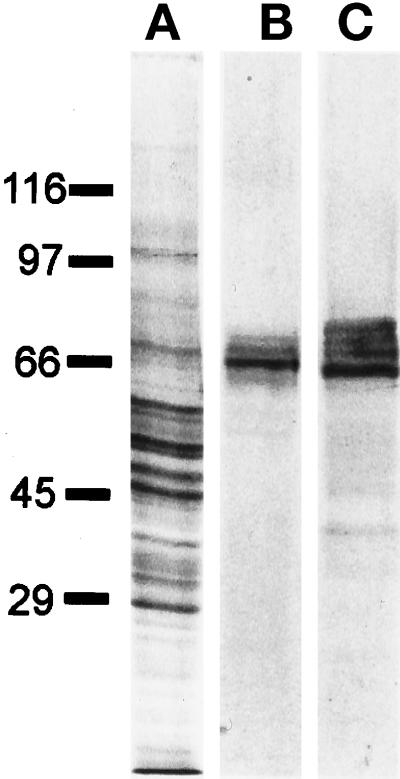
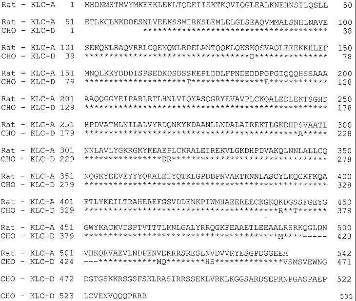
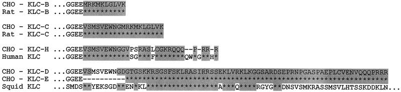
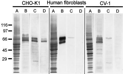
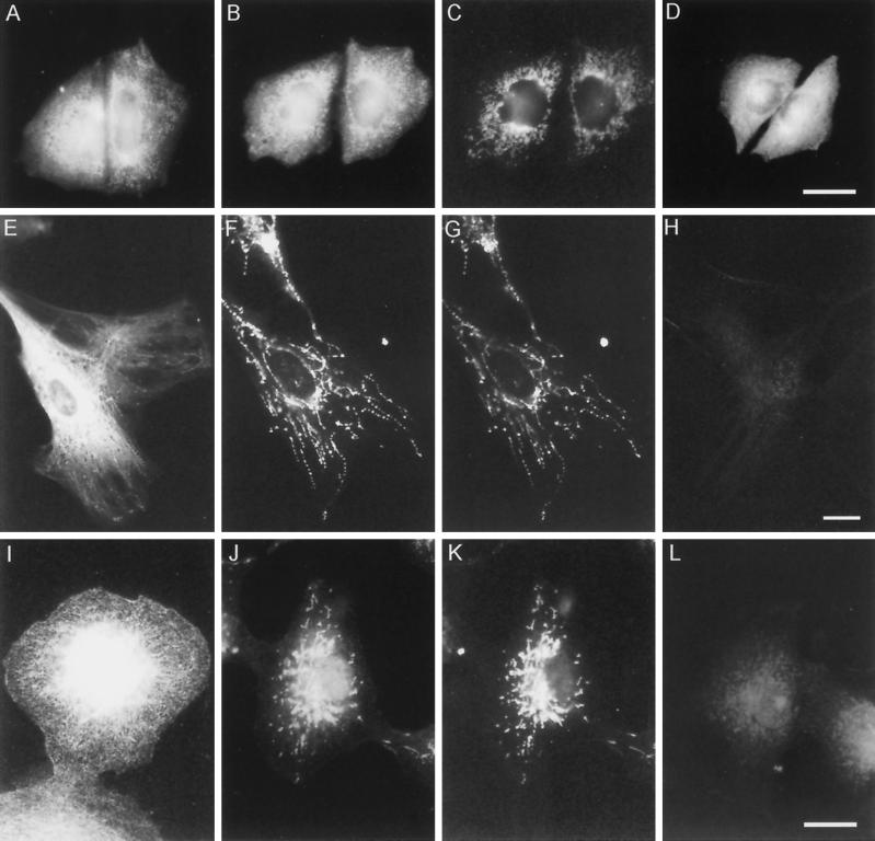
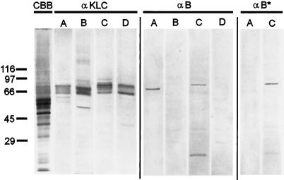
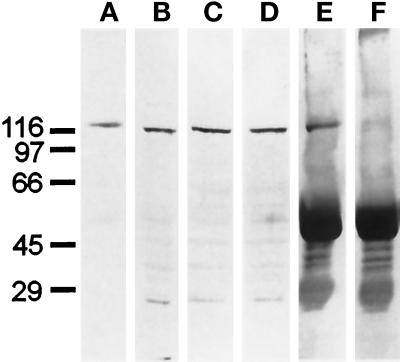
The motor protein kinesin is implicated in the intracellular transport of organelles along microtubules. Kinesin light chains (KLCs) have been suggested to mediate the selective binding of kinesin to its cargo. To test this hypothesis, we isolated KLC cDNA clones from a CHO-K1 expression library. Using sequence analysis, they were found to encode five distinct isoforms of KLCs. The primary region of variability lies at the carboxyl termini, which were identical or highly homologous to carboxyl-terminal regions of rat KLC B and C, human KLCs, sea urchin KLC isoforms 1-3, and squid KLCs. To examine whether the KLC isoforms associate with different cytoplasmic organelles, we made an antibody specific for a 10-amino acid sequence unique to B and C isoforms. In an indirect immunofluorescence assay, this antibody specifically labeled mitochondria in cultured CV-1 cells and human skin fibroblasts. On Western blots of total cell homogenates, it recognized a single KLC isoform, which copurified with mitochondria. Taken together, these data indicate a specific association of a particular KLC (B type) with mitochondria, revealing that different KLC isoforms can target kinesin to different cargoes.
Figures
Similar articles
-
An isoform of kinesin light chain specific for the Golgi complex.J Cell Sci. 2000 Jun;113 ( Pt 11):2047-54. doi: 10.1242/jcs.113.11.2047. J Cell Sci. 2000. PMID: 10806115
-
Kinesin's light chains inhibit the head- and microtubule-binding activity of its tail.Proc Natl Acad Sci U S A. 2010 Jun 29;107(26):11781-6. doi: 10.1073/pnas.1005854107. Epub 2010 Jun 14. Proc Natl Acad Sci U S A. 2010. PMID: 20547877 Free PMC article.
-
Kinesin light-chain KLC3 expression in testis is restricted to spermatids.Biol Reprod. 2001 May;64(5):1320-30. doi: 10.1095/biolreprod64.5.1320. Biol Reprod. 2001. PMID: 11319135 Free PMC article.
-
Expression cloning with pan kinesin antibodies.Methods Mol Biol. 2001;164:21-41. doi: 10.1385/1-59259-069-1:21. Methods Mol Biol. 2001. PMID: 11217609 Review. No abstract available.
-
Microtubules, mitochondria, and molecular chaperones: a new hypothesis for in vivo assembly of microtubules.Biochem Cell Biol. 1990 Dec;68(12):1352-63. doi: 10.1139/o90-198. Biochem Cell Biol. 1990. PMID: 1982213 Review.
Cited by
-
Mitochondrial dynamics.New Phytol. 2003 Dec;160(3):463-478. doi: 10.1046/j.1469-8137.2003.00918.x. Epub 2003 Nov 6. New Phytol. 2003. PMID: 33873653 Review.
-
Transport of Drosophila fragile X mental retardation protein-containing ribonucleoprotein granules by kinesin-1 and cytoplasmic dynein.Proc Natl Acad Sci U S A. 2004 Dec 14;101(50):17428-33. doi: 10.1073/pnas.0408114101. Epub 2004 Dec 6. Proc Natl Acad Sci U S A. 2004. PMID: 15583137 Free PMC article.
-
A human dynamin-related protein controls the distribution of mitochondria.J Cell Biol. 1998 Oct 19;143(2):351-8. doi: 10.1083/jcb.143.2.351. J Cell Biol. 1998. PMID: 9786947 Free PMC article.
-
Ginsenoside Re rescues methamphetamine-induced oxidative damage, mitochondrial dysfunction, microglial activation, and dopaminergic degeneration by inhibiting the protein kinase Cδ gene.Mol Neurobiol. 2014 Jun;49(3):1400-21. doi: 10.1007/s12035-013-8617-1. Epub 2014 Jan 16. Mol Neurobiol. 2014. PMID: 24430743
-
Association of kinesin light chain with outer dense fibers in a microtubule-independent fashion.J Biol Chem. 2003 May 2;278(18):16159-68. doi: 10.1074/jbc.M213126200. Epub 2003 Feb 19. J Biol Chem. 2003. PMID: 12594206 Free PMC article.
References
-
- Alberts B, Bray D, Lewis J, Raff M, Roberts K, Watson JD. Molecular Biology of the Cell. 3rd ed. New York: Garland Publishing; 1994.
-
- Beushausen S, Kladakis A, Jaffe H. Kinesin light chains: identification and characterization of a family of proteins from the optic lobe of the squid Loligo pealii. DNA Cell Biol. 1993;12:901–910. - PubMed
-
- Bloom GS, Endow SA. Motor proteins 1: kinesins. Protein Profile. 1995;1:1059–1088. - PubMed
-
- Bloom GS, Wagner MC, Pfister KK, Brady ST. Native structure and physical properties of bovine brain kinesin and identification of the ATP-binding subunit polypeptide. Biochemistry. 1988;27:3409–3416. - PubMed
Publication types
MeSH terms
Substances
Associated data
- Actions
Grants and funding
LinkOut - more resources
Full Text Sources
Other Literature Sources
Molecular Biology Databases
Miscellaneous
