. 1998 Feb;9(2):345-54.
doi: 10.1091/mbc.9.2.345.

14-3-3 proteins act as negative regulators of the mitotic inducer Cdc25 in Xenopus egg extracts

Affiliations
Free PMC article

14-3-3 proteins act as negative regulators of the mitotic inducer Cdc25 in Xenopus egg extracts

A Kumagai et al. Mol Biol Cell. 1998 Feb.
Free PMC article

Abstract

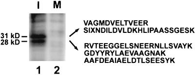
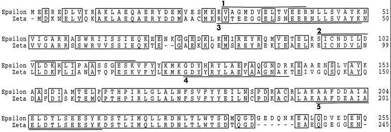
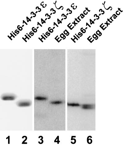
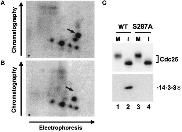
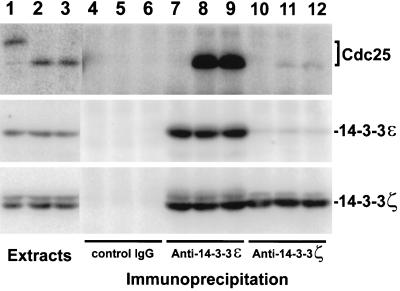
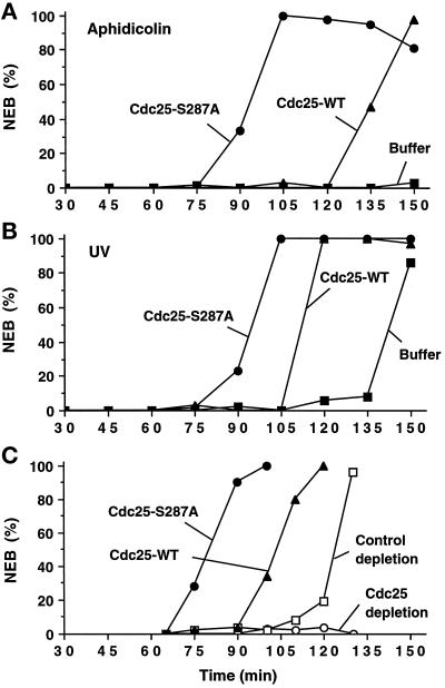
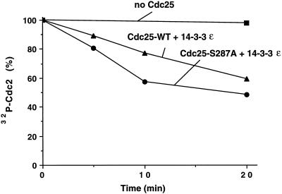
Cdc25, the dual-specificity phosphatase that dephosphorylates the Cdc2-cyclin B complex at mitosis, is highly regulated during the cell cycle. In Xenopus egg extracts, Cdc25 is associated with two isoforms of the 14-3-3 protein. Cdc25 is complexed primarily with 14-3-3epsilon and to a lesser extent with 14-3-3zeta. The association of these 14-3-3 proteins with Cdc25 varies dramatically during the cell cycle: binding is high during interphase but virtually absent at mitosis. Interaction with 14-3-3 is mediated by phosphorylation of Xenopus Cdc25 at Ser-287, which resides in a consensus 14-3-3 binding site. Recombinant Cdc25 with a point mutation at this residue (Cdc25-S287A) is incapable of binding to 14-3-3. Addition of the Cdc25-S287A mutant to Xenopus egg extracts accelerates mitosis and overrides checkpoint-mediated arrests of mitotic entry due to the presence of unreplicated and damaged DNA. These findings indicate that 14-3-3 proteins act as negative regulators of Cdc25 in controlling the G2-M transition.


Figures

Figure 1
Figure 1
The 28-kDa and 31-kDa proteins bind to the interphase form of Xenopus Cdc25. Nickel-agarose beads containing His6-Cdc25 protein were incubated in interphase (lane 1) or M-phase (lane 2) egg extracts. Bound proteins were eluted with 150 mM imidazole in HBS, separated by SDS-PAGE, and silver stained. From the 31-kDa protein, we obtained the following peptide sequences: 1, VAGMDVELTVEER; 2, SIXNDILDVLDKHLIPAASSGESK. From the 28-kDa protein, we obtained the following three sequences: 3, RVTEEGGELSNEERNLLSVAYK; 4, GDYYRYLAEVAAGNAK; 5, AAFDEAIAELDTLSEESYK. X denotes unreadable residues.
Figure 2
Figure 2
Comparison of the amino acid sequences of the Xenopus 14-3-3ε and 14-3-3ζ proteins. Peptides from the amino acid sequencing analysis in Figure 1 are underlined. The GenBank accession numbers for the DNA sequences of Xenopus 14-3-3ε and 14-3-3ζ are AF033311 and AF033312, respectively.
Figure 3
Figure 3
The proteins 14-3-3ε and 14-3-3ζ are abundant in Xenopus egg extracts. Bacterially expressed His6-14-3-3ε (lane 1, 2.5 μg of protein; lane 3, 40 ng of protein), His6-14-3-3ζ (lane 2, 2.5 μg of protein; lane 5, 40 ng of protein), and 1 μl of interphase egg extract (lanes 4 and 6) were subjected to SDS-PAGE and either stained with Coomassie blue (lanes 1 and 2) or immunoblotted with anti-14-3-3ε antibodies (lanes 3 and 4) or anti-14-3-3ζ antibodies (lanes 5 and 6). Note that the electrophoretic mobilities of the recombinant 14-3-3 proteins are reduced due to the presence of a six-histidine tag.
Figure 4
Figure 4
Phosphorylation of Ser-287 in Xenopus Cdc25 is essential for the binding of 14-3-3 proteins. (A and B) Tryptic phosphopeptide mapping of His6-Cdc25 (A) and His6-Cdc25-S287A (B) that had been radiolabeled in interphase extracts. Arrows indicate the expected position of the phosphopeptide containing Ser287. Origins are indicated by a dot in the lower left of each map. (C) The His6-Cdc25-S287A mutant does not bind to 14-3-3ε in egg extracts. His6-Cdc25 (lanes 1 and 2) and His6-Cdc25-S287A (lanes 3 and 4) were incubated in either M-phase (lanes 1 and 3) or interphase (lanes 2 and 4) extracts. The proteins were reisolated as described in MATERIALS AND METHODS and subjected to SDS-PAGE and immunoblotting with anti-Cdc25 antibodies (top) or anti-14-3-3ε antibodies (bottom).
Figure 5
Figure 5
Xenopus Cdc25 binds mainly to 14-3-3ε in interphase extracts. Two microliters of M-phase extract (lane 1), interphase extract containing no sperm nuclei (lane 2), and interphase extract containing 3000 UV-damaged sperm nuclei per μl (lane 3) were subjected to SDS-PAGE and immunoblotted with anti-Cdc25 antibodies (top), anti-14-3-3ε antibodies (middle), or anti-14-3-3ζ antibodies (bottom). One hundred microliters of M-phase extract (lanes 4, 7, and 10), interphase extract containing no sperm nuclei (lanes 5, 8, and 11), and interphase extract containing 3000 UV-damaged sperm nuclei/μl (lanes 6, 9, and 12) were immunoprecipitated with control antibodies (lanes 4–6), anti-14-3-3ε antibodies (lanes 7–9), or anti-14-3-3ζ antibodies (lanes 10–12). Immunoprecipitated proteins were separated by SDS-PAGE and immunoblotted with anti-Cdc25 antibodies (top), anti-14-3-3ε antibodies (middle), or anti-14–3-3ζ antibodies (bottom). All extracts contained 100 μg/ml cycloheximide.
Figure 6
Figure 6
Cdc25-S287A mutant induces mitosis more efficiently than the wild-type Cdc25 protein. His6-Cdc25 (▴), His6-Cdc25-S287A (•), or buffer alone (▪) were added to the interphase egg extracts containing either aphidicolin (A) or UV-damaged sperm nuclei (B). (C) Xenopus egg extracts were immunodepleted with anti-Cdc25 antibodies (▴, •, or ○) or control rabbit anti-mouse IgG antibodies (□). We verified by immunoblotting with anti-Cdc25 antibodies that Cdc25 was quantitatively removed by this procedure. To the Cdc25-depleted extracts, we added His6-Cdc25 (▴), His6-Cdc25-S287A (•), or buffer alone (○). Buffer alone was added to the control-depleted extract (□). (AC) Recombinant His6-Cdc25 and His6-Cdc25-S287A were added to a final concentration of 10 μg/ml (0.14 μM). Aliquots were taken at the indicated times and NEB was monitored by microscopy.
Figure 7
Figure 7
Effect of 14-3-3 proteins on Cdc25 activity. Nickel-agarose beads containing His6-Cdc25 protein and the His6-Cdc25-S287A mutant protein were incubated in interphase extracts. Beads were isolated and bound proteins were eluted with 150 mM imidazole in HBS. A 32P-phosphorylated Cdc2–cyclin B complex phosphorylated in vitro by Myt1 was mixed with His6-Cdc25 (▴), His6-Cdc25-S287A (•), or control buffer (▪) in the presence of 50 μg/ml His6-14-3-3ε protein. Aliquots were taken at the times indicated and subjected to SDS-PAGE. 32P remaining on Cdc2 was quantitated by using a PhosphorImager. In control experiments, His6-Cdc25 and His6-Cdc25-S287A purified directly from baculovirus-infected insect cells showed very similar Cdc2-specific phosphatase activity.

References

    1. Aitken A. 14-3-3 and its possible role in co-ordinating multiple signalling pathways. Trends Cell Biol. 1996;6:341–347. - PubMed
    1. al-Khodairy F, Carr AM. DNA repair mutants defining G2 checkpoint pathways in Schizosaccharomyces pombe. EMBO J. 1992;11:1343–1350. - PMC - PubMed
    1. Blasina A, Paegle ES, McGowan CH. The role of inhibitory phosphorylation of CDC2 following DNA replication block and radiation-induced damage in human cells. Mol Biol Cell. 1997;8:1013–1023. - PMC - PubMed
    1. Boyle WJ, van der Geer P, Hunter T. Phosphopeptide mapping and phosphoamino acid analysis by two-dimensional separation on thin-layer cellulose plates. Methods Enzymol. 1991;201:110–149. - PubMed
    1. Carpenter PB, Mueller PR, Dunphy WG. Role for a Xenopus Orc2-related protein in controlling DNA replication. Nature. 1996;379:357–360. - PubMed

Publication types

MeSH terms

Associated data