. 1998 Feb;9(2):403-19.
doi: 10.1091/mbc.9.2.403.

RhoA-dependent phosphorylation and relocalization of ERM proteins into apical membrane/actin protrusions in fibroblasts

Affiliations
Free PMC article

RhoA-dependent phosphorylation and relocalization of ERM proteins into apical membrane/actin protrusions in fibroblasts

R J Shaw et al. Mol Biol Cell. 1998 Feb.
Free PMC article

Abstract

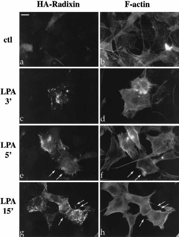
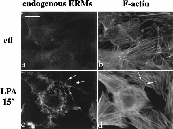
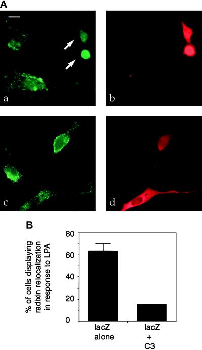
The ERM proteins (ezrin, radixin, and moesin) are a group of band 4. 1-related proteins that are proposed to function as membrane/cytoskeletal linkers. Previous biochemical studies have implicated RhoA in regulating the association of ERM proteins with their membrane targets. However, the specific effect and mechanism of action of this regulation is unclear. We show that lysophosphatidic acid stimulation of serum-starved NIH3T3 cells resulted in relocalization of radixin into apical membrane/actin protrusions, which was blocked by inactivation of Rho by C3 transferase. An activated allele of RhoA, but not Rac or CDC42Hs, was sufficient to induce apical membrane/actin protrusions and localize radixin or moesin into these structures in both Rat1 and NIH3T3 cells. Lysophosphatidic acid treatment led to phosphorylation of radixin preceding its redistribution into apical protrusions. Significantly, cotransfection of RhoAV14 or C3 transferase with radixin and moesin revealed that RhoA activity is necessary and sufficient for their phosphorylation. These findings reveal a novel function of RhoA in reorganizing the apical actin cytoskeleton and suggest that this function may be mediated through phosphorylation of ERM proteins.


Figures

Figure 1
Figure 1
LPA-induced relocalization of radixin into apical membrane/actin protrusions in NIH3T3 cells. (A) Indirect immunofluorescence was performed on NIH3T3 cells stably expressing HA-epitope–tagged radixin. These cells were serum starved, and then treated with vehicle for 15 min (a and b), or with LPA (6 μM) for 3 min (c and d), 5 min (e and f), or 15 min (g and h). HA-radixin localization (using the 12CA5 mAb against the HA epitope) is shown in panels a, c, e, and g, and F-actin localization (using rhodamine phalloidin) is shown in b, d, f, and h. Starting 3–5 min after LPA treatment, radixin was relocalized into peripheral and apical membrane protrusions that also contain actin. These results were observed in three independent stable cell lines. Arrows denote colocalization of HA-radixin and actin in LPA-induced membrane protrusions. (B) NIH3T3 cells were serum starved and then treated with vehicle (a and b) or LPA for 15 min (c and d) and visualized for endogenous ERM proteins (a and c) using the CR22 polyclonal antibody or F-actin using rhodamine phalloidin (b and d). Arrows indicate the colocalization of ERM proteins and actin in membrane protrusions. Similar results were observed when a radixin-specific antibody was used as well. These results demonstrate that endogenous ERM proteins are similarly relocalized after LPA treatment as the exogenous epitope-tagged form of radixin. Bar, 10 μm.
Figure 1
Figure 1
LPA-induced relocalization of radixin into apical membrane/actin protrusions in NIH3T3 cells. (A) Indirect immunofluorescence was performed on NIH3T3 cells stably expressing HA-epitope–tagged radixin. These cells were serum starved, and then treated with vehicle for 15 min (a and b), or with LPA (6 μM) for 3 min (c and d), 5 min (e and f), or 15 min (g and h). HA-radixin localization (using the 12CA5 mAb against the HA epitope) is shown in panels a, c, e, and g, and F-actin localization (using rhodamine phalloidin) is shown in b, d, f, and h. Starting 3–5 min after LPA treatment, radixin was relocalized into peripheral and apical membrane protrusions that also contain actin. These results were observed in three independent stable cell lines. Arrows denote colocalization of HA-radixin and actin in LPA-induced membrane protrusions. (B) NIH3T3 cells were serum starved and then treated with vehicle (a and b) or LPA for 15 min (c and d) and visualized for endogenous ERM proteins (a and c) using the CR22 polyclonal antibody or F-actin using rhodamine phalloidin (b and d). Arrows indicate the colocalization of ERM proteins and actin in membrane protrusions. Similar results were observed when a radixin-specific antibody was used as well. These results demonstrate that endogenous ERM proteins are similarly relocalized after LPA treatment as the exogenous epitope-tagged form of radixin. Bar, 10 μm.
Figure 2
Figure 2
C3 pretreatment blocks LPA-induced relocalization of radixin in NIH3T3 cells. (A) NIH3T3 cells stably expressing HA-radixin were transfected with expression plasmids encoding lacZ and C3 transferase (a and b) or lacZ alone (c and d) and then serum-starved and treated with LPA for 15 min. LacZ (b and d) localization (red) is shown to indicate transfected cells, and HA-radixin localization (green) is shown in panels a and c. C3-transfected cells (arrows in panel a) show a lack of radixin localization into apical protrusions after LPA treatment. Bar, 10 μm. (B) Percentage of cells in lacZ only or lacZ plus C3 transfections displaying relocalization of radixin to apical protrusions after LPA treatment. Results of three independent experiments are shown. One hundred transfected cells were counted for each sample. Errors bars denote SD of the mean.
Figure 3
Figure 3
Cotransfection of RhoAV14, but not RhoAN19, RacV12, or CDC42V12, with radixin is sufficient to cause relocalization to apical membrane/actin protrusions in NIH3T3 cells. NIH3T3 cells were transiently transfected with radixin (a and b), radixin and RhoAV14 (c and d), radixin and RhoAN19 (e and f), radixin and RacV12 (g and h), and radixin and CDC42V12 (b). All GTPase constructs were myc epitope tagged. HA-radixin (a, b, c, e, and g) and myc-tagged GTPases (d, f, and h) localizations are shown. Cotransfections followed by immunolocalization were performed in 12 independent experiments. Radixin localization in the presence of activated RhoA was further verified by transfection of radixin into two independent NIH3T3 cell lines stably expressing the RhoAV14 allele. In all experiments, at least 100 transfected cells were examined for each condition, and cells shown are representative of > 80% of the transfected cell population. In the presence of activated RhoA, but not dominant negative RhoA, activated Rac, or activated CDC42, radixin localized into apical membrane protrusions. α-myc and actin staining were used in all experiments to verify functional expression of RhoAV14. Bar, 10 μm.
Figure 4
Figure 4
Cotransfection with RhoV14 relocalizes moesin, but not merlin, to apical membrane protrusions in R12 cells. Cells were transiently transfected with moesin (a), moesin and RhoAV14 (b and c), moesin and RhoAN19 (d), moesin and RacV12 (e), moesin and CDC42v12 (f), merlin (g), or merlin and RhoAV14 (h). Moesin and merlin were tagged with HA-epitope tag at their C termini. HA-moesin (a–f) or HA-merlin (g and h) immunolocalization using the 12CA5 mAb is shown. Panels b and c represent two different focal planes of the same cell to better illustrate the nature of the apical protrusions. All cells presented here were also shown to be positive for GTPase expression (where applicable) by α-myc immunostaining. These transfections were performed seven independent times, and at least 100 transfected cells were examined for each condition. Cells shown are representative of >90% of the transfected cell population. Bar, 10 μm.
Figure 5
Figure 5
SEM of RhoAV14-induced apical membrane structures and CDC42-induced filopodia. SEM was performed on serum-starved vector control (a), RhoAV14-transfected (b), and CDC42V12-transfected (c) NIH3T3 cells. Note the number and length of the apical membrane protrusions induced by the presence of activated RhoA. Three independent transfections were examined by SEM with more than 200 cells examined per transfection. The percentage of cells demonstrating the phenotype as shown for RhoAV14 or CDC42V12 was nearly identical to the percentage shown to be transfected by either GTPase when a parallel coverslip was analyzed by indirect immunofluorescence. Furthermore, <2% of vector-only transfectants had apical structures similar to those seen with RhoAV14, and they were highly reduced in number and size (ca. 20 per cell as opposed to 100–200). Bar, 10 μm.
Figure 6
Figure 6
Modulation of RhoA-dependent apical protrusions by a radixin carboxyl-terminal domain mutant. R12 cells were transfected with the RADC mutant alone (a and b), or RADC and RhoAV14 (c and d). HA-RADC localization shown in panels a and c, and F-actin as visualized by rhodamine phalloidin are shown in b and d. Note colocalization of RADC with stress fibers in absence of RhoAV14 (arrows in a and b) and localization of actin into profuse stress fibers and long apical processes containing RADC in the presence of RhoAV14 (arrows in c and d). NIH3T3 cells were transfected with RhoAV14 and full-length radixin (e) or RhoAV14 and RADC mutant (f) and then serum-starved and subjected to confocal microscopy analysis. Panels e and f represent combined optical sections of HA-radixin (e) or HA-RADC (f) immunolocalization. NIH3T3 cells were transfected with lacZ and RhoAV14 (g) or lacZ, RhoAV14, and RADC (h) and then serum-starved and subjected to double immunostaining for lacZ and another marker of RhoA-induced apical structures, CD44. CD44 immunolocalization is shown in g and h. LacZ-positive cells (i.e., transfectants) are indicated by arrows. Transfections were performed in four independent experiments. Twenty-five percent of the cells transfected with RADC and RhoAV14 displayed the extremely long apical protrusions as seen in panels c, f, and h, whereas cells transfected with RhoA alone or RhoA plus full-length radixin never showed this phenotype. These results suggest that the presence of a mutant form of radixin can directly alter the size and number of RhoA-induced apical protrusions, but that additional factors (e.g., cell cycle) may control the full expressivity of this phenotype. Bar, 10 μm.
Figure 7
Figure 7
LPA treatment of NIH3T3 cells induces phosphorylation of radixin within 1 min. Metabolically labeled NIH3T3 cells stably expressing HA-radixin or parental (mock) NIH3T3 cells were serum-starved, then treated with LPA (6 μM) for various timepoints as indicated. Cell lysates were immunoprecipitated with 12CA5 (α-HA) mAb. Radixin is indicated by arrowhead at left. Radixin phosphorylation was increased threefold within 1 min of LPA treatment and remained constant for 15 min. Note background band (*) whose orthophosphate content was not apparently modulated by LPA. Molecular size standards are indicated at right. The data shown are representative of four independent experiments.
Figure 8
Figure 8
RhoA activity is necessary and sufficient for phosphorylation of moesin. R12 or NIH3T3 cells were transfected with vector only, HA-moesin only, HA-moesin and RhoAV14, or HA-moesin and C3 transferase, as indicated. Cells were then serum starved and metabolically labeled with [32P]orthophosphate; cell lysates were immunoprecipitated as in Figure 7. To ensure that equivalent levels of HA-moesin were expressed and immunoprecipitated, immunoprecipitated samples were transferred to PVDF after SDS-PAGE and immunoblotted with 12CA5 antibody; signal was detected by enhanced chemiluminescence. Moesin is indicated by arrowhead at left. The presence of RhoA increased the basal level of moesin phosphorylation by 7-fold in R12 cells and 2.5-fold in NIH3T3 cells, while C3 treatment reduced the basal level of phosphorylation by at least 50% in both cell types (top panel). Protein levels were approximately equivalent in all conditions tested (bottom panel). The data shown are representative of three independent experiments.
Figure 9
Figure 9
32P-labeled moesin partitions with the Triton X-100 insoluble fraction. NIH3T3 cells were transfected with HA-moesin only or HA-moesin and RhoAV14 as indicated. Cells were then serum starved and metabolically labeled with [32P]orthophosphate or [35S]methionine/cysteine as described in Figure 8, and then subjected to 0.5% Triton X-100 detergent extraction. Soluble (S) and insoluble (I) fractions were immunoprecipitated with 12CA5 antibody. In contrast to total (35S) moesin, 32P-labeled moesin was found almost exclusively in the insoluble fraction. Also, coexpression of RhoAV14 increases the percentage of moesin in the insoluble fraction. The data shown are representative of three independent experiments.

Similar articles

Cited by

References

    1. Aktories K, Braun U, Rosener S, Just I, Hall A. The rho gene product expressed in E. coli is a substrate of ADP-ribosyltransferase C3. Biochem Biophys Res Commun. 1989;158:209–213. - PubMed
    1. Amano M, Chihara K, Kimura K, Fukata Y, Nakamura N, Matsuura Y, Kaibuchi K. Formation of actin stress fibers and focal adhesions enhanced by Rho- kinase. Science. 1997;275:1308–1311. - PubMed
    1. Amano M, Mukai H, Ono Y, Chihara K, Matsui T, Hamajima Y, Okawa K, Iwamatsu A, Kaibuchi K. Identification of a putative target for Rho as the serine-threonine kinase protein kinase N. Science. 1996;271:648–650. - PubMed
    1. Amieva M, Furthmayr H. Subcellular localization of moesin in dynamic filopodia, retraction fibers, and other structures involved in substrate exploration, attachment, and cell-cell contacts. Exp Cell Res. 1995;219:180–196. - PubMed
    1. Berryman M, Franck Z, Bretscher A. Ezrin is concentrated in the apical microvilli of a wide variety of epithelial cells whereas moesin is found primarily in endothelial cells. J Cell Sci. 1993;105:1025–1043. - PubMed

MeSH terms