. 1998 Feb;9(2):451-67.
doi: 10.1091/mbc.9.2.451.

Activation of the p42 mitogen-activated protein kinase pathway inhibits Cdc2 activation and entry into M-phase in cycling Xenopus egg extracts

Affiliations
Free PMC article

Activation of the p42 mitogen-activated protein kinase pathway inhibits Cdc2 activation and entry into M-phase in cycling Xenopus egg extracts

J C Bitangcol et al. Mol Biol Cell. 1998 Feb.
Free PMC article

Abstract

We have added constitutively active MAP kinase/ERK kinase (MEK), an activator of the mitogen-activated protein kinase (MAPK) signaling pathway, to cycling Xenopus egg extracts at various times during the cell cycle. p42MAPK activation during entry into M-phase arrested the cell cycle in metaphase, as has been shown previously. Unexpectedly, p42MAPK activation during interphase inhibited entry into M-phase. In these interphase-arrested extracts, H1 kinase activity remained low, Cdc2 was tyrosine phosphorylated, and nuclei continued to enlarge. The interphase arrest was overcome by recombinant cyclin B. In other experiments, p42MAPK activation by MEK or by Mos inhibited Cdc2 activation by cyclin B. PD098059, a specific inhibitor of MEK, blocked the effects of MEK(QP) and Mos. Mos-induced activation of p42MAPK did not inhibit DNA replication. These results indicate that, in addition to the established role of p42MAPK activation in M-phase arrest, the inappropriate activation of p42MAPK during interphase prevents normal entry into M-phase.


Figures

Figure 1
Figure 1
Recombinant constitutively active MEK1 protein phosphorylates ERK2 in vitro. Left, recombinant GST-MEK fusion proteins (wild-type = WT; kinase mutant = KM; and a constitutively active mutant in which Gln-56 was mutated to Pro = QP) were produced in E. coli and purified as described in MATERIALS AND METHODS. These proteins were then tested for activity in an in vitro kinase assay with [γ-32P]ATP and a recombinant kinase-inactive ERK2 (ERK2KR) as a substrate. Right, quantitation of ERK2KR phosphorylation in the above assay by phosphorimage analysis.
Figure 2
Figure 2
MEK(QP) Activates p42MAPK and inhibits entry into M-phase in cycling Xenopus egg extracts. (A) Cycling egg extracts were prepared and buffer or constitutively active MEK(QP) was added. Reactions were incubated at room temperature and samples were taken for analysis by immunoblotting with Cdc25C antibodies (top) or anti-phosphotyrosine antibodies (middle) and for assay of histone H1 kinase activity (bottom). Bold lines under H1 kinase assays indicate time points when NEBD and chromosome condensation (CC), markers of M-phase, were observed. Bottom, quantitation of H1 kinase activity. (B) Samples were taken for cytology from control cycling extracts or extracts to which MEK(QP) had been added at the start of incubation. Bars, 10 μm. In double adjoining panels, the left image is Hoechst fluorescence and the right image, denoted with ′, is a phase-contrast image. (a–g) Control cycling extracts. (a) Sperm preparation. (b) Sperm nucleus at 10 min. (c and c′) Decondensed nucleus at 20 min. (d and d′) Nucleus just before M-phase at 40 min. (e and e′) NEBD and CC at 50 min. (f and f′) Multiple decondensing nuclei at 70 min. (g and g′) Decondensed nucleus with nuclear envelope at 70. (h and i) Extracts inhibited from entering M-phase by p42MAPK activation in interphase. (h and h′) Decondensed nucleus at 30 min. (i and i′) Nucleus with enlarged intact nuclear envelope at 90 min.
Figure 2
Figure 2
MEK(QP) Activates p42MAPK and inhibits entry into M-phase in cycling Xenopus egg extracts. (A) Cycling egg extracts were prepared and buffer or constitutively active MEK(QP) was added. Reactions were incubated at room temperature and samples were taken for analysis by immunoblotting with Cdc25C antibodies (top) or anti-phosphotyrosine antibodies (middle) and for assay of histone H1 kinase activity (bottom). Bold lines under H1 kinase assays indicate time points when NEBD and chromosome condensation (CC), markers of M-phase, were observed. Bottom, quantitation of H1 kinase activity. (B) Samples were taken for cytology from control cycling extracts or extracts to which MEK(QP) had been added at the start of incubation. Bars, 10 μm. In double adjoining panels, the left image is Hoechst fluorescence and the right image, denoted with ′, is a phase-contrast image. (a–g) Control cycling extracts. (a) Sperm preparation. (b) Sperm nucleus at 10 min. (c and c′) Decondensed nucleus at 20 min. (d and d′) Nucleus just before M-phase at 40 min. (e and e′) NEBD and CC at 50 min. (f and f′) Multiple decondensing nuclei at 70 min. (g and g′) Decondensed nucleus with nuclear envelope at 70. (h and i) Extracts inhibited from entering M-phase by p42MAPK activation in interphase. (h and h′) Decondensed nucleus at 30 min. (i and i′) Nucleus with enlarged intact nuclear envelope at 90 min.
Figure 3
Figure 3
MEK(QP)-induced p42MAPK activation during entry into M-phase in cycling egg extracts leads to a Ca2+-sensitive metaphase arrest. (A) Cycling egg extracts were incubated for 40 min before buffer, MEK(WT), or MEK(QP) was added. Reactions were incubated further and samples were taken for analysis as in Figure 2A. Bold lines under blots indicate time points when M-phase (NEBD and CC) was observed. (B) Samples were taken for cytology as described in Figure 2B after MEK(QP) protein had been added to extracts at 40 min of incubation to activate p42MAPK during entry into M-phase. Extracts arrested in metaphase by MEK(QP) induced activation of p42MAPK during entry into M-phase (sample from A above). (a°, a, and a′) Metaphase spindle at 100 min [60 min after MEK(QP) addition]. Bar, 10 μm. Extracts were arrested in M-phase by the addition of MEK(QP) protein as described above. Thirty minutes later, samples were taken, then Ca2+ (b–f) or double distilled H2O (g) was added, and samples were taken for cytology during further incubation. The samples noted below indicate the duration of incubation after the addition of Ca2+ or double distilled H2O. Bar, 10 μm. Letter alone denotes fluorescence image (Hoechst staining); ′ denotes phase/contrast image; ° denotes fluorescence/phase double exposure. (b and b°) Anaphase at 10 min. (c and c′) Decondensing nuclei at 30 min. (d and d′) Decondensed nucleus at 30 min. (e and e′) Decondensed nuclei at 40 min. (f) Enlarging nucleus at 70 min. (g°, g, and g′) Metaphase spindles at 70 min.
Figure 3
Figure 3
MEK(QP)-induced p42MAPK activation during entry into M-phase in cycling egg extracts leads to a Ca2+-sensitive metaphase arrest. (A) Cycling egg extracts were incubated for 40 min before buffer, MEK(WT), or MEK(QP) was added. Reactions were incubated further and samples were taken for analysis as in Figure 2A. Bold lines under blots indicate time points when M-phase (NEBD and CC) was observed. (B) Samples were taken for cytology as described in Figure 2B after MEK(QP) protein had been added to extracts at 40 min of incubation to activate p42MAPK during entry into M-phase. Extracts arrested in metaphase by MEK(QP) induced activation of p42MAPK during entry into M-phase (sample from A above). (a°, a, and a′) Metaphase spindle at 100 min [60 min after MEK(QP) addition]. Bar, 10 μm. Extracts were arrested in M-phase by the addition of MEK(QP) protein as described above. Thirty minutes later, samples were taken, then Ca2+ (b–f) or double distilled H2O (g) was added, and samples were taken for cytology during further incubation. The samples noted below indicate the duration of incubation after the addition of Ca2+ or double distilled H2O. Bar, 10 μm. Letter alone denotes fluorescence image (Hoechst staining); ′ denotes phase/contrast image; ° denotes fluorescence/phase double exposure. (b and b°) Anaphase at 10 min. (c and c′) Decondensing nuclei at 30 min. (d and d′) Decondensed nucleus at 30 min. (e and e′) Decondensed nuclei at 40 min. (f) Enlarging nucleus at 70 min. (g°, g, and g′) Metaphase spindles at 70 min.
Figure 4
Figure 4
Tyrosine phosphorylation of Cdc2 increases in extracts inhibited from entering M-phase by MEK(QP). (A) Buffer or MEK(QP) was added to cycling extracts, and then samples were taken during incubation for analysis by immunoblotting with anti-phosphotyrosine antibodies. (B) MEK(QP) was added to cycling extracts, and then samples were taken at 0, 60, or 80 min of incubation and immunoblotted with anti-phosphotyrosine (top) and anti-Cdc2 antibodies (bottom) along with samples from G2-arrested germinal vesicle stage oocytes (GV).
Figure 5
Figure 5
Activation of Cdc2 by exogenous cyclin BΔ90 triggers entry into M-phase in MEK(QP) interphase extracts. (A) MEK(QP) was added to cycling extracts, and then reactions were incubated at room temperature. At 40 min, samples were taken, then 1 min later a buffer or cyclin BΔ90 was added, and samples were taken during further incubation. Samples were analyzed by immunoblotting with anti-Cdc25 antibodies (top), anti-phosphotyrosine antibodies (middle), and anti-cyclin B2 antibodies (bottom). Bold line indicates time points when NEBD and CC were observed. (B) Samples from the experiment in A were immunoblotted with anti-p90rsk antibodies. Bold line indicates time points when NEBD and CC were observed. (C) Extracts were prepared from cycloheximide-treated activated eggs that lack mitotic cyclins (see MATERIALS AND METHODS), cyclin BΔ90 protein was added, and then samples were taken during further incubation and immunoblotted with anti-p90rsk antibodies. Bold line indicates time points when NEBD and CC were observed.
Figure 6
Figure 6
MEK(QP) inhibits activation of Cdc2 by cyclin BΔ90 in CHX extracts. Extracts were prepared from cycloheximide-treated activated eggs that lack endogenous mitotic cyclins (see MATERIALS AND METHODS). Either buffer or MEK(QP) was added to extracts, after incubation for 20 min. Samples were taken, then increasing concentrations of recombinant cyclin BΔ90 protein were added, and samples were taken as described above. Samples were analyzed by immunoblotting with anti-Cdc25C antibodies (top) and anti-phosphotyrosine antibodies (middle) and assayed for histone H1 kinase activity (bottom). Bold lines below H1 kinase data indicate the time points when NEBD and CC were observed. (A) Sham. Buffer added to reactions, then incubated for 20 min before cyclin BΔ90 addition. (B) MEK(QP) was added to reactions and then incubated for 20 min before cyclin BΔ90 addition. (C–F) Phosphorimager quantitation of histone H1 kinase assay data in A and B above.
Figure 6
Figure 6
MEK(QP) inhibits activation of Cdc2 by cyclin BΔ90 in CHX extracts. Extracts were prepared from cycloheximide-treated activated eggs that lack endogenous mitotic cyclins (see MATERIALS AND METHODS). Either buffer or MEK(QP) was added to extracts, after incubation for 20 min. Samples were taken, then increasing concentrations of recombinant cyclin BΔ90 protein were added, and samples were taken as described above. Samples were analyzed by immunoblotting with anti-Cdc25C antibodies (top) and anti-phosphotyrosine antibodies (middle) and assayed for histone H1 kinase activity (bottom). Bold lines below H1 kinase data indicate the time points when NEBD and CC were observed. (A) Sham. Buffer added to reactions, then incubated for 20 min before cyclin BΔ90 addition. (B) MEK(QP) was added to reactions and then incubated for 20 min before cyclin BΔ90 addition. (C–F) Phosphorimager quantitation of histone H1 kinase assay data in A and B above.
Figure 7
Figure 7
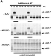
MEK(QP) and Mos-induced inhibition of Cdc2 activation occurs by specific activation of the p42MAPK signaling pathway. (A) PD inhibits phosphorylation of p42MAPK by MEK(WT) and MEK(QP) in vitro. Left, MEK(WT) was preincubated with buffer, DMSO, or PD (100 μM), and then activity was assayed in vitro by using recombinantXenopus Mos as an activator and recombinant kinase-inactive Xenopus p42MAPK as a substrate. Right, MEK(QP) was preincubated with buffer, DMSO, or PD (100 μM), and then activity was assayed in vitro by using the p42MAPK protein used above as a substrate (see MATERIALS AND METHODS for details). (B) PD prevents the Mos-induced inhibition of Cdc2 activation by cyclin B. DMSO or PD (250 μM) was added to aliquots of CHX extracts on ice, then Mos(WT) or Mos(KM) were added, and reactions were incubated at room temperature for 45 min before the addition of cyclin BΔ90 to 25 nM. Samples were taken for analysis by immunoblotting and H1 kinase assay. (C) PD prevents the MEK(QP)-induced inhibition of Cdc2 activation by cyclin B. DMSO or PD (250 μM) was added to aliquots of CHX extracts on ice, MEK(WT) or MEK(QP) were added, and then reactions were incubated at room temperature for 20 min prior to the addition of cyclin BΔ90 to 25 nM. Samples were taken for analysis by immunoblotting and H1 kinase assay.
Figure 7
Figure 7
MEK(QP) and Mos-induced inhibition of Cdc2 activation occurs by specific activation of the p42MAPK signaling pathway. (A) PD inhibits phosphorylation of p42MAPK by MEK(WT) and MEK(QP) in vitro. Left, MEK(WT) was preincubated with buffer, DMSO, or PD (100 μM), and then activity was assayed in vitro by using recombinantXenopus Mos as an activator and recombinant kinase-inactive Xenopus p42MAPK as a substrate. Right, MEK(QP) was preincubated with buffer, DMSO, or PD (100 μM), and then activity was assayed in vitro by using the p42MAPK protein used above as a substrate (see MATERIALS AND METHODS for details). (B) PD prevents the Mos-induced inhibition of Cdc2 activation by cyclin B. DMSO or PD (250 μM) was added to aliquots of CHX extracts on ice, then Mos(WT) or Mos(KM) were added, and reactions were incubated at room temperature for 45 min before the addition of cyclin BΔ90 to 25 nM. Samples were taken for analysis by immunoblotting and H1 kinase assay. (C) PD prevents the MEK(QP)-induced inhibition of Cdc2 activation by cyclin B. DMSO or PD (250 μM) was added to aliquots of CHX extracts on ice, MEK(WT) or MEK(QP) were added, and then reactions were incubated at room temperature for 20 min prior to the addition of cyclin BΔ90 to 25 nM. Samples were taken for analysis by immunoblotting and H1 kinase assay.
Figure 8
Figure 8
Mos-induced activation of p42MAPK does not inhibit DNA replication in Xenopus egg extracts. Buffer, Mos(KM), or Mos(WT) was added to CHX extracts and then incubated at room temperature for 40 min. Demembranated sperm nuclei (600 nuclei/μl) and [α-32P]dATP (0.2 μCi/μl) were added to each reaction, and samples taken at 0, 0.5, 1, and 2 h of incubation at room temperature. Samples were analyzed by electrophoresis on 0.8% agarose gels (see MATERIALS AND METHODS).

Similar articles

Cited by

References

    1. Alessi DR, Cuenda A, Cohen P, Dudley DT, Saltiel AR. PD 098059 is a specific inhibitor of the activation of mitogen-activated protein kinase kinase in vitro and in vivo. J Biol Chem. 1995;270:27489–27494. - PubMed
    1. Barth H, Hoffmann I, Klein S, Kaszkin M, Richards J, Kinzel V. Role of Cdc25-C phosphatase in the immediate G2 delay induced by the exogenous factors epidermal growth factor and phorbol ester. J Cell Physiol. 1996;168:589–599. - PubMed
    1. Boulton TG, Cobb MH. Identification of multiple extracellular signal-regulated kinases (ERKs) with anti-peptide antibodies. Cell Reg. 1991;2:357–371. - PMC - PubMed
    1. Bottorff D, Stang S, Agellon S, Stone JC. RAS signalling is abnormal in c-raf MEK1 double mutant. Mol Cell Biol. 1995;5:5113–5122. - PMC - PubMed
    1. Cobb MH, Boulton TG, Robbins DJ. Extracellular signal-regulated kinases: ERKs in progress. Cell Reg. 1991;2:965–978. - PMC - PubMed

Publication types

MeSH terms

LinkOut - more resources