. 1998 Feb;9(2):483-96.
doi: 10.1091/mbc.9.2.483.

Wortmannin-sensitive phosphorylation, translocation, and activation of PLCgamma1, but not PLCgamma2, in antigen-stimulated RBL-2H3 mast cells

Affiliations
Free PMC article

Wortmannin-sensitive phosphorylation, translocation, and activation of PLCgamma1, but not PLCgamma2, in antigen-stimulated RBL-2H3 mast cells

S A Barker et al. Mol Biol Cell. 1998 Feb.
Free PMC article

Abstract

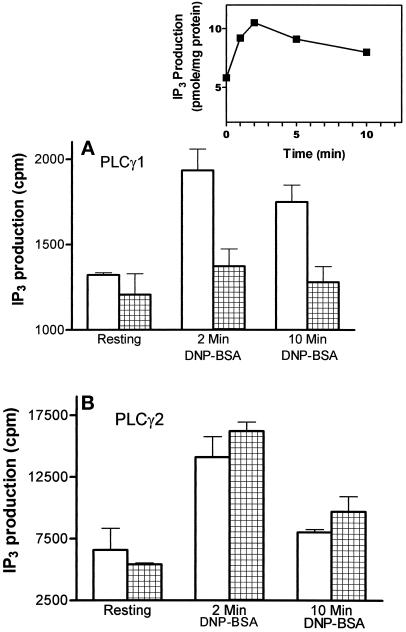
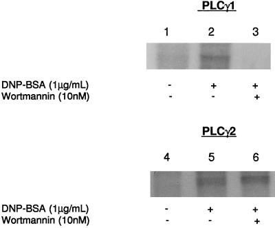
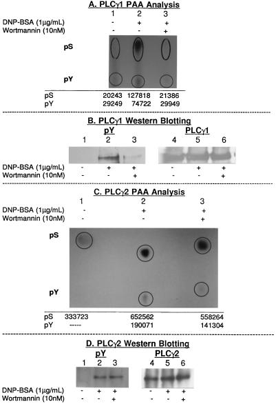
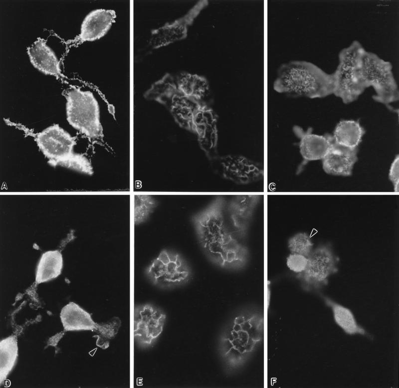
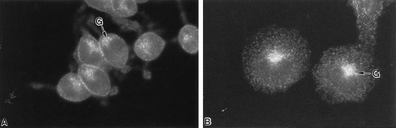
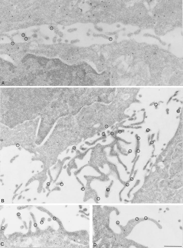
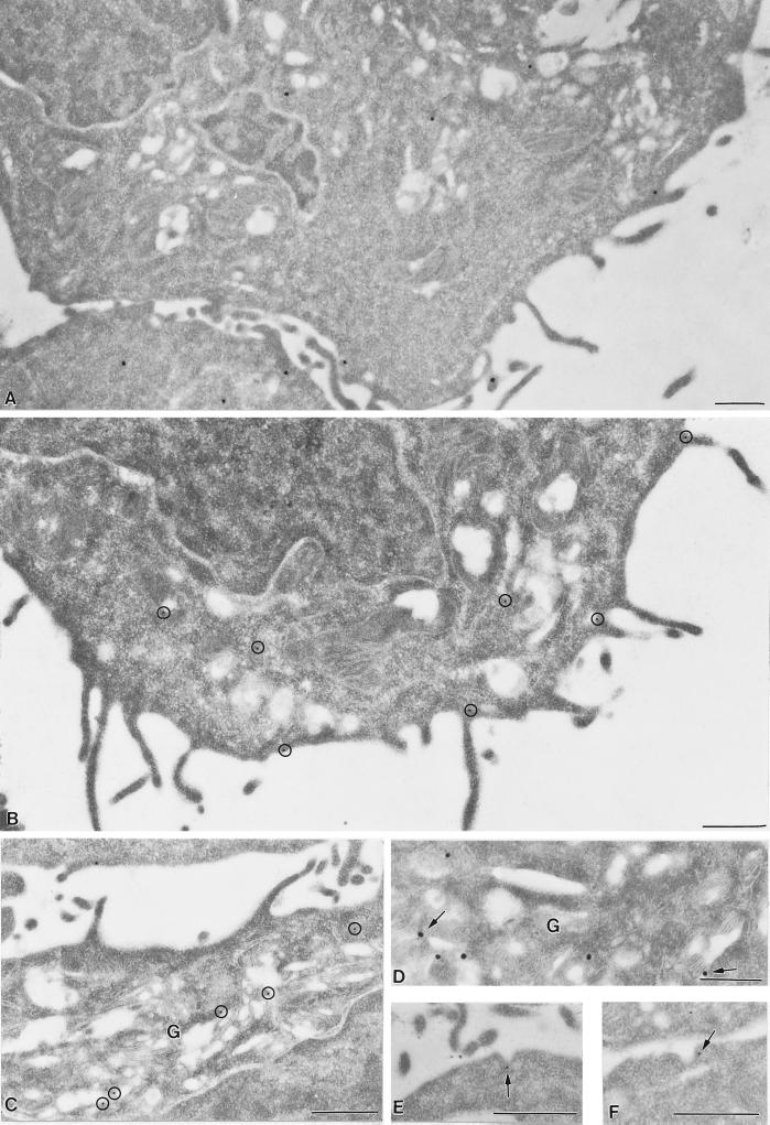
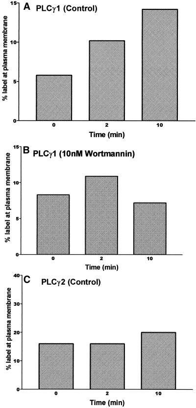
In RBL-2H3 tumor mast cells, cross-linking the high affinity IgE receptor (FcepsilonRI) with antigen activates cytosolic tyrosine kinases and stimulates Ins(1,4,5)P3 production. Using immune complex phospholipase assays, we show that FcepsilonRI cross-linking activates both PLCgamma1 and PLCgamma2. Activation is accompanied by the increased phosphorylation of both PLCgamma isoforms on serine and tyrosine in antigen-treated cells. We also show that the two PLCgamma isoforms have distinct subcellular localizations. PLCgamma1 is primarily cytosolic in resting RBL-2H3 cells, with low levels of plasma membrane association. After antigen stimulation, PLCgamma1 translocates to the plasma membrane where it associates preferentially with membrane ruffles. In contrast, PLCgamma2 is concentrated in a perinuclear region near the Golgi and adjacent to the plasma membrane in resting cells and does not redistribute appreciably after FcepsilonRI cross-linking. The activation of PLCgamma1, but not of PLCgamma2, is blocked by wortmannin, a PI 3-kinase inhibitor previously shown to block antigen-stimulated ruffling and to inhibit Ins(1,4,5)P3 synthesis. In addition, wortmannin strongly inhibits the antigen-stimulated phosphorylation of both serine and tyrosine residues on PLCgamma1 with little inhibition of PLCgamma2 phosphorylation. Wortmannin also blocks the antigen-stimulated translocation of PLCgamma1 to the plasma membrane. Our results implicate PI 3-kinase in the phosphorylation, translocation, and activation of PLCgamma1. Although less abundant than PLCgamma2, activated PLCgamma1 may be responsible for the bulk of antigen-stimulated Ins(1,4,5)P3 production in RBL-2H3 cells.


Figures

Figure 1
Figure 1
FcεRI cross-linking activates both isoforms of PLCγ and PLCγ1 activation is selectively blocked by wortmannin. PLC activity was measured in anti-PLCγ immune complexes prepared from resting and antigen-stimulated RBL-2H3 cells from the hydrolysis of [3H]-PtdIns(4,5)P2 in the Triton-based mixed micelle assay described in MATERIALS AND METHODS. Results show activation of PLCγ1(A) and PLCγ2 (B) at 2 and 10 min after addition of cross-linking reagent (1 μg/ml DNP-BSA) to control cells (open bars). PLCγ1 activation, but not that of PLCγ2, is inhibited in cells pretreated for 15 min with 10 nM wortmannin (hatched bars). Results are the average of duplicate samples in one of at least two similar experiments. Inset: The time course of antigen-induced Ins(1,4,5)IP3 production in control cells, measured with the radioreceptor assay described in Deanin et al. (1991).
Figure 2
Figure 2
A). Wortmannin inhibits the antigen-stimulated phosphorylation of PLCγ1 but not PLCγ2. IgE-primed, [32P]orthophosphate-labeled cells (4 × 10⁷) were incubated for 15 min with or without 10 nM wortmannin, followed by 0 or 2 min with 1 μg/ml DNP-BSA. In lanes 1–3, PLCγ1 was immunoprecipitated from lysis supernatants, separated by SDS-PAGE and detected by autoradiography. PLCγ1 phosphorylation is not detected in resting cells (lane 1), although longer exposures to film can show low levels of phosphate incorporation (unpublished observation). FcεRI cross-linking causes the phosphorylation of PLCγ1 (lane 2). Wortmannin treatment blocks the phosphorylation of PLCγ1 (lane 3). Lanes 4 to 6 show that phosphorylation of PLCγ2 is weakly detectable in resting cells (lane 4), and increases in response to antigen (lane 5). Lane 6 shows that the increase in PLCγ2 phosphorylation is unaffected by the presence of wortmannin. Similar results were obtained in three experiments.
Figure 3
Figure 3
Phosphoamino acid analysis of PLCγ isoforms. Phosphate-labeled bands corresponding to PLCγ1 and PLCγ2 were excised from PVDF membranes after transfer from SDS-PAGE. Each band was digested to constituent amino acids and the entire sample was used for one-dimensional TLC analysis as described in MATERIALS AND METHODS. Mobilities of phosphoserine and phosphotyrosine were determined on the basis of authentic standards; neither isoform had detectable levels of phosphothreonine. The numbers under each lane represent the relative PhosphorImager units corresponding to phosphoserine and phosphotyrosine, adjusted for background. A) Phosphoamino acid content of PLCγ1 in resting cells (lane 1), after 2 min (lane 2) of cross-linking FcεRI with antigen and after 10 nM wortmannin treatment followed by 2 min with antigen (lane 3). B+D). Western blot analysis of PLCγ1(B) or PLCγ2(D) immunoprecipitated from resting and 2 min antigen-activated cells (with and without wortmannin pretreatment) probed with anti-pY (B+D; lanes 1–3), stripped and reprobed with anti-PLCγ1 antibodies (B; lanes 4–6) or anti-PLCγ2 (D; lanes 4–6). C) Phosphoamino acid content of PLCγ2 in resting cells (lane 4), in control cells after 2 min of antigen (lane 5) or in 10 nM wortmannin-treated cells after 2 min of antigen (lane 6).
Figure 4
Figure 4
PLCγ1 translocates to actin-rich membrane ruffles in antigen-stimulated cells. Filamentous actin was labeled with rhodamine phalloidin in fixed and permeablized cells before and after FcεRI cross-linking. The actin stain shows the transformation from microvillous surface architecture in resting cells (A) to ruffles at 10 min of exposure to antigen (B); the majority of wortmannin-treated cells fail to make surface ruffles in response to antigen (C). PLCγ1 was localized by immunofluorescence microscopy in unstimulated RBL-2H3 cells (D) and antigen-stimulated (1 μg/ml DNP-BSA, 10 min) cells without (E) and with (F) wortmannin treatment. The anti-PLCγ1 stain shows a redistribution of enzyme from a generally diffuse distribution in resting cells to an association with membrane ruffles in antigen-stimulated cells. The presence of wortmannin inhibits membrane labeling of antigen-stimulated cells. The arrowheads in D and F point to occasional ruffles that form in resting or wortmannin-treated cells and label with PLCγ1 antibodies. Magnification 750×.
Figure 5
Figure 5
Immunoelectron microscopy of PLCγ1. Resting (A) and antigen-activated (B-D) (1 μg/ml DNP-BSA, 10 min) RBL-2H3 cells were embedded in LR White and thin sections labeled sequentially with anti-PLCγ1 and 15 nm Protein A-gold particles. Membrane-associated gold particles marking the location of PLCγ1 are circled in all panels to emphasize the concentration of membrane-associated PLCγ1 in membrane projections. Panels A and B show the presence of additional gold particles in the cell interior, including the nucleus. Micrographs are representative of results in three different experiments. Bar = 0.5 μm.
Figure 6
Figure 6
PLCγ2 distribution is not markedly altered after antigen stimulation. PLCγ2 was localized by immunofluorescence microscopy in unstimulated (A) and antigen-stimulated (B) (1 μg/ml DNP-BSA, 10 min) RBL-2H3 cell monolayers. PLCγ2 is distributed in small clusters associated with the Golgi region (G) and plasma membrane under both conditions. Magnification 750×.
Figure 7
Figure 7
Immunoelectron microscopy of PLCγ2. anti-PLCγ2 was localized in thin sections of LR White-embedded resting (A) and antigen-activated (B-F) RBL-2H3 cells by labeling with anti-PLCγ2 followed by 15 nm Protein A-gold with a rabbit anti-mouse bridge (B,C,E,F) or with 30 nm anti-mouse IgG-colloidal gold particles (A,D). 15 nm gold particles in B,C are circled for emphasis. Gold particles marking PLCγ2 associate with the plasma membrane and also prominently label vesicles in the Golgi region (G) in panels C,D. Occasional gold particles label Golgi stacks (arrows, D). Gold labeling was also found in association with coated pits (E,F). Micrographs are representative of results in 3 different experiments. Bar = 0.5 μm.
Figure 8
Figure 8
Morphometric measurements of gold-labeled PLCγ isoforms show wortmannin-sensitive translocation of PLCγ1, but not PLCγ2, to the plasma membrane. A) Percent of anti-PLCγ1 gold label within 60 nm of the plasma membrane of resting, control RBL-2H3 cells, followed by 2 or 10 min of antigen stimulation. B) Percent of anti-PLCγ1 gold label at the plasma membrane after 15 min wortmannin treatment, followed by 2 or 10 min of antigen stimulation. C) Percent of anti-PLCγ2 gold label at the plasma membrane of resting, control RBL-2H3 cells, followed by 2 or 10 min of antigen. Micrographs were blind-coded before counting; each experimental point represents the percent of total gold counts from replicate thin sections cut from blocks generated in 2 separate experiments. At least 1,000 gold particles were counted for each experimental condition.

Similar articles

Cited by

References

    1. Atkinson TP, Kaliner MA, Hohman RJ. Phospholipase Cγ1 is translocated to the membrane of rat basophilic leukemia cells in response to aggregation of IgE receptors. J Immunol. 1992;148:2194–2200. - PubMed
    1. Atkinson TP, Lee C-W, Rhee SG, Hohman RJ. Orthovanadate induces translocation of phospholipase C-γ1 and -γ2 in permeabilized mast cells. J Immunol. 1993;151:1448–1455. - PubMed
    1. Barker SA, Caldwell KK, Hall A, Martinez AM, Pfeiffer JR, Oliver JM, Wilson BS. Wortmannin blocks lipid and protein kinase activities associated with PI 3-kinase and inhibits a specific subset of responses induced by FcεRI cross-linking. Mol Biol Cell. 1995;6:1145–1158. - PMC - PubMed
    1. Bar-Sagi D, Suhan JP, McCormick F, Feramisco JR. Localization of phospholipase A2 in normal and ras-transformed cells. J Cell Biol. 1988;106:1649–1658. - PMC - PubMed
    1. Bar-Sagi D, Rotin D, Batzer A, Mandiya V, Schlessinger J. SH3 domains direct cellular localization of signaling molecules. Cell. 1993;74:83–91. - PubMed

Publication types

MeSH terms