. 1998 Feb;9(2):497-511.
doi: 10.1091/mbc.9.2.497.

Proteolytic processing and Ca2+-binding activity of dense-core vesicle polypeptides in Tetrahymena

Affiliations
Free PMC article

Proteolytic processing and Ca2+-binding activity of dense-core vesicle polypeptides in Tetrahymena

J W Verbsky et al. Mol Biol Cell. 1998 Feb.
Free PMC article

Abstract

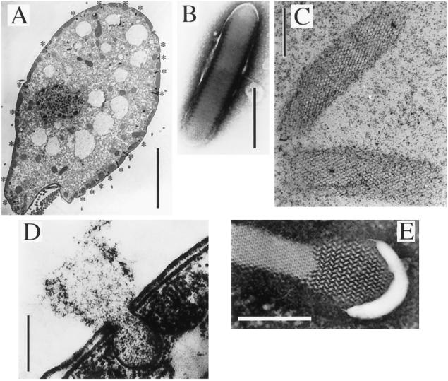
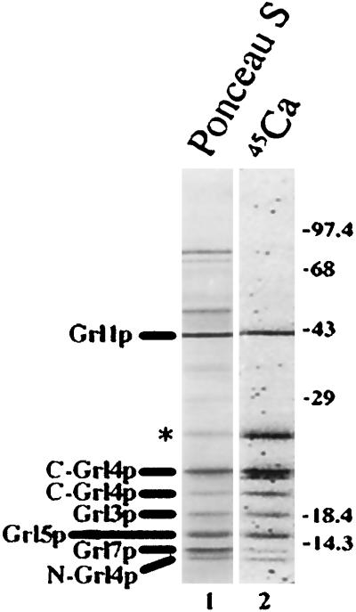
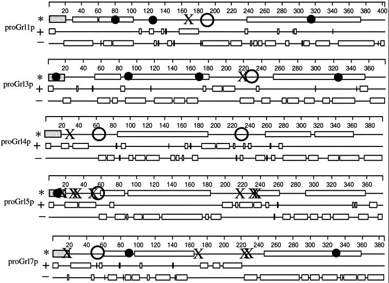
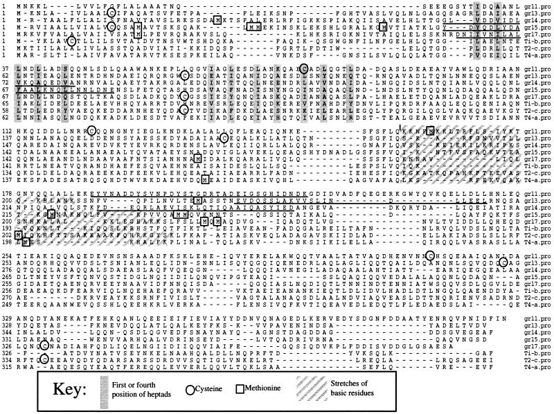
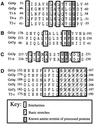
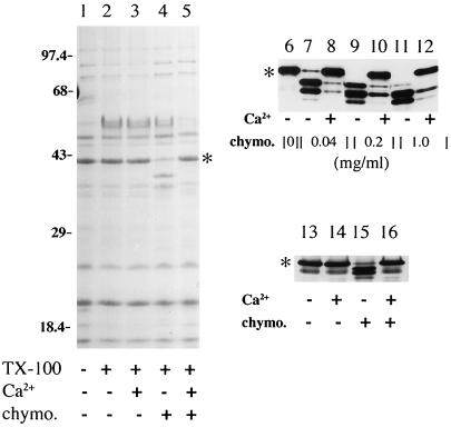
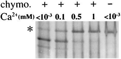
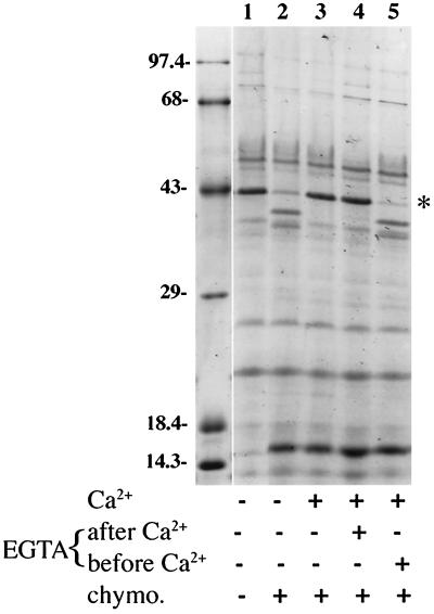
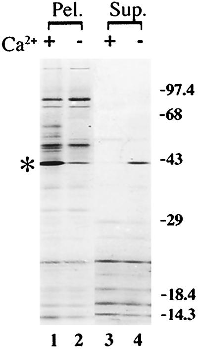
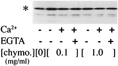
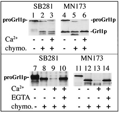
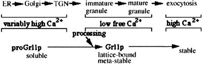
Formation and discharge of dense-core secretory vesicles depend on controlled rearrangement of the core proteins during their assembly and dispersal. The ciliate Tetrahymena thermophila offers a simple system in which the mechanisms may be studied. Here we show that most of the core consists of a set of polypeptides derived proteolytically from five precursors. These share little overall amino acid identity but are nonetheless predicted to have structural similarity. In addition, sites of proteolytic processing are notably conserved and suggest that specific endoproteases as well as carboxypeptidase are involved in core maturation. In vitro binding studies and sequence analysis suggest that the polypeptides bind calcium in vivo. Core assembly and postexocytic dispersal are compartment-specific events. Two likely regulatory factors are proteolytic processing and exposure to calcium. We asked whether these might directly influence the conformations of core proteins. Results using an in vitro chymotrypsin accessibility assay suggest that these factors can induce sequential structural rearrangements. Such progressive changes in polypeptide folding may underlie the mechanisms of assembly and of rapid postexocytic release. The parallels between dense-core vesicles in different systems suggest that similar mechanisms are widespread in this class of organelles.


Figures

Figure 1
Figure 1
Electron micrographs. (A) Thin section of Tetrahymena thermophila. Dense core secretory vesicles docked at the cell surface are marked by ∗. The larger bodies lying near the cell surface are mitochondria. Bar = 10 μm. (B) Isolated vesicle, in which the granule lattice core has been visualized by negative staining by phosphotungstate. Bar = 0.4 μm. (C) Two expanded granules, negatively stained with uranyl acetate. Note difference in scale between B and C: the expanded lattices are >sixfold longer than in the condensed state. The particulate matter is residual Percoll. Bar = 2 μm. (D) Fusion of a secretory vesicle and release of contents. Such fusion can be triggered by glutaraldehyde during cell fixation, producing such exocytic Figures. Bar = 0.8 μm. (E) Isolated granule in a state of partial expansion, negatively stained with phosphotungstate. Bar = 0.4 μm.
Figure 2
Figure 2
In vitro calcium binding to proteins in granule cores. Proteins in isolated granules were resolved by SDS-PAGE (17.5% polyacrylamide, 40 μg/lane) and transferred to nitrocellulose. Lane 1 was stained for protein with Ponceau S; lane 2 was incubated with 45Ca, and bands were visualized after overnight exposure to a PhosphorImager screen. Identies of characterized species are indicated on the left. A prominent calcium-binding band, which appears to be a minor species by protein staining, is marked by ∗. Molecular weight standards are indicated on the right.
Figure 3
Figure 3
Predicted features of proGrlps. On the basis of the deduced amino acid sequences and the known N-termini of isolated polypeptides, a number of features are predicted. Features of each Grlp are shown on three consecutive lines. On the first of each set of three lines are shown the regions forming coiled-coils (as open rectangles), sites corresponding to known amino termini of processed polypeptides (as large open circles), cysteine residues (as small solid circles), methionines (as X’s), and signal sequences (as shaded rectangles). On the second line, stretches of basic residues are shown as open rectangles. On the third line, stretches of acidic residues are shown as open rectangles.
Figure 4
Figure 4
Deduced amino acid sequences from GRL3, GRL4, GRL5 and GRL7. For the purpose of comparison, GRL1 and Paramecium TMPs (called Ti-b, T2-c, and T4-a) have been included. Sequences were aligned to illustrate alignment of several heptad repeats. The majority of the heptad repeats are not labeled since it was not possible to select a single best alignment. In each of the deduced Grlp sequences, the experimentally determined polypeptide sequence is underlined. Cysteines, methionines, and the basic stretches diagrammed in Figure 3 are indicated. Both nucleotide and amino acid sequences are deposited under the following GenBank acquisition numbers: GRL3: AF031319; GRL4: AF031320; GRL5: AF031321; GRL7: AF031322.
Figure 5
Figure 5
Evidence for an internal repeat in proGrl5p. Sequences from the amino- and carboxyl-terminal halves of proGrl5p have been aligned to show amino acid conservation.
Figure 6
Figure 6
Conserved motifs in proGrlps and Tmps. (A) Alignment of sequences at which cleavage occurs following a Gly. The amino-terminal residue of the processed peptide is shaded in A–C. (B) Alignment of sequences at which cleavage occurs following a single basic residue. (C) Alignment of sequences at which cleavage occurs following a single asparagine residue. (D) Alignment of sequences illustrating a tetrapeptide motif that lies amino-terminal to the central basic stretch.
Figure 7
Figure 7
Chymotryptic digestion of Grl1p in isolated granules and cell homogenates. In all three panels, ∗ indicates the position of mature Grl1p. Several of the smaller Grlps cannot be seen on this gel; their chymotrypsin insensitivity was noted in other experiments using higher percentage gels. Lanes 1–5: Aliquots of isolated granules were treated as described in the table beneath the lanes; ∼40 μg of granule protein was loaded per lane of a 15% polyacrylamide gel. In the presence of Triton X-100, chymotrypsin (1 mg/ml) generated distinct fragments of Grl1p (lane 4). Incubation with calcium (1 mM) before addition of chymotrypsin resulted in protection of Grl1p (lane 5). Samples were visualized with Coomassie blue. Lanes 6–12. Isolated granules were incubated with 1% Triton X-100, and aliquots containing 8 μg of protein, were treated as indicated in the table beneath the lanes. After SDS-PAGE, proteins were transferred to nitrocellulose and antibody blotted with antiGrl1p. A ladder of proteolytic products was generated with increasing concentrations of chymotrypsin (lanes 7, 9, 11). At all chymotrypsin concentrations, preincubation with 1 mM calcium provided extensive protection (lanes 8, 10, 12). Lanes 13–16. Aliquots of a crude particulate fraction from MN173 cells were treated as indicated. One hundred and fifty micrograms of total protein was loaded per lane; the chymotrypsin concentration was 0.5 mg/ml. Proteins were visualized by antibody blotting with anti-Grl1p.
Figure 8
Figure 8
Calcium titration. Aliquots of isolated granules in 1% Triton X-100 were incubated with the indicated calcium concentrations, followed by addition of chymotrypsin. 60 μg of protein were loaded per lane, and samples were visualized with Coomassie blue. Grl1p is marked by ∗.
Figure 9
Figure 9
Nonreversibility of calcium effect. Lanes 1–5: Isolated granules were treated as in Figure 7, with two additional variables. In lane 4, 10 mM EGTA was added after the calcium incubation, to reduce free Ca2+ to <1 μM. In lane 5, 10 mM EGTA was added before calcium. Samples were then incubated with chymotrypsin. Gel samples contained ∼40 μg of protein, and were visualized with Coomassie blue. Grl1p is marked by ∗; the prominent band migrating near 15 kDa in lanes 2–5 is chymotrypsin. Molecular weight standards are indicated.
Figure 10
Figure 10
Heat solubilization of Grl1p. Isolated granules (30 μg protein/aliquot) in 1% TX-100 were incubated at 95°C for 30 min, with or without previous exposure to 1 mM calcium. Samples were then centrifuged at 17,000 × g for 30 min. Fifty percent of the supernatant fractions and 100% of the pellets were analyzed by SDS-PAGE (17.5% polyacrylamide) and visualized by Coomassie blue.
Figure 11
Figure 11
Chymotrypsin resistance of soluble Grl1p. Isolated granules were incubated at 95°C for 30 min, and then sedimented at 150,000 × g for 2 h. Aliquots of the supernatant, containing ∼7 μg of protein, were incubated with or without 1 mM calcium, followed by 10 mM EGTA, as indicated. Chymotrypsin was then added at the indicated concentrations. Samples were visualized by antibody blotting after SDS-PAGE. Grl1p is marked by ∗.
Figure 12
Figure 12
Effect of calcium on proGrl1p. Crude membrane fractions of either SB281 or MN173 were prepared, incubated with calcium as noted, and digested with 1 mg/ml chymotrypsin. Approximately 100 μg of protein were loaded per lane. In lanes 1–3, chymotrypsin digestion resulted in disappearance of proGrl1p and appearance of Grl1p-sized fragments. Comparison of lanes 2 and 3 with 5 and 6 indicates that exposure to calcium inhibited digestion of Grl1p, but not proGrl1p. At lower chymotrypsin concentrations (0.1 mg/ml), intermediate digestion products of proGrl1p are visible in membrane fractions from both SB281 (lane 8) and MN173 (lane 12). These appeared to be reversibly destabilized by addition of calcium (lanes 9, 10 and 13, 14). The difference between the sizes of the proteolytic fragments (lanes 8 and 12) was variable; both MN173 and SB281 showed fragments of both sizes in other experiments. Seventy-five micrograms of protein per lane was loaded. Samples in Figure 7 were visualized by antibody blotting.
Figure 13
Figure 13
Model for compartmental regulation of Grl1p and core dynamics. The activity of proteases in maturing dense-core secretory granules cleaves the protein proregion. Low calcium may contribute to their activation. At least one domain of the processed protein, Grl1p, undergoes refolding to form a metastable structure that is an essential element of the granule lattice. Upon exocytosis, Grl1p is exposed to high extracellular calcium, inducing refolding to a fully stable structure.

Similar articles

Cited by

References

    1. Adoutte A. Exocytosis: Biogenesis, Transport and Secretion of Trichocysts. In: Görtz HD, editor. Paramecium. Berlin: Springer-Verlag; 1988. pp. 325–362.
    1. Adoutte A, Garreau de Loubresse N, Beisson J. Proteolytic cleavage and maturation of the crystalline secretion products of Paramecium. J Mol Biol, 1984;180:1065–1081. - PubMed
    1. Anderer R, Hausmann K. Properties and structure of isolated extrusive organelles. J Ultrastruct Res, 1977;60:21–26. - PubMed
    1. Arvan P, Castle JD. Protein sorting and secretion granule formation in regulated secretory cells. Trends Cell Biol, 1992;2:327–331. - PubMed
    1. Attanoos RL, Allen AK. The characterization of the proteins which are secreted by the mucocysts of Tetrahymena thermophila. Biochim Biophys Acta, 1987;924:154–158.

MeSH terms

LinkOut - more resources