. 1998 Feb;66(2):528-39.
doi: 10.1128/IAI.66.2.528-539.1998.

Diffusely adhering Escherichia coli strains induce attaching and effacing phenotypes and secrete homologs of Esp proteins

Affiliations

Diffusely adhering Escherichia coli strains induce attaching and effacing phenotypes and secrete homologs of Esp proteins

C Beinke et al. Infect Immun. 1998 Feb.

Abstract

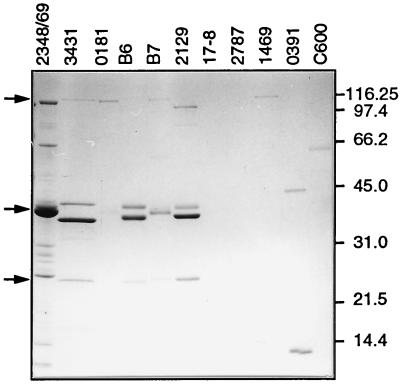
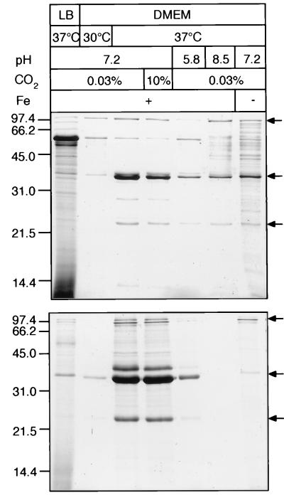
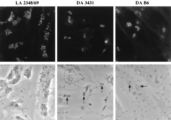
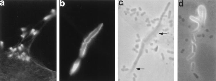
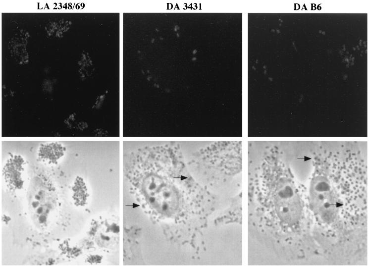
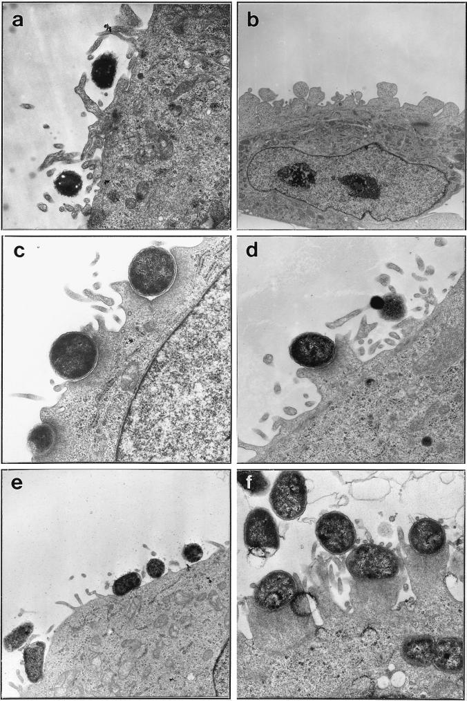
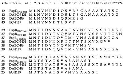
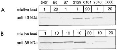
Recent epidemiological studies indicate that Escherichia coli strains which exhibit the diffuse-adherence phenotype (DAEC strains) represent a potential cause of diarrhea in infants. We investigated the interaction of DAEC strains isolated from diarrhea patients in Brazil and in Germany with epithelial cells in tissue culture. The investigated strains were identified as DAEC strains by (i) their attachment pattern, (ii) presence of genes associated with the Dr family of adhesins, and (iii) lack of genetic markers for other diarrhea-associated E. coli categories. Several clinical DAEC isolates were shown to secrete similar patterns of proteins into tissue culture medium. Protein secretion was found to be regulated by environmental parameters, namely, medium, temperature, pH, and iron concentration. DAEC strains secreting these proteins induced accumulation of actin and tyrosine-phosphorylated proteins at sites of bacterial attachment, leading to the formation of pedestals and/or extended surface structures. These changes were phenotypically similar to the attaching and effacing (A/E) lesions observed with enteropathogenic and some enterohemorrhagic E. coli strains carrying the locus of enterocyte effacement (LEE) pathogenicity island. Proteins homologous to the EspA, EspB, and EspD proteins, necessary for signal transduction events inducing A/E lesions, were identified by sequence analysis and cross-reaction of specific antibodies. However, initially nonadhering strains secreting these proteins induced signal transduction events only after prolonged infection. These results indicate that secretion of the Esp proteins alone is not sufficient for efficient signal transduction. This study further shows that some DAEC strains are likely to contain a homolog(s) of the LEE locus which may contribute to the pathogenic potential of DAEC.


Figures

FIG. 1
FIG. 1
Secretion of characteristic proteins of DAEC strains into tissue culture medium. Bacteria were grown as static cultures in DMEM for 12 h at 37°C. Bacteria were removed from the supernatants by centrifugation; proteins were precipitated, separated by gradient SDS-PAGE (9 to 16% gel), and visualized by Coomassie blue staining. Protein samples loaded had been normalized based on the optical density at 600 nm of the corresponding bacterial cultures. The bacterial strains used are indicated at the top. EPEC strain 2348/69 and the laboratory K-12 strain C600 are shown for comparison. Bands of similar-size secreted proteins of several strains are marked by arrows on the left. Molecular mass markers in kilodaltons are indicated on the right.
FIG. 2
FIG. 2
Regulation of protein secretion by different growth conditions. Profiles of proteins secreted into the culture supernatant by EPEC strain 2348/69 (top) and DAEC strain 3431 (bottom) are shown. Bacterial strains were incubated for 12 h in LB at 37°C or in DMEM at 30 or 37°C at pH 7.2, 5.8, or 8.5. Growth was in air containing 0.03 or 10% CO2 and in the presence or absence of iron. Growth conditions with respect to medium, temperature, pH, CO2 concentration, and presence of iron are indicated at the top. Proteins were isolated as described in the legend to Fig. 1. Loaded protein samples were normalized according to the number of bacteria in the corresponding cultures. The characteristic pattern of secreted proteins is marked by arrows on the right. Optimal conditions for protein secretion were observed in DMEM in the presence of iron at 37°C and pH 7.2. Molecular mass markers are indicated in kilodaltons on the left.
FIG. 3
FIG. 3
Accumulation of actin at sites of adherent DAEC. HeLa cells were infected for 3 h with DAEC strain 3431 or B6 or the localized adhering (LA) EPEC strain 2348/69 and stained for filamentous actin with FITC-phalloidin. Fluorescence (top) and corresponding phase-contrast (bottom) micrographs are shown. Bacteria are detectable in the phase-contrast micrographs as enlarged, darker, plaque-like structures (indicated by arrows). Bacteria showing actin staining were often seen in small groups. Magnification, ×165.
FIG. 4
FIG. 4
Bacteria induce complex extended structures of polymerized actin. HeLa cells were infected with DAEC strain 3431 for 6 h (a) or 3 h (b and c) or with DAEC strain B6 for 3 h (d) and processed as for Fig. 3. Fluorescence (a and b), phase-contrast (c), and simultaneous fluorescence and phase-contrast (d) micrographs are shown. Micrographs b and c are taken from identical areas. Actin accumulates in long horn-like structures protruding from the surfaces of the HeLa cells with a single bacterium on the top (a) or in long tubes associated with bacteria (b to d). Accumulated actin can be detected in phase-contrast micrographs as darker areas (indicated by arrows in panel c). Magnifications: a, ×260; b to d, ×410.
FIG. 5
FIG. 5
Tyrosine-phosphorylated proteins accumulate underneath adherent bacteria. HeLa cells were infected for 3 h with DAEC strain 3431 or B6 or with EPEC strain 2348/69, washed, fixed, labeled with antiphosphotyrosine monoclonal antibody PT-66, and examined by immunofluorescence microscopy. Fluorescence (top) and corresponding phase-contrast (bottom) micrographs are shown. Concentrations of host tyrosine-phosphorylated proteins can be seen as bright spots or rings at sites of adherent bacteria. Fewer bacteria with fluorescence spots are observed for DAEC strains 3431 and B6 than for strain 2348/69. Magnification, ×165.
FIG. 6
FIG. 6
DAEC strains induce morphological changes on HeLa cells. Transmission electron micrographs show adherence phenotypes after a 3-h incubation of DAEC strains secreting characteristic proteins with HeLa cells. DAEC strains 3431 (a and b), B6 (c and d), and 0181 (e), and EPEC strain 2348/69 (f) are shown. Magnifications: a, d, and f, ×14,400; b, ×4,400; c, ×16,900; e, ×7,100.
FIG. 7
FIG. 7
Amino-terminal sequence homologies of secreted proteins of DAEC strains 3431 and B6 and E. coli 2129 to EspD (top), EspB (middle), and EspA (bottom) proteins. Amino-terminal sequences of various lengths for the proteins were aligned with sequences of EPEC strain 2348/69 (30, 32), EHEC strain EDL933 (15), and EHEC strain 413.89-1 (15). The apparent molecular masses, E. coli strains, and positions of amino acids (aa) are indicated. Amino acids are abbreviated by the single-letter code, and those which could not be determined are indicated by X’s. Gaps were allowed for optimal alignment.
FIG. 8
FIG. 8
Recognition of secreted proteins by antisera directed at the secreted 43-kDa (A) and 38-kDa (B) proteins of DAEC strain 3431. Secreted proteins of the indicated strains were prepared as described in the legend to Fig. 1 and separated by SDS-PAGE (13% gel). Samples were analyzed by immunoblotting with the appropriate antisera. No signals besides those shown could be detected. Low cross-reactivity was detected by loading larger amounts of the secreted proteins for the corresponding strain as indicated in relative numbers at the top.

References

    1. Aleksic S, Karch H, Bockemuhl J. A biotyping scheme for Shiga-like (Vero) toxin-producing Escherichia coli O157 and a list of serological cross-reactions between O157 and other gram-negative bacteria. Int J Med Microbiol Virol Parasitol Infect Dis. 1992;276:221–230. - PubMed
    1. Baqui A H, Sack R B, Black R E, Haider K, Hossain A, Alim A R, Yunus M, Chowdhury H R, Siddique A K. Enteropathogens associated with acute and persistent diarrhea in Bangladeshi children less than 5 years of age. J Infect Dis. 1992;166:792–796. - PubMed
    1. Benz I, Schmidt M A. AIDA-I, the adhesin involved in diffuse adherence of the diarrhoeagenic Escherichia coli strain 2787 (O126:H27), is synthesized via a precursor molecule. Mol Microbiol. 1992;6:1539–1546. - PubMed
    1. Benz I, Schmidt M A. Cloning and expression of an adhesin (AIDA-I) involved in diffuse adherence of enteropathogenic Escherichia coli. Infect Immun. 1989;57:1506–1511. - PMC - PubMed
    1. Benz I, Schmidt M A. Isolation and serologic characterization of AIDA-I, the adhesin mediating the diffuse adherence phenotype of the diarrhea-associated Escherichia coli strain 2787 (O126:H27) Infect Immun. 1992;60:13–18. - PMC - PubMed

Publication types

LinkOut - more resources