. 1998 Feb;66(2):587-93.
doi: 10.1128/IAI.66.2.587-593.1998.

Partial purification and characterization of biological effects of a lipid toxin produced by Mycobacterium ulcerans

Affiliations

Partial purification and characterization of biological effects of a lipid toxin produced by Mycobacterium ulcerans

K M George et al. Infect Immun. 1998 Feb.

Abstract

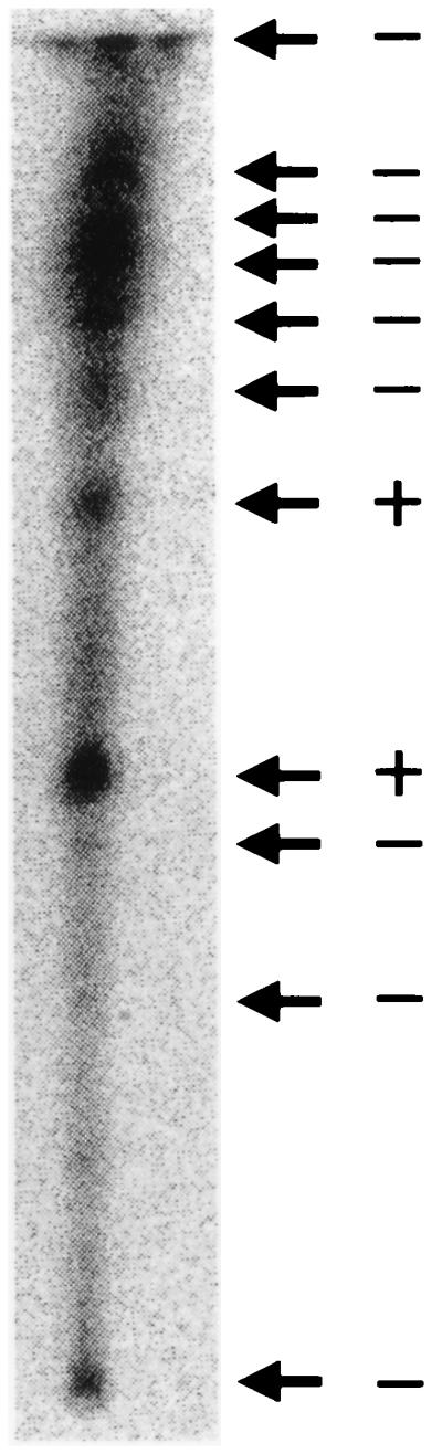
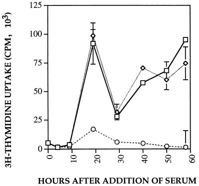
Organisms in the genus Mycobacterium cause a variety of human diseases. One member of the genus, M. ulcerans, causes a necrotizing skin disease called Buruli ulcer. Buruli ulcer is unique among mycobacterial diseases in that the organisms at the site of infection are extracellular and there is little acute inflammatory response. Previous literature reported the presence of a toxin in the culture supernatant of M. ulcerans which causes a cytopathic effect on the mouse fibroblast cell line L929 in which the adherent cells round up and detach from the tissue culture plate. Here we report partial purification of a lipid toxin from the culture supernatant of M. ulcerans which is capable of causing the cytopathic effect on L929 cells. We also show that this cytopathic effect is a result of cytoskeletal rearrangement. The M. ulcerans toxin does not cause cell death but instead arrests cells in the G1 phase of the cell cycle.


Figures

FIG. 1
FIG. 1
TLC of acetone-soluble lipids from M. ulcerans. M. ulcerans culture was radiolabeled with [1-14C]acetate, and acetone-soluble lipids were prepared. The lipids were resolved on a glass-backed TLC plate in chloroform-methanol (2:1, vol/vol) and detected on a PhosphorImager. Individual lipids were scraped off the glass backing, eluted with methanol, and tested for cytopathic activity. Arrows indicate lipids which were scraped off the TLC plate, and the results of the cytopathic assay are noted with plus and minus signs.
FIG. 2
FIG. 2
Effect of toxin on cytoskeleton of L929 cells. L929 cells were exposed to either M7H9 medium or M. ulcerans SF and stained with the actin-specific stain phalloidin-red. (A) L929 cells exposed to M7H9 medium for 4 h show normal actin structure with stress fibers. (B) L929 cells exposed to M. ulcerans SF for 4 h show some rearrangement of actin. (C) M7H9-treated L929 cells at 10 h. (D) SF-treated L929 cells at 10 h show dramatic cytoskeleton rearrangements.
FIG. 3
FIG. 3
Flow cytometry analysis of toxin-treated L929 cells. L929 cells were treated with M7H9 medium (A), M. marinum SF (B), M. ulcerans SF (C), and M. ulcerans acetone-soluble lipid fraction (D) for 48 h. Floating and attached cells were harvested, lysed, and stained with propidium iodide. After 48 h of treatment, there was a significant shift in the percent of M. ulcerans-treated cells (C and D) in the G0/G1-phase of the cell cycle compared to the medium- and M. marinum-treated (A and B) controls.
FIG. 4
FIG. 4
Kinetics of DNA and protein synthesis. (A) DNA synthesis was measured at various times after treatment with M7H9 medium (squares), M. marinum SF (diamonds), M. ulcerans SF (circles), or DMEM alone (triangles). (B) Protein synthesis was measured at various hours after treatment with M7H9 medium (squares), M. marinum SF (diamonds), M. ulcerans s SF (circles), or DMEM alone (triangles). All time points and samples were done in quadruplicate, and standard deviations are indicated with bars.
FIG. 5
FIG. 5
Ability of M. ulcerans SF to inhibit G1 progression in quiescent L929 cells stimulated by FCS. L929 cells were synchronized in G0 by incubation in serum-free DMEM for 5 days. After 5 days, the cells were stimulated by addition of DMEM–10% FCS (squares), and either M7H9 medium (diamonds) or M. ulcerans SF (circles) was added. DNA synthesis was measured at various times. All time points and samples were done in quadruplicate, and standard deviations are indicated with bars.
FIG. 6
FIG. 6
Determination of restriction point of toxin’s action in growth-arrested L929 cells. Growth-arrested L929 cells were stimulated with DMEM–10% FCS, and at various times after addition, M7H9 medium (hatched bars) or M. ulcerans SF (solid bars) was added. DNA synthesis was measured at 20 h poststimulation. All time points and samples were done in quadruplicate, and standard deviations are indicated with bars.

References

    1. Adams J L, Czuprynski C J. Mycobacterial cell wall components induce the production of TNF-α, IL-1, and IL-6 by bovine monocytes and the murine macrophage cell line RAW 264.7. Microb Pathog. 1995;16:401–411. - PubMed
    1. Aepfelbacher M, Essler M, Luber De Quintana K, Weber P C. ADP-ribosylation of the GTP-binding protein rhoA blocks cytoplasmic division in human myelomonocytic cells. Biochem J. 1995;308:853–858. - PMC - PubMed
    1. Aktories K. Rho proteins: targets for bacterial toxins. Trends Microbiol. 1997;5:282–288. - PubMed
    1. Barker L P, George K M, Falkow S, Small P L C. Differential trafficking of live and dead Mycobacterium marinum organisms in macrophages. Infect Immun. 1997;65:1497–1504. - PMC - PubMed
    1. Behling C A, Perex R L, Kidd M R, Staton G W, Jr, Hunter R L. Induction of pulmonary granulomas, macrophage procoagulant activity, and tumor necrosis factor-alpha by trehalose glycolipids. Ann Clin Lab Sci. 1993;23:256. - PubMed

LinkOut - more resources