. 1998 Feb;66(2):741-6.
doi: 10.1128/IAI.66.2.741-746.1998.

Identification of a putative precursor to the major surface glycoprotein of Pneumocystis carinii

Affiliations

Identification of a putative precursor to the major surface glycoprotein of Pneumocystis carinii

S M Sunkin et al. Infect Immun. 1998 Feb.

Abstract

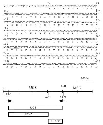
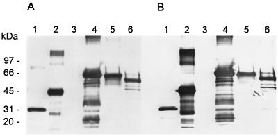
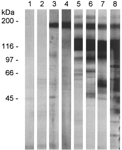
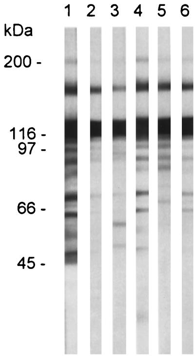
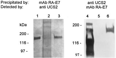
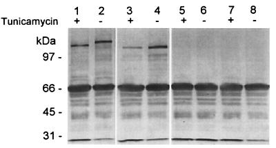
The major surface glycoprotein (MSG) of Pneumocystis carinii f. sp. carinii is a family of proteins encoded by a family of heterogeneous genes. Messenger RNAs encoding different MSGs each begin with the same 365-bp sequence, called the Upstream Conserved Sequence (UCS), which is in frame with the contiguous MSG sequence. The UCS contains several potential start sites for translation. To determine if translation of MSG mRNAs begins in the UCS, polyclonal antiserum was raised against the 123-amino-acid peptide encoded by the UCS. The anti-UCS serum reacted with a P. carinii protein that migrated at 170 kDa; however, it did not react with the mature MSG protein, which migrates at 116 kDa. A 170-kDa protein was immunoprecipitated with anti-UCS serum and shown to react with a monoclonal antibody against a conserved MSG epitope. To explore the functional role of the UCS in the trafficking of MSG, the nucleotide sequence encoding the UCS peptide was ligated to the 5' end of an MSG gene and incorporated into a recombinant baculovirus. Insect cells infected with the UCS-MSG hybrid gene expressed a 160-kDa protein which was N-glycosylated. By contrast, insect cells infected with a baculovirus carrying an MSG gene lacking the UCS expressed a nonglycosylated 130-kDa protein. These data suggest that in P. carinii, translation begins in the UCS to produce a pre-MSG protein, which is subsequently directed to the endoplasmic reticulum and processed to the mature form by proteolytic cleavage.


Figures

FIG. 1
FIG. 1
Sequences and map of the UCS. In the nucleotide sequence, the uppercase letters show the putative coding sequences, and the lowercase letters indicate the 5′ untranslated region of the UCS-MSG cDNA clone 1 (47). The numerals in the right margin above the nucleotide sequence starting at 60 are aligned with the corresponding nucleotide. Numbers on the left starting with +1 and ending with +132 are encoded amino acid residues, which are shown in single-letter code below the nucleotide sequence, with the amino acid aligned with the second nucleotide of each codon. The SalI and EagI sites are underlined. The underlined KR is a potential site for proteolytic cleavage. The drawing below the sequences is a map of the UCS-MSG locus. Crosshatches mark 100-bp increments. The UCS region extends from +1 to +418. The MSG region begins at +419. The location and orientation of the two primers utilized in the initial PCR are indicated by arrows. The regions expressed as fusion proteins are represented by rectangles labeled UCS, UCS5′, and UCS3′.
FIG. 2
FIG. 2
Antiserum reactivity with UCS fusion proteins. E. coli lysates containing either GST, G9, or these proteins fused to UCS protein were separated on duplicate 8 to 16% Novex SDS-PAGE gels. Resolved proteins were transferred to a nitrocellulose membrane which was incubated with either α-UCS2 (A) or α-UCS3 (B) serum. Bound rabbit antibodies were detected by reaction with goat anti-rabbit IgG conjugated to alkaline phosphatase. Phosphatase activity was detected by incubation with BCIP-NBT. Lanes: 1, GST; 2, GST-UCS; 3, G9; 4, G9-UCS; 5, G9-UCS-5′; 6, G9-UCS-3′. The positions of Bio-Rad high-molecular-mass markers are indicated to the left of lane 1.
FIG. 3
FIG. 3
α-UCS sera recognize a 170-kDa protein in P. carinii f. sp. carinii. Proteins in a lysate from P. carinii f. sp. carinii organisms were separated by electrophoresis through an 8 to 16% Novex SDS-PAGE gel. Each lane contained lysate from approximately 107 organisms. After electrophoresis, proteins were transferred to a nitrocellulose sheet, strips of which were incubated with the following: lane 1, prebleed serum 2; lane 2, prebleed serum 3; lane 3, α-UCS2 serum; lane 4, α-UCS3 serum; lane 5, MAb RB-E3; lane 6, MAb RA-E7; lane 7, anti-MSG serum; lane 8, anti-P. carinii serum. Strips 1 to 4, 7, and 8 were reacted with goat anti-rabbit IgG phosphatase-labeled conjugate. Strips 5 and 6 were reacted with goat anti-mouse IgG phosphatase-labeled conjugate. All strips were developed in BCIP-NBT. The positions of the Bio-Rad high-molecular-mass markers are indicated to the left of lane 1.
FIG. 4
FIG. 4
Reactivity of MSG-specific MAbs with P. carinii f. sp. carinii proteins. P. carinii f. sp. carinii proteins were separated by electrophoresis through an 8 to 16% Novex SDS-PAGE gel. Each lane contained lysate from approximately 107 organisms. Proteins were transferred to a nitrocellulose sheet, strips of which were incubated with the following mouse MAbs: lane 1, RA-E7; lane 2, RA-C1; lane 3, RA-C6; lane 4, RA-C7; lane 5, RB-C8; lane 6, RA-C11. Bound antibodies were detected as described in the legend to Fig. 3. Positions of molecular mass markers are indicated to the left of lane 1.
FIG. 5
FIG. 5
The 170-kDa protein immunoprecipitates with UCS and MSG antibodies. The UCS-MSG protein was immunoprecipitated with the α-UCS2 serum and MAb RA-E7. The resulting precipitate was subjected to electrophoresis in either a 6% Novex gel for the MAb RA-E7 precipitate (lanes 1 to 3) or an 8 to 16% Novex gel for the α-UCS2 precipitate (lanes 4 to 6). Each gel was blotted to nitrocellulose and reacted to either the α-UCS2 serum (lanes 1 to 3) or MAb RA-E7 (lanes 4 to 6). The blots were developed as described in the legend to Fig. 3. Lanes 1 and 4 contain an aliquot of the immunoprecipitation-starting material. Lanes 2 and 5 contain the precipitates produced in the preclearing step from the MAb RA-E7 and α-UCS2 immunoprecipitations, respectively. Lanes 3 and 6 contain the immunoprecipitates obtained with MAb RA-E7 and α-UCS2 serum, respectively. The positions of the molecular mass markers are indicated to the left of lanes 1 and 4.
FIG. 6
FIG. 6
The UCS is required for N-glycosylation of MSG in insect cells. Insect cells were infected with recombinant baculoviruses containing genes encoding UCS-MSG (lanes 1 and 2), MSG (lanes 3 and 4), or SP-A, an unrelated protein (lanes 5 and 6). Lysates from uninfected cells are in lanes 7 and 8. The cells were incubated in the absence (−) or presence (+) of 5 μg of tunicamycin per ml for 24 h and harvested by scraping. Cell lysates were fractionated on a 6% Novex gel, transferred to nitrocellulose, and reacted with MAb RA-E7. The blot was developed by incubation with antimouse IgG horseradish peroxidase-labeled conjugate and horseradish peroxidase-dependent oxidation of o-phenylenediamine. The positions of Bio-Rad low-molecular-mass markers are indicated to the left of lane 1.

Similar articles

Cited by

References

    1. Angus C W, Tu A, Vogel P, Qin M, Kovacs J A. Expression of variants of the major surface glycoprotein of Pneumocystis carinii. J Exp Med. 1996;183:1229–1234. - PMC - PubMed
    1. Cushion M T, Kaselis M, Stringer S L, Stringer J R. Genetic stability and diversity of Pneumocystis carinii infecting rat colonies. Infect Immun. 1993;61:4801–4813. - PMC - PubMed
    1. Dei-Cas E, Mazars E, Ferragut C O, Durand I, Aliouat E-M, Dridba M, Palluault F, Cailliez J-C, Seguy N, Tibayrenc M, Mullet C, Creusy C, Camus D. Ultrastructural, genomic, isoenzymatic and biological features make it possible to distinguish rabbit Pneumocystis from other mammal Pneumocystis strains. J Eukaryot Microbiol. 1994;41:84S. - PubMed
    1. De Luca A, Ortona E, Margutti P, Visconti E, Tamburrini E, Siracusano A. Different amplification efficiency and nucleotide sequence variation in various Pneumocystis isolates from humans and rats. J Eukaryot Microbiol. 1994;41:85S. - PubMed
    1. Edlind T D, Bartlett M S, Weinberg G A, Prah G N, Smith J W. The beta-tubulin gene from rat and human isolates of Pneumocystis carinii. Mol Microbiol. 1992;6:3365–3373. - PubMed

Publication types

LinkOut - more resources