. 1998 Feb;66(2):747-55.
doi: 10.1128/IAI.66.2.747-755.1998.

Comprehensive study of the intestinal stage of listeriosis in a rat ligated ileal loop system

Affiliations

Comprehensive study of the intestinal stage of listeriosis in a rat ligated ileal loop system

B Pron et al. Infect Immun. 1998 Feb.

Abstract

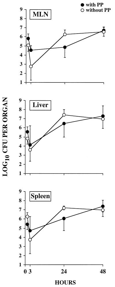
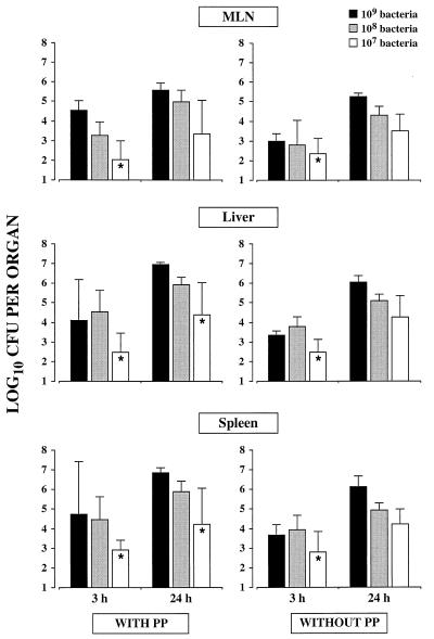
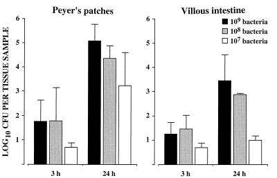
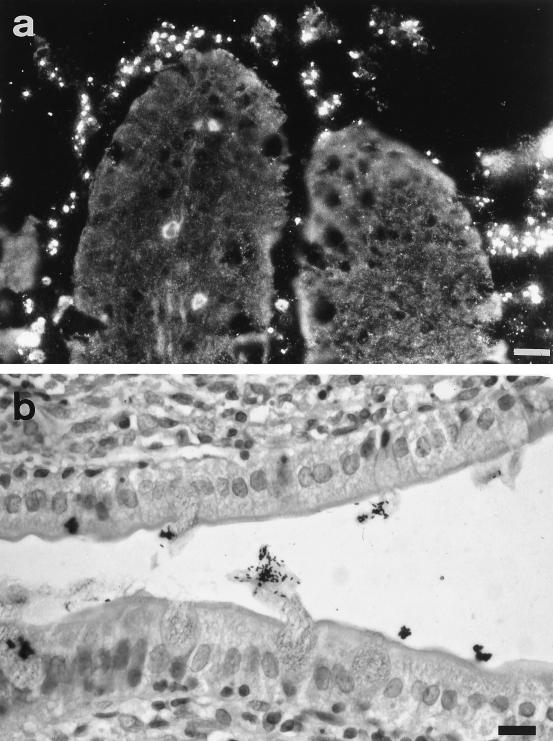
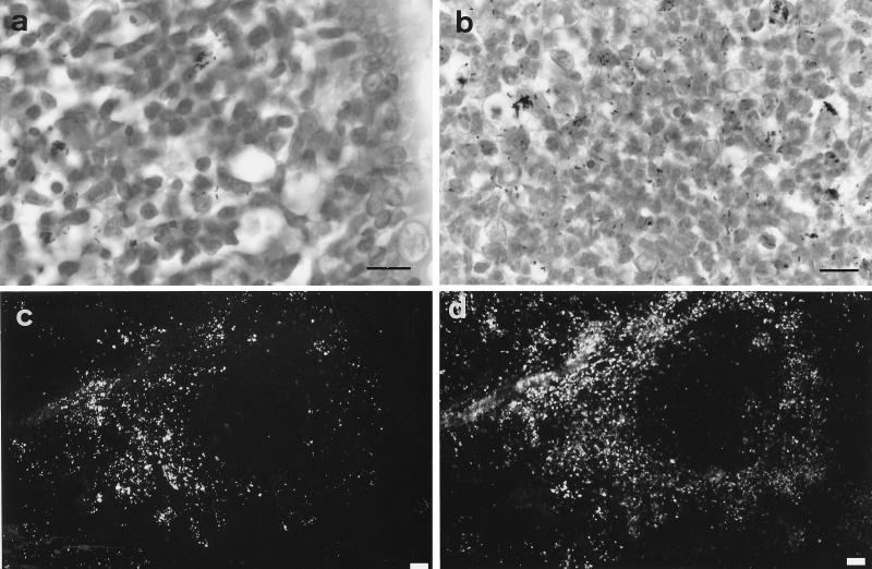
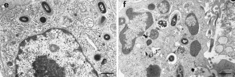
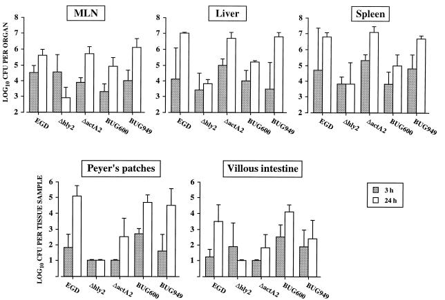
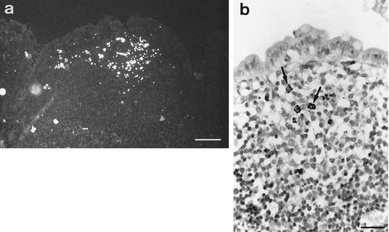
The intestinal stage of listeriosis was studied in a rat ligated ileal loop system. Listeria monocytogenes translocated to deep organs with similar efficiencies after inoculation of loops with or without Peyer's patches. Bacterial seeding of deep organs was demonstrated as early as 15 min after inoculation. It was dose dependent and nonspecific, as the delta inlAB, the delta hly, and the delta actA L. monocytogenes mutants and the nonpathogenic species, Listeria innocua, translocated similarly to wild-type L. monocytogenes strains. The levels of uptake of listeriae by Peyer's patches and villous intestine were similar and low, 50 to 250 CFU per cm2 of tissue. No listeria cells crossing the epithelial sheet of Peyer's patches and villous intestine were observed by transmission electron microscopy. The lack of significant interaction of listeriae and the follicle-associated epithelium of Peyer's patches was confirmed by scanning electron microscopy. The follicular tissue of Peyer's patches was a preferential site of Listeria replication. With all doses tested, the rate of bacterial growth was 10 to 20 times higher in Peyer's patches than in villous intestine. At early stages of Peyer's patch infection, listeriae were observed inside mononuclear cells of the dome area. Listeriae then disseminated throughout the follicular tissue except for the germinal center. The virulence determinants hly and, to a lesser extent, actA, but not inlAB, were required for the completion of this process. This study suggests that Peyer's patches are preferential sites for replication rather than for entry of L. monocytogenes, due to the presence of highly permissive mononuclear cells whose nature remains to be defined.


Figures

FIG. 1
FIG. 1
Infection of MLN, liver, and spleen after inoculation of L. monocytogenes EGD into loops with or without Peyer’s patches (PP). Loops were inoculated with ca. 109 CFU/loop. After 1 h of contact, they were deligated (time zero) and treated intraluminally with gentamicin (1 mg/ml). The number of viable bacteria was determined by killing groups of rats at intervals. Data points and error bars represent the mean and standard deviation of log10 CFU per organ (mean of four rats for each point).
FIG. 2
FIG. 2
Effect of amount of inoculum on L. monocytogenes translocation from loops with or without Peyer’s patches (PP). Loops were inoculated with EGD at doses of ca. 109, 108, and 107 CFU/loop. After 1 h of contact, they were deligated (time zero) and treated intraluminally with gentamicin (1 mg/ml). The number of viable bacteria was determined by killing groups of rats at intervals. Data points and error bars represent the mean and standard deviation of log10 CFU per organ (mean of four rats for each point). ∗, bacteria recovered from three of four animals only.
FIG. 3
FIG. 3
Listeria infection of Peyer’s patches and villous intestine. Loops were inoculated with EGD at doses of ca. 109, 108, and 107 CFU/loop. Intestinal tissue samples (Peyer’s patches or pieces of villous intestine of similar size) were recovered at 3 and 24 h postinoculation and processed to determine the number of gentamicin-protected bacteria. Data points and error bars represent the mean and standard deviation of log10 CFU per sample (mean of four rats for each point).
FIG. 4
FIG. 4
Histological and immunohistological analysis of villous intestine 1 h after inoculation with EGD. (a) Immunofluorescence labeling of listeriae. Bacteria are abundant in the intestinal lumen, apparently embedded in mucus; note that very few bacteria are close to the epithelial cell surface. (b) Gram-Weigert staining. Aggregates of bacteria are stuck to mucus released by goblet cells; no bacteria are seen associated with the epithelial cell surface. Bars = 10 μm.
FIG. 5
FIG. 5
Histological, immunohistological, and TEM analysis of Peyer’s patch infection with EGD. (a and b) Gram-Weigert staining. (a) Six-hour infection (a few bacteria are seen in the dome area); (b) 24-h infection (many bacteria can be seen in the follicular tissue; note the major inflammatory reaction consisting of both mononuclear cells and neutrophils); bars = 5 μm. (c and d) Twenty-four-hour infection, double-fluorescence labeling of listeriae (c) and the macrophage-related antigen ED1 (d). A very large number of bacteria infect the follicular tissue except the germinal center; bars = 20 μm. (e and f) Twenty-four-hour infection, TEM of listeriae located within a mononuclear cell (e) and listeriae inside a neutrophil (f); bars = 1 μm.
FIG. 5
FIG. 5
Histological, immunohistological, and TEM analysis of Peyer’s patch infection with EGD. (a and b) Gram-Weigert staining. (a) Six-hour infection (a few bacteria are seen in the dome area); (b) 24-h infection (many bacteria can be seen in the follicular tissue; note the major inflammatory reaction consisting of both mononuclear cells and neutrophils); bars = 5 μm. (c and d) Twenty-four-hour infection, double-fluorescence labeling of listeriae (c) and the macrophage-related antigen ED1 (d). A very large number of bacteria infect the follicular tissue except the germinal center; bars = 20 μm. (e and f) Twenty-four-hour infection, TEM of listeriae located within a mononuclear cell (e) and listeriae inside a neutrophil (f); bars = 1 μm.
FIG. 6
FIG. 6
Role of Listeria virulence factors. Loops were inoculated with the mutants and their parental strains at a dose of ca. 109 CFU/loop, and the numbers of bacteria in Peyer’s patches, villous intestine, MLN, liver, and spleen were determined at 3 and 24 h postinoculation. The mean and standard deviation of log10 CFU per organ are shown (mean of four rats for each point).
FIG. 7
FIG. 7
Histology and immunohistology of Peyer’s patches 24 h after infection with the actA mutant. (a) Immunofluorescence labeling of bacteria. The foci of infection are less numerous and smaller than with EGD; they are restricted to the dome area. (b) Gram-Weigert staining. Discrete foci of bacterial replication inside mononuclear cells can be seen (arrows). Bars = 20 μm.

References

    1. Amerongen H, Weltzin R, Farnet C, Michetti P, Haseltine W, Neutra M. Transepithelial transport of HIV-1 by intestinal M cells: a mechanism for transmission of AIDS. J Acquired Immune Defic Syndr. 1991;4:760–765. - PubMed
    1. Amerongen H, Wilson G, Fields B, Neutra M. Proteolytic processing of reovirus is required for adherence to intestinal M cells. J Virol. 1994;68:8428–8432. - PMC - PubMed
    1. Barza M. Listeriosis and milk. N Engl J Med. 1985;312:438–440. - PubMed
    1. Berg R D. Bacterial translocation from the gastrointestinal tract. Trends Microbiol. 1995;3:149–154. - PubMed
    1. Berg R D, Owens W E. Inhibition of translocation of viable Escherichia coli from the gastrointestinal tract of mice by bacterial antagonism. Infect Immun. 1979;25:820–827. - PMC - PubMed

Publication types