. 1998 Feb 15;18(4):1440-8.
doi: 10.1523/JNEUROSCI.18-04-01440.1998.

Synergistic effects of schwann- and muscle-derived factors on motoneuron survival involve GDNF and cardiotrophin-1 (CT-1)

Affiliations

Synergistic effects of schwann- and muscle-derived factors on motoneuron survival involve GDNF and cardiotrophin-1 (CT-1)

V Arce et al. J Neurosci. .

Abstract

The survival of central neurons depends on multiple neurotrophic factors produced by different cell types. We demonstrate that media conditioned by muscle and Schwann cell lines show strong synergistic effects on survival of purified embryonic day 14.5 rat motoneurons in culture. Different lines of evidence implicate glial cell line-derived neurotrophic factor (GDNF) and cardiotrophin-1 (CT-1) in this synergy. Their expression in the environment of the motoneuron is compartmentalized: gdnf transcripts are expressed principally in Schwann cell lines, whereas ct-1 mRNA is present in myotubes. Blocking antibodies to GDNF inhibit the trophic activity of Schwann cell line-conditioned media by 75%, whereas CT-1 antibodies diminish the myotube-derived activity by 46%. CT-1 and GDNF act synergistically to enhance motoneuron survival in vitro. In vivo, individual motoneurons coexpress both GDNF and CT-1 receptor components. GDNF and CT-1, therefore, are major components of the trophic support provided by the Schwann and muscle cells, respectively. The possibility that they act together on individual motoneurons suggests that the motoneuron must integrate distinct signals from different cellular partners when deciding whether to die or to survive.


Figures

Fig. 1.
Fig. 1.
Synergy between Schwann cell- and myotube-conditioned media on motoneuron survival. C2 myotube-conditioned medium (C2-CM) and MSC80 Schwann cell-conditioned medium (SC-CM) were tested alone or in combination after 6 d (A) and 10 d (B) of culture. Each medium was used at a concentration of 10% (v/v) in neurobasal (NB) medium. The number of motoneurons per field was determined and expressed relative to the number of motoneurons surviving in 100 pg/ml GDNF determined after 24 hr of culture and taken as 100%. Means ± SEM for duplicate dishes are shown. Strong synergy is observed between C2-CM and SC-CM.
Fig. 2.
Fig. 2.
Expression of gdnf andct-1 mRNAs is compartmentalized in the environment of the motoneuron. RT-PCR analysis of RNA extracted from Schwann cell lines [TSC2 (lane 1) and MSC80 (lane 2)] and from muscle cells [C2/C7 myoblasts (lane 3) and C2/C7 myotubes (lane 4)]. Signal for ct-1 was detected only in mRNA from muscle cells, whereas gdnf mRNA was more abundant in Schwann cells than in myoblasts or myotubes. gapdh was used as a positive control. PCR reactions were performed on the same RNA samples incubated with (+) or without (−) reverse transcriptase.
Fig. 3.
Fig. 3.
GDNF and CT-1 are major components of the trophic activity of Schwann cell- and myotube-conditioned medium, respectively.A, Blocking antibodies inhibit the activity of the relevant factor and show no toxic activity in the presence of irrelevant factors. B, Blocking antibodies toGDNF strongly inhibit the trophic activity of SC-CM.C, Blocking antibodies toCT-1 diminish the activity ofC2-CM. The effects of antibody depletion were measured using purified motoneurons. Antibodies (20 μg/ml) were preincubated for 1 hr at 37°C in the presence of the indicated factor or conditioned medium: GDNF (100 pg/ml), CT-1 (10 ng/ml), neurturin (NTN, 100 pg/ml), CNTF (10 ng/ml). The number of motoneurons surviving after 3 d in culture is expressed as actual motoneuron counts per diameter of a 16 mm well; survival values in neurobasal medium (NB) have been subtracted. Means ± SEM in AC are from the same experiment but have been separated for clarity.
Fig. 4.
Fig. 4.
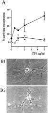
GDNF and CT-1 act synergistically to promote long-term survival of motoneurons. A, Increasing concentrations of CT-1 were tested for their long-term motoneuron survival activity in the presence (•) or absence (○) of a fixed concentration of 100 pg/ml GDNF. The number of motoneurons per field was determined after 14 d in culture and is expressed relative to the number of motoneurons surviving in 100 pg/ml GDNF determined after 24 hr of culture and taken as 100%. Means ± SEM for duplicate dishes, after correction for survival in basal medium (taken as 0%), are shown. B, Phase-contrast micrographs of motoneurons maintained for 14 d in culture in the presence of 100 pg/ml GDNF supplemented with 5 ng/ml CT1 (B1) or 500 pg/ml CNTF (B2). Note the large multipolar cell body morphology.
Fig. 5.
Fig. 5.
GDNF acts synergistically with two cytokines of the IL-6 family that use α-subunits. Increasing concentrations of CNTF (A) were added to motoneurons in the presence (•) or absence (○) of a fixed concentration of GDNF (100 pg/ml). The dashed line indicates the value obtained using GDNF alone. B, Comparison of motoneuron survival in the presence of cytokines alone and with combinations of these cytokines taken 2 × 2 as indicated. Survival values were determined after 10 d (A) or 6 d (B) in culture and are expressed as described in the legend to Figure 4. All cytokines (CNTF, CT-1, and LIF) showed significant trophic activity at shorter survival times (not shown).
Fig. 6.
Fig. 6.
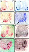
Double-labeling in situhybridization detects expression of components of two different receptors in individual motoneurons. Single sections of E12.5 (AD) mouse and E14.5 (EH) rat brachial spinal cords were hybridized with two probes: DIG-labeled lifrβ and Fluo-labeled ret (AD) or DIG-labeled cntfrα and Fluo-labeledgdnfrα (EH). Anti-DIG antibodies were applied first and stained using Fast Red to reveal cells expressing lifrβ (A,C) and cntfrα (E,G). Anti-Fluo antibodies were then applied and detected using NBT/BCIP to reveal cells expressing ret(B, D) and gdnfrα(F, H) after removal of the first red reaction product. Most motoneurons express components of two receptor complexes (examples indicated by arrows). Compare enlargements of one mouse ventral horn in C(lifrβ) and D (ret) and enlargements of one rat ventral horn in G(cntfrα) and H(gdnfrα). Arrowheads indicate motoneurons that show a strong signal for ret(D) and very low signal forlifrβ (C) or that are positive for cntfrα (G) and nearly negative for gdnfrα (H). Loss of lifrβ signal from roof plate (asterisk in A and B) demonstrates lack of interference between color reactions.
Fig. 7.
Fig. 7.
Diagram showing compartmentalized expression of GDNF and CT1 and their potential synergy. Two signaling pathways are activated: one by CT-1 (dashed line) produced by the muscle and the other byGDNF (solid line) coming from the Schwann cells. Successful activation of both pathways allows the long-term survival of motoneurons.

References

    1. Boutry JM, Hauw JJ, Gansmuller A, Di BN, Pouchelet M, Baron-Van Evercooren A. Establishment and characterization of a mouse Schwann cell line which produces myelin in vivo. J Neurosci Res. 1992;32:15–26. - PubMed
    1. Catala F, Wanner R, Barton P, Cohen A, Wright W, Buckingham M. A skeletal muscle-specific enhancer regulated by factors binding to E and CArG boxes is present in the promoter of the mouse myosin light-chain 1A gene. Mol Cell Biol. 1995;15:4585–4596. - PMC - PubMed
    1. Chandler CE, Parsons LM, Hosang M, Shooter EM. A monoclonal antibody modulates the interaction of nerve growth factor with PC12 cells. J Biol Chem. 1984;259:6882–6889. - PubMed
    1. Davis S, Aldrich TH, Valenzuela DM, Wong VV, Furth ME, Squinto SP, Yancopoulos GD. The receptor for ciliary neurotrophic factor. Science. 1991;253:59–63. - PubMed
    1. DeChiara TM, Vejsada R, Poueymirou WT, Acheson A, Suri C, Conover JC, Friedman B, McClain J, Pan L, Stahl N, Ip NY, Kato A, Yancopoulos GD. Mice lacking the CNTF receptor, unlike mice lacking CNTF, exhibit profound motor neuron deficits at birth. Cell. 1995;83:313–322. - PubMed

Publication types

MeSH terms

LinkOut - more resources