. 1998 Jan;5(1):50-7.
doi: 10.1128/CDLI.5.1.50-57.1998.

Expression, structure, and location of epitopes of the major surface glycoprotein of Pneumocystis carinii f. sp. carinii

Affiliations

Expression, structure, and location of epitopes of the major surface glycoprotein of Pneumocystis carinii f. sp. carinii

M J Linke et al. Clin Diagn Lab Immunol. 1998 Jan.

Abstract

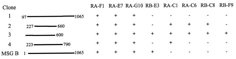
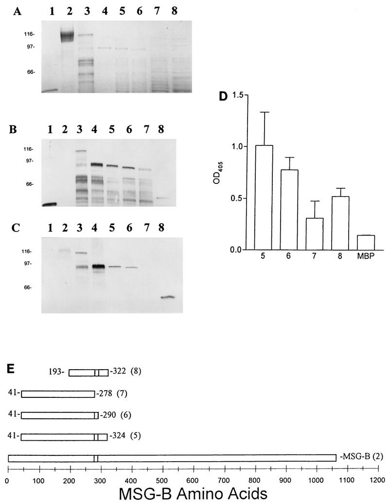
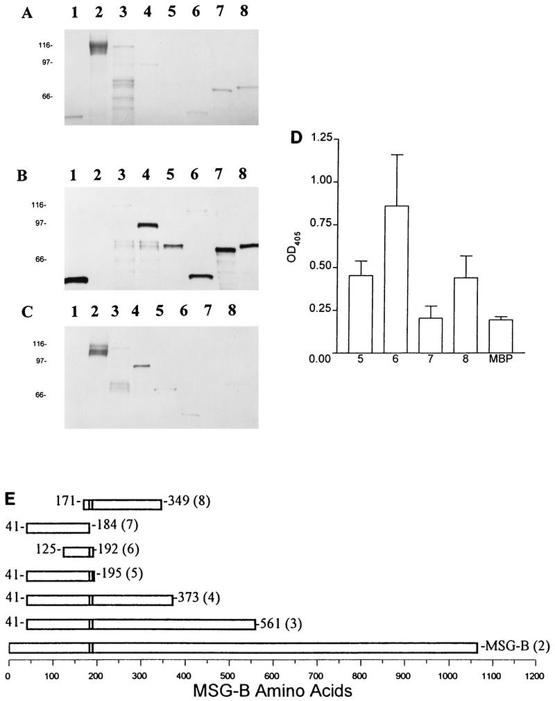
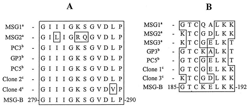
The major surface glycoprotein (MSG) of Pneumocystis carinii f. sp. carinii consists of a heterogeneous family of proteins that are encoded by approximately 100 unique genes. A genomic expression library was screened with a panel of MSG-specific monoclonal antibodies (MAbs) to identify conserved and rare epitopes. All of the antibodies reacted with epitopes that are encoded within the 5' end of MSG. The results from the expression screening identified antibodies that recognize highly conserved, moderately conserved, and rare epitopes. Four MAbs (MAbs RA-F1, RA-E7, RA-G10, and RB-E3) reacted with a maltose binding protein-MSG-B fusion protein ([MBP]MSG-B41-1065) by immunoblotting and enzyme-linked immunosorbent assay. Three of the MAbs (MAbs RA-F1, RA-G10, and RA-E7) reacted with the same continuous epitope that was localized to amino acids 278 to 290 of MSG-B. Comparison of the sequence of the RA-F1-, RA-G10-, and RA-E7-reactive epitope to the deduced amino acid sequences of multiple MSGs demonstrated that it is highly conserved. The reactivity of RB-E3 with MSG-B was shown to be dependent on amino acids 184 to 192, which may comprise a portion of a discontinuous epitope.


Figures

FIG. 1
FIG. 1
Physical maps and MAb reactivities of four λgt11 clones each carrying a fragment from a different MSG gene. The lines show the sizes of the inserts carried in the four phage clones compared to the size of a previously characterized cloned msg gene (msg-b). The numbers at the ends of each line indicate the amino acid residues encoded by each msg gene fragment. The reactivity (+) or nonreactivity (−) of the plaques produced by each clone is indicated for each of eight MAbs.
FIG. 2
FIG. 2
Mapping of MAb RA-E7 reactivity with MBPMSG truncated fusion proteins by SDS-PAGE and immunoblotting. (A) Coomassie blue-stained gel. (B) Immunoblot with anti-MBP polyclonal antisera. (C) Immunoblot with RB-E7. Lanes 1, MBP; lanes 2, native MSG; lanes 3, MBPMSG41–561; lanes 4, MBPMSG41–373; lanes 5, MBPMSG41–324; lanes 6, MBPMSG41–290; lanes 7, MBPMSG41–278; lanes 8, MBPMSG193–322. (D) ELISA analysis of MAb RA-E7 reactivity with MBPMSG-B truncated fusion proteins. Wells of 96-well microtiter plates were coated with the fusion proteins or MBP alone. The numbers correspond to the lanes described above. (E) Schematic of mapping of the epitope reactive with MAb RA-E7. The numbers in parentheses correspond to the lanes described above.
FIG. 3
FIG. 3
Mapping of MAb RB-E3 reactivity with MBPMSG truncated fusion proteins by SDS-PAGE, immunoblotting, and ELISA. (A) Coomassie blue-stained gel. (B) Immunoblot with anti-MBP polyclonal antisera. (C) Immunoblot with RB-E3. Lanes 1, MBP; lanes 2, native MSG; lanes 3, MBPMSG41–561; lanes 4, MBPMSG41–373; lanes 5, MBPMSG41–195; lanes 6, MBPMSG125–192; lanes 7, MBPMSG41–184; lanes 8, MBPMSG171–349. (D) ELISA analysis of MAb RB-E3 reactivity with MBPMSG-B truncated fusion proteins. Wells of 96-well microtiter plates were coated with the fusion proteins or MBP alone. The numbers correspond to the lanes described above. (E) Schematic of mapping of the epitope reactive with MAb RB-E3. The numbers in parentheses correspond to the lanes described above.
FIG. 4
FIG. 4
Alignment of MSG-B epitope region reactive with MAbs RA-E7 and RB-E3 with seven additional deduced MSG amino acid sequences. The deduced amino acid sequence of the epitope regions from five previously described P. carinii f. sp. carinii MSGs and from the four phage clones were visually aligned with the MAb-reactive epitopes identified on MSG-B. (A) RA-E7 epitope; (B) RB-E3 epitope. a, from reference ; b, from reference ; c, deduced amino acids from λgt11 clones.

References

    1. Angus C W, Tu A, Vogel P, Qin M, Kovacs J A. Expression of variants of the major surface glycoprotein of Pneumocystis carinii. J Exp Med. 1996;183:1229–1234. - PMC - PubMed
    1. Ausubel F M, Brent B, Kingston R E, Moore D D, Seidman J G, Smith J A, Struhl K, editors. Current protocols in molecular biology. New York, N.Y: Greene Publishing Associates and Wiley-Interscience; 1991.
    1. Barlow D J, Edwards M S, Thornton J M. Continuous and discontinuous protein antigenic determinants. Nature. 1986;322:747–748. - PubMed
    1. Cushion M T, Ruffolo J J, Walzer P D. Analysis of the developmental stages of Pneumocystis carinii, in vitro. Lab Invest. 1988;58:324–331. - PubMed
    1. Cushion M T, Zhang J, Kaselis M, Giuntoli D, Stringer S L, Stringer J R. Evidence for two genetic variants of Pneumocystis carinii coinfecting laboratory rats. J Clin Microbiol. 1993;31:1217–1223. - PMC - PubMed

Publication types

MeSH terms