. 1998 Feb 9;140(3):525-40.
doi: 10.1083/jcb.140.3.525.

A pore-forming toxin interacts with a GPI-anchored protein and causes vacuolation of the endoplasmic reticulum

Affiliations

A pore-forming toxin interacts with a GPI-anchored protein and causes vacuolation of the endoplasmic reticulum

L Abrami et al. J Cell Biol. .

Abstract

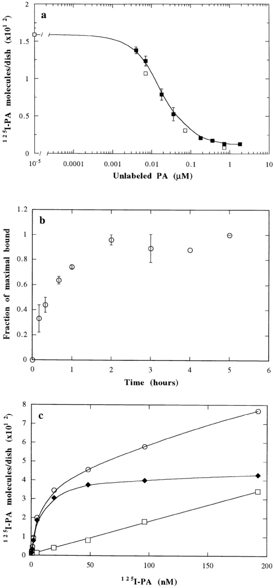
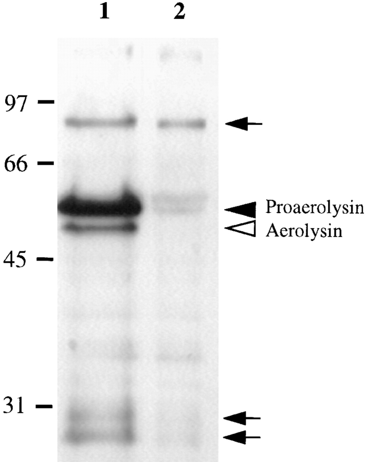
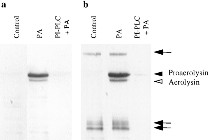
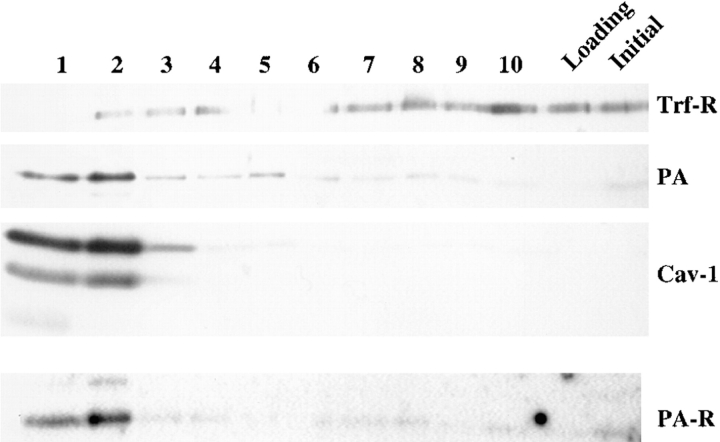
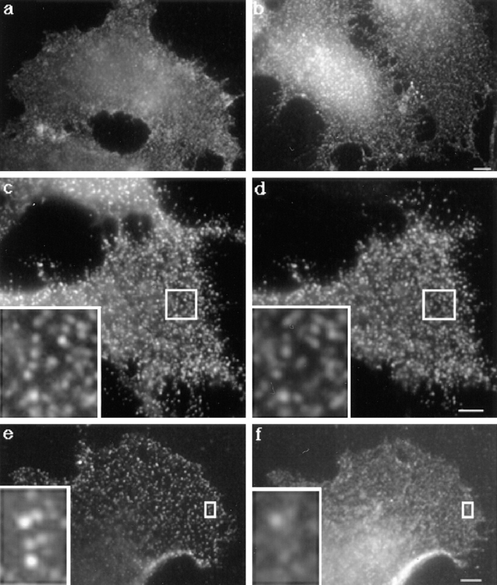
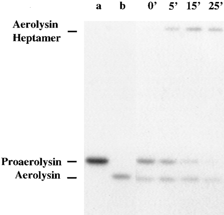
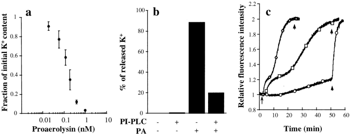
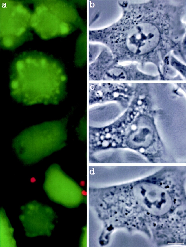
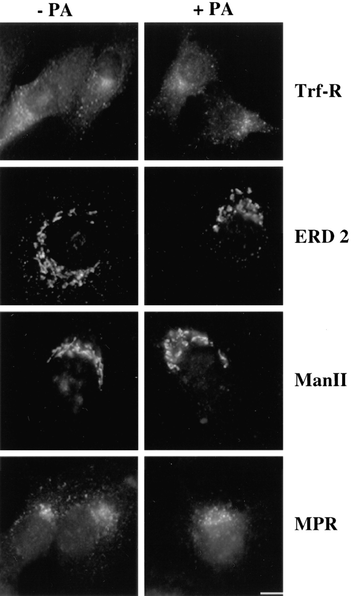
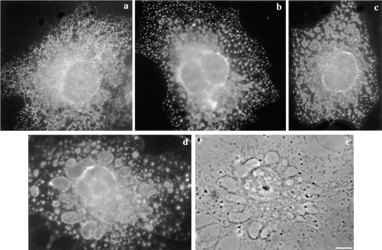
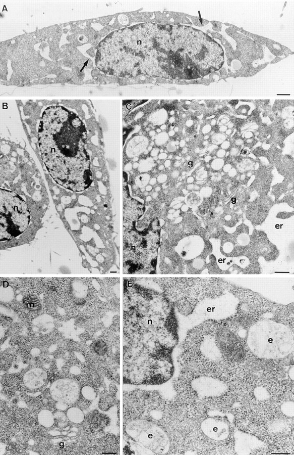
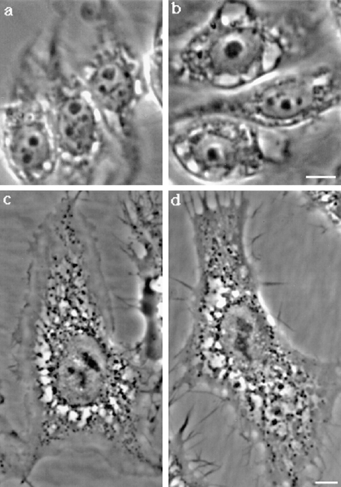
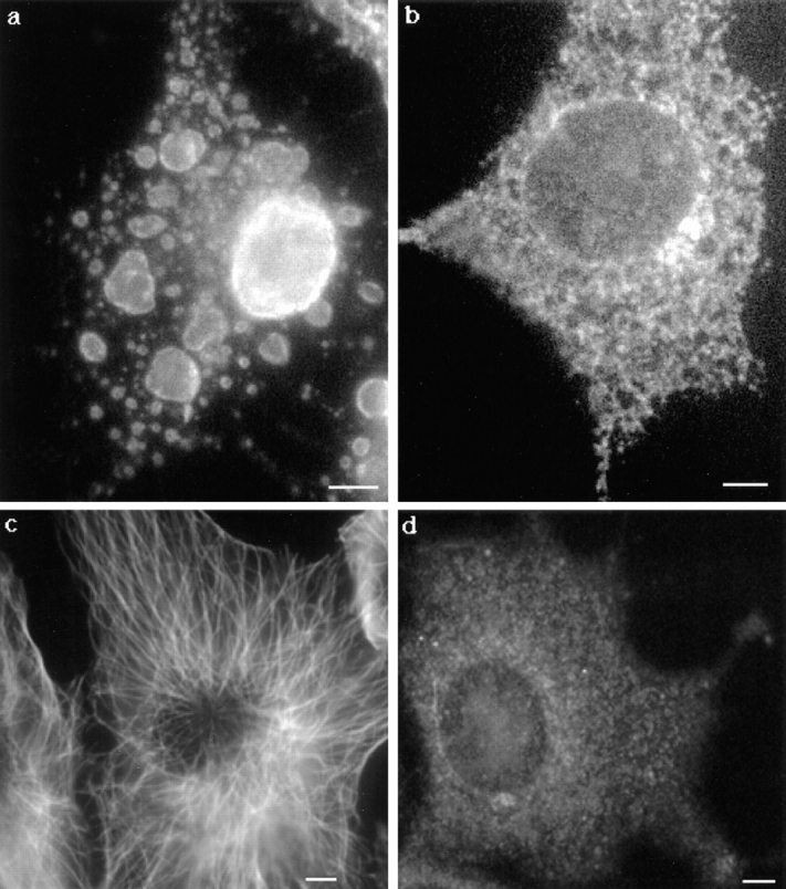
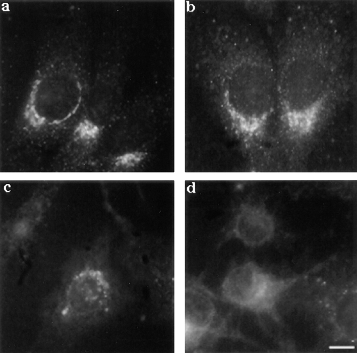
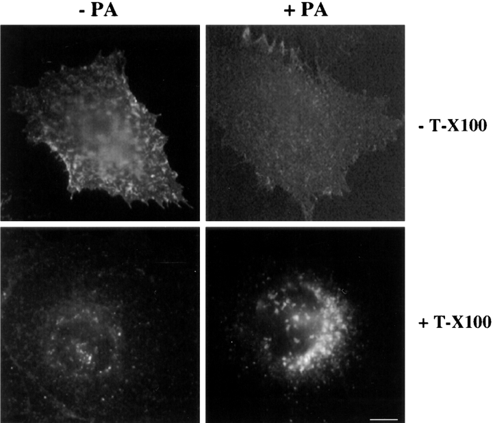
In this paper, we have investigated the effects of the pore-forming toxin aerolysin, produced by Aeromonas hydrophila, on mammalian cells. Our data indicate that the protoxin binds to an 80-kD glycosyl-phosphatidylinositol (GPI)-anchored protein on BHK cells, and that the bound toxin is associated with specialized plasma membrane domains, described as detergent-insoluble microdomains, or cholesterol-glycolipid "rafts." We show that the protoxin is then processed to its mature form by host cell proteases. We propose that the preferential association of the toxin with rafts, through binding to GPI-anchored proteins, is likely to increase the local toxin concentration and thereby promote oligomerization, a step that it is a prerequisite for channel formation. We show that channel formation does not lead to disruption of the plasma membrane but to the selective permeabilization to small ions such as potassium, which causes plasma membrane depolarization. Next we studied the consequences of channel formation on the organization and dynamics of intracellular membranes. Strikingly, we found that the toxin causes dramatic vacuolation of the ER, but does not affect other intracellular compartments. Concomitantly we find that the COPI coat is released from biosynthetic membranes and that biosynthetic transport of newly synthesized transmembrane G protein of vesicular stomatitis virus is inhibited. Our data indicate that binding of proaerolysin to GPI-anchored proteins and processing of the toxin lead to oligomerization and channel formation in the plasma membrane, which in turn causes selective disorganization of early biosynthetic membrane dynamics.


Figures

Figure 1
Figure 1
Binding of 125I-proaerolysin to BHK cells. (a) Inhibition of 125I-proaerolysin binding to BHK cells by unlabeled toxin. Cells were incubated for 1 h with 4 nM of 125I-proaerolysin plus the indicated amount of unlabeled wild-type toxin (▪). 50% inhibition occurred in the presence of 16 nM of unlabeled proaerolysin. Bars indicate the standard deviation (n = 3). Binding of wild-type 125I-proaerolysin could also be competed with an excess of unlabeled G202C-I445C mutant proaerolysin (□). (b) Kinetics of specific binding of 125I-proaerolysin to BHK cells at 4°C. Cells were incubated with 4 nM 125I-proaerolysin and washed at the indicated times. Specifically bound proaerolysin was determined by subtracting the values obtained in the presence of 50-fold excess of unlabeled toxin. Bars indicate the standard deviation (n = 3). (c) Concentration dependence of proaerolysin binding to BHK cells. Cells were incubated for 2.5 h at 4°C with 125I-proaerolysin in the absence (○) or in the presence of 50-fold excess unlabeled toxin (□). Specific binding (♦) was calculated by subtraction of the unspecific bound toxin (□) from the total bound toxin (○). The results represent the mean of two independent experiments. Maximal errors were <16%.
Figure 2
Figure 2
Proaerolysin binds to an 80-kD protein on BHK cells. Cells were treated or not with 2 nM proaerolysin for 1 h at 4°C, thoroughly washed, homogenized, and a PNS was prepared. 20 μg of PNS of toxin-treated cells (lane 1) and control cells (lane 2) were submitted to the proaerolysin overlay assay as described in Materials and Methods. The overlay was performed with unlabeled proaerolysin and revealed with anti-aerolysin antibodies. Arrowheads indicate proaerolysin and aerolysin (that were bound to the plasma membrane, only in lane 1) migrating at their expected molecular weights. Arrows indicate proaerolysin that bound to specific proteins on the nitrocellulose membrane during the overlay procedure.
Figure 4
Figure 4
PI-PLC inhibits the binding of proaerolysin to BHK cells. Cells were incubated with or without 6 U/ml of PI-PLC for 1 h at 37°C in the presence of 10 μg/ml cycloheximide. The cells were then incubated in presence or absence of 2 nM proaerolysin for 1 h at 4°C, thoroughly washed, and homogenized. (a) The PNSs were probed for the presence of proaerolysin by Western blotting. (b) The PNSs were analyzed by proaerolysin overlay for the presence of proaerolysin binding proteins. Lane 1, control cells; lanes 2, proaerolysin-treated cells; lane 3, PI-PLC and proaerolysin-treated cells. Arrowheads indicate proaerolysin and aerolysin (that were bound to the plasma membrane) migrating at their expected molecular weights. Arrows indicate proaerolysin that bound to specific proteins on the nitrocellulose membrane.
Figure 3
Figure 3
Proaerolysin binds to detergent-insoluble microdomains of the plasma membrane of BHK cells. Cells treated with proaerolysin (4°C) were solubilized in 1% Triton X-100 and detergent-insoluble fractions were floated on a step sucrose gradient. The 10 fractions from the gradient (lanes 1 to 10; top to bottom of the gradient), the loading region (3 ml) and the starting homogenate were analyzed by Western Blot analysis for the presence of proaerolysin (PA), of caveolin-1 (Cav-1), and of the transferrin receptor (Trf-R). As is often the case, two forms of caveolin-1 could be observed. The nitrocellulose membrane was also subjected to the proaerolysin overlay assay, using radiolabeled toxin, to localize the 80-kD proaerolysin-binding protein (PA-R). 18 μg of protein was loaded in each lane except in lane 1, which contained 13 μg.
Figure 5
Figure 5
Distribution of proaerolysin at the plasma membrane of BHK cells. Cells were incubated with proaerolysin (2 nM) for 1 h at 4°C. (a) Cells were fixed with paraformaldehyde and further processed for immunofluorescence as described in Materials and Methods. (b) Cells were incubated for 15 min at 37°C, and then processed as in a. (c and d) BHK cells were transiently transfected with PLAP cDNA as described in Materials and Methods. Cells were kept at 4°C and incubated for 1 h with proaerolysin (2 nM), 1 h with anti-proaerolysin monoclonal and antialkaline phosphatase polyclonal antibodies, 1 h with the corresponding secondary antibodies. Cells were then fixed with paraformaldehyde. The same cell is shown stained for both proaerolysin (c) and PLAP (d). (e and f) BHK cells were kept at 4°C and incubated for 1 h with proaerolysin (2 nM), 1 h with anti-proaerolysin monoclonal antibody, 1 h with FITC-conjugated goat anti–rabbit, cells were then fixed and permeabilized with methanol (e). The cells were fixed with methanol and incubated 30 min at room temperature with anti–caveolin-1 antibody and for 30 min at room temperature with lissamine rhodamine sulfonyl chloride–conjugated goat anti–mouse IgG (f). Bars, 6.7 μm.
Figure 6
Figure 6
Proaerolysin is processed by host cell proteases. BHK cells were incubated with 0.95 nM 125I-proaerolysin for 1 h at 4°C, thoroughly washed and incubated with a toxin-free medium at 37°C. After defined times, cells were homogenized, PNS was prepared and analyzed by SDS-PAGE (10% gel), followed by autoradiography. Lane a, proaerolysin marker; lane b, trypsin-activated aerolysin marker; all other lanes are labeled according to the incubation time at 37°C. 15 μg of total PNS proteins was loaded per lane. Note that the aerolysin heptamer although not covalent is not disassembled by boiling in SDS and can therefore be readily seen at the top of the gel.
Figure 7
Figure 7
Effect of proaerolysin on the intracellular potassium concentration and the membrane potential of BHK cells. (a) Cells were incubated with different concentrations of proaerolysin for 1 h at 4°C, thoroughly washed and incubated with a toxin-free medium for 15 min at 37°C. Potassium contents were determined by flame photometry. Experiments were done in triplicate, and the standard deviation was calculated. (b) Cells were incubated with or without 6 U/ml of PI-PLC for 1 h at 37°C in the presence of 10 μg/ml cycloheximide, treated with or without 0.38 nM proaerolysin for 1 h at 4°C, thoroughly washed and incubated with a toxin-free medium for 15 min at 37°C. The potassium content was then determined by flame photometry. (c) Trypsinized-BHK cells were incubated with the membrane potential–sensitive dye DiS-C3(5) as described in Materials and Methods. The arrow indicates the time at which proaerolysin was added. Maximal depolarization was obtained at the end of each experiment by adding 1 μg/ml (final concentration) of trypsin-activated aerolysin (arrowheads). ♦, 100 ng/ml wild-type proaerolysin; □, 20 ng/ml wild-type proaerolysin; ○, 100 ng/ml G202C-I445C proaerolysin. The slight increase in fluorescence observed in the cells treated with the G202C-I445C mutant proaerolysin was not significant since a similar drift was observed in the absence of toxin.
Figure 8
Figure 8
Effects of proaerolysin on the viability and morphology of BHK cells. (a) Cells were incubated with 0.38 nM proaerolysin at 37°C for 75 min, and then submitted to the DEAD/LIVE viability assay (see text). At this time point, all cells excluded ethidium dimer–1 and all retained cellular esterases as witnessed by the conversion of Calcein-AM to fluorescent calcein. Phase contrast image of control cells (b), cells incubated with 0.34 nM proaerolysin (c) for 1 h at 37°C. In d, cells were incubated with 6 U/ml of PI-PLC for 1 h at 37°C in the presence of 10 μg/ml cycloheximide, and then incubated with 0.38 nM proaerolysin for 1 h at 4°C, washed and incubated in a toxin-free medium for 1 h at 37°C. Bar, 10.5 μm.
Figure 10
Figure 10
The distributions of the transferrin receptor (Trf-R), the KDEL receptor ERD2 (ERD2), mannosidase II (Man II), and the mannose-6-phosphate receptor (MPR) are not affected by proaerolysin in BHK cells. Cells were incubated in the presence or absence of 0.38 nM proaerolysin either for 50 min (MPR) or 1 h (Trf-R, ERD2, and Man II) at 37°C, and then processed for immunofluorescence as described in Materials and Methods. Bar: (Trf-R) 20 μm; (ERD2, Man II, and MPR) 13 μm.
Figure 9
Figure 9
Effect of proaerolysin on the distribution of calnexin in BHK cells. Cells were incubated in the presence of 0.38 nM proaerolysin for various times at 37°C, immediately fixed with methanol, and then processed for immunofluorescence as described in Materials and Methods. Cells incubated without toxin (a) and with the toxin for 20 min (b), 60 min (c), and 90 min (d). e, phase contrast image of the cell shown in d. Bar: (a) 15 μm; (b) 17 μm; (c) 14 μm; (d and e) 12 μm.
Figure 11
Figure 11
Ultrastructural analysis of aerolysin-treated BHK cells. Cells were incubated with 0.38 nM proaerolysin for 1 h at 37°C, fixed, and then processed for embedding in Epon and sectioning. A and B are low magnification overviews showing the typical morphology of the cells. Large electron lucent vacuoles are the striking characteristic of the treated cells. In many sections the vacuoles are clearly in continuity with the nuclear envelope (A, arrows; also see E). Together with the presence of membrane-associated ribosomes this identifies the vacuoles as part of the ER. Despite the drastic effects on the ER morphology the general ultrastructure of the cells is well preserved. Note that the cytoplasm is dense, mitochondria (m; e.g., D) are not swollen, and endosomes (e) show normal morphology (C–E). Golgi complexes are slightly swollen but still recognizable (g; C and D). n, nucleus. Bars: (A and B) 0.5 μm; (C–E): 0.25 μm.
Figure 12
Figure 12
Degradation of ε-COP and BFA does not affect aerolysin-induced vacuolation. ldlF cells were incubated at 34° (a) and 40°C (b) for 6 h in culture medium. Cells were then shifted to 37°C and incubated for 60 min with 20 ng/ml proaerolysin. BHK cells were incubated with (d) or without (c) BFA for 45 min at 37°C before toxin addition. Images were taken on living cells 60 min after toxin addition. In d, BFA was also present during toxin treatment. Bar, 10.5 μm.
Figure 13
Figure 13
Proaerolysin-induced vacuolation is inhibited upon depolymerization of the microtubule network by nocodazole. Cells were treated with (b and d) or without (a and c) 10 μM of nocodazole for 1 h at 37°C before the addition of the toxin. The microtubule-depolymerizing drug remained present during the toxin treatment. After 60 min, cells were fixed with methanol and stained with anti-calnexin antibodies (a and b) or anti-tubulin antibodies. Bar, 6.7 μm
Figure 14
Figure 14
Proaerolysin leads to the progressive release of β-COP into the cytosol. Cells were incubated in the presence of 0.38 nM proaerolysin for various times at 37°C, immediately fixed with methanol, and then processed for immunofluorescence as described in Materials and Methods. Cells incubated without toxin (a) and with the toxin for 20 min (b), 60 min (c), and 90 min (d). Bar, 16 μm.
Figure 15
Figure 15
Transport of newly synthesized VSV-G to the plasma membrane is inhibited in proaerolysin-treated cell. Cells were infected with tsO45-VSV for 3 h 10 min at 39.5°C in the absence of toxin. Cells were incubated an additional 20 min in IM with or without 0.38 nM proaerolysin. Cycloheximide was added to the cells before shifting them to 31°C for 45 min. Cells were then fixed with paraformaldehyde and either directly decorated with antibodies for plasma membrane staining or permeabilized with Triton X-100 (T-X100) to visualized intracellular staining. Processing for immunofluorescence was performed as described in Materials and Methods. Bar, 15 μm.

References

    1. Anderson HA, Chen Y, Norkin LC. Bound simian virus 40 translocates to caveolin-enriched membrane domains, and its entry is inhibited by drugs that selectively disrupt caveolae. Mol Biol Cell. 1996;7:1825–1834. - PMC - PubMed
    1. Anderson RG. Caveolae: where incoming and outgoing messengers meet. Proc Natl Acad Sci USA. 1993;90:10909–10913. - PMC - PubMed
    1. Asao T, Kinoshita Y, Kozaki S, Uemura T, Sakaguchi G. Purification and some properties of Aeromonas hydrophilahemolysin. Infect Immun. 1984;46:122–127. - PMC - PubMed
    1. Bergeron JJ, Brenner MB, Thomas DY, Williams DB. Calnexin: a membrane-bound chaperone of the endoplasmic reticulum. Trends Biochem Sci. 1994;19:124–128. - PubMed
    1. Bernheimer A, Avigad L, Avigad G. Interactions between aerolysin, erythrocytes and erythrocyte membranes. Infect Immun. 1975;11:1312–1319. - PMC - PubMed

Publication types

MeSH terms