. 1998 Feb 9;140(3):647-57.
doi: 10.1083/jcb.140.3.647.

Rho-kinase phosphorylates COOH-terminal threonines of ezrin/radixin/moesin (ERM) proteins and regulates their head-to-tail association

Affiliations

Rho-kinase phosphorylates COOH-terminal threonines of ezrin/radixin/moesin (ERM) proteins and regulates their head-to-tail association

T Matsui et al. J Cell Biol. .

Abstract

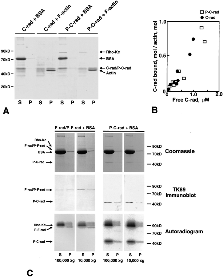
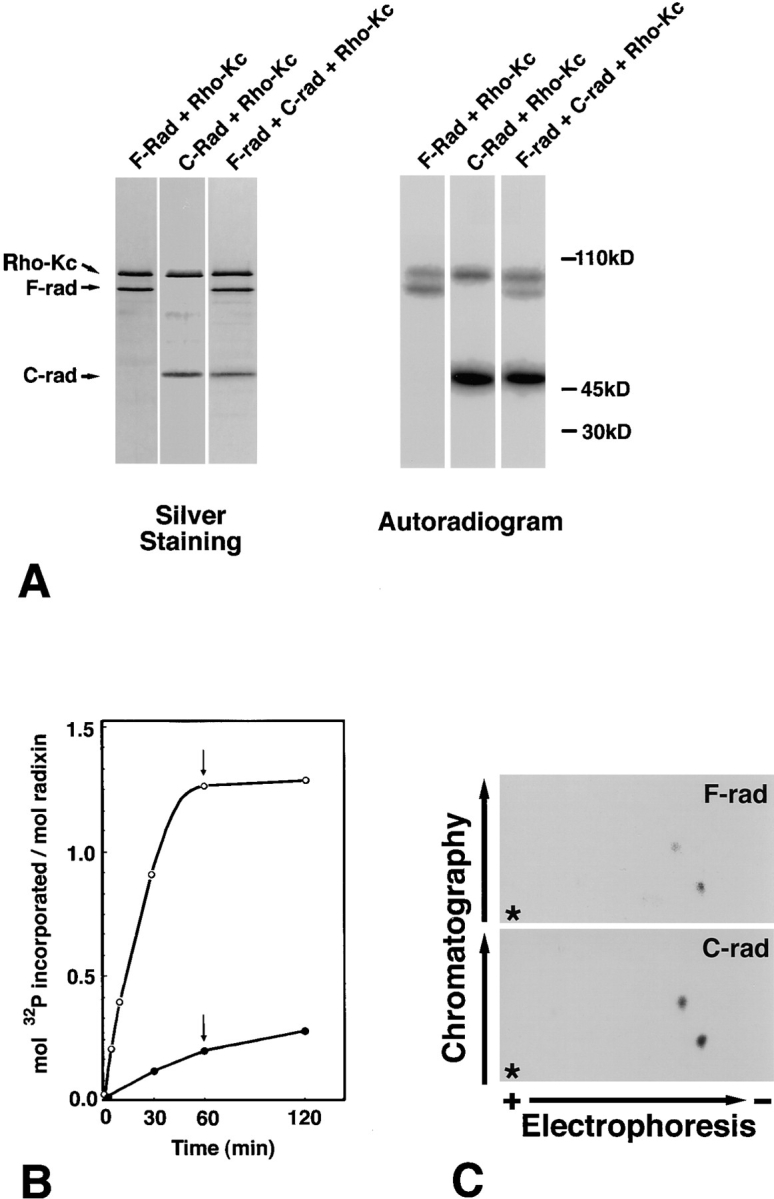
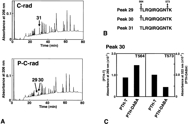
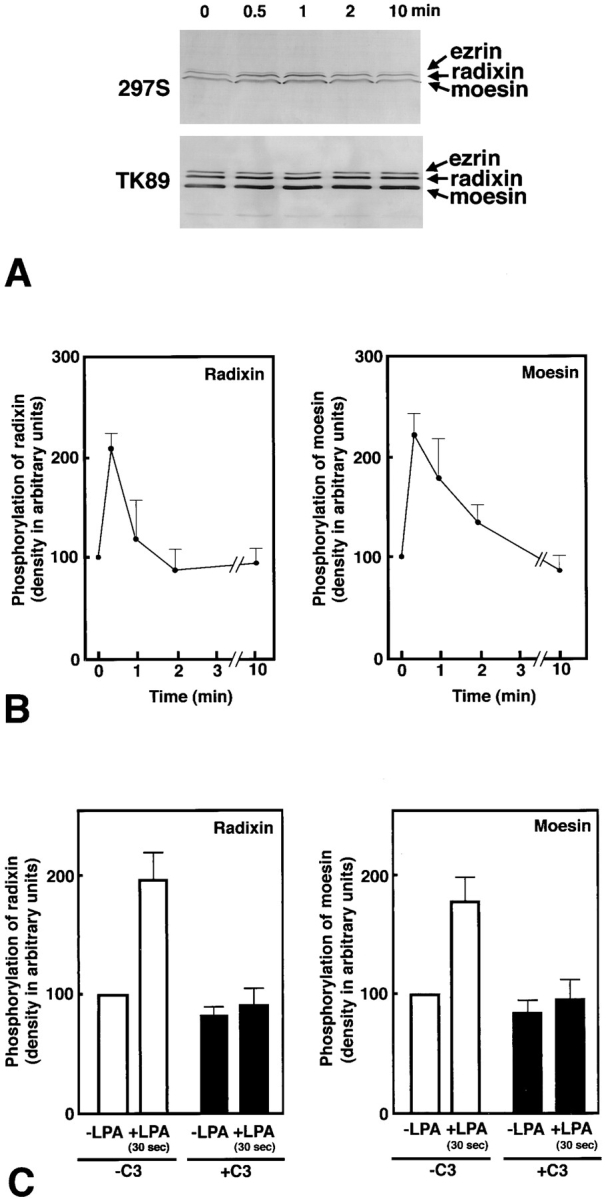
The ezrin/radixin/moesin (ERM) proteins are involved in actin filament/plasma membrane interaction that is regulated by Rho. We examined whether ERM proteins are directly phosphorylated by Rho-associated kinase (Rho-kinase), a direct target of Rho. Recombinant full-length and COOH-terminal half radixin were incubated with constitutively active catalytic domain of Rho-kinase, and approximately 30 and approximately 100% of these molecules, respectively, were phosphorylated mainly at the COOH-terminal threonine (T564). Next, to detect Rho-kinase-dependent phosphorylation of ERM proteins in vivo, we raised a mAb that recognized the T564-phosphorylated radixin as well as ezrin and moesin phosphorylated at the corresponding threonine residue (T567 and T558, respectively). Immunoblotting of serum-starved Swiss 3T3 cells with this mAb revealed that after LPA stimulation ERM proteins were rapidly phosphorylated at T567 (ezrin), T564 (radixin), and T558 (moesin) in a Rho-dependent manner and then dephosphorylated within 2 min. Furthermore, the T564 phosphorylation of recombinant COOH-terminal half radixin did not affect its ability to bind to actin filaments in vitro but significantly suppressed its direct interaction with the NH2-terminal half of radixin. These observations indicate that the Rho-kinase-dependent phosphorylation interferes with the intramolecular and/ or intermolecular head-to-tail association of ERM proteins, which is an important mechanism of regulation of their activity as actin filament/plasma membrane cross-linkers.


Figures

Figure 5
Figure 5
Effects of T564 phosphorylation in radixin on the actin filament binding ability of C-rad. (A) Cosedimentation of non- or T564-phosphorylated C-rad with actin filaments. Non- (C-rad) or T564-phosphorylated C-rad (P-C-rad) was incubated with BSA (BSA) or with actin filaments (F-actin), then centrifuged at 100,000 g. Supernatant (S) and pellet (P) were resolved by SDS-PAGE followed by Coomassie brilliant blue staining. Both non- and T564-phosphorylated C-rad (C-rad/ P-C-rad) were cosedimented with actin filaments (Actin) to the same extent, but not with BSA (BSA). (B) Quantitative analysis. 2 μM F-actin was incubated with various amounts of non- or T564-phosphorylated C-rad and centrifuged, then the amounts of cosedimented non- (C-rad; filled circles) and T564-phosphorylated C-rad (P-C-rad; open squares) were quantified by densitometric scanning of Coomassie brilliant blue–stained gels. (C) Sedimentation of F-rad. Partially [32P]-phosphorylated F-rad (P-F-rad) and fully [32P]- phosphorylated C-rad (P-C-rad) were centrifuged in the presence of BSA at 100,000 or 10,000 g for 30 min. Supernatant (S) and pellet (P) were resolved by SDS-PAGE followed by Coomassie brilliant blue staining (Coomassie), immunoblot with TK89 (TK89 Immunoblot), or autoradiography (Autoradiogram). In the absence of actin filaments, F-rad, especially P-F-rad, were mostly recovered in pellet even at 10,000 g, whereas P-C-rad was mainly recovered in supernatant at 100,000 g as well as 10,000 g.
Figure 1
Figure 1
In vitro phosphorylation of the full-length radixin (F-rad) and the COOH-terminal half radixin (C-rad) by the constitutively active catalytic domain of Rho-kinase (Rho-Kc). (A) SDS-PAGE banding pattern of the reaction mixture (Silver Staining) and its accompanying autoradiogram (Autoradiogram). Purified 3 pmol F-rad (F-rad + Rho-Kc), C-rad (C-rad + Rho-Kc), or their mixture (F-rad + C-rad + Rho-Kc) was incubated with 2 pmol Rho-Kc for 10 min at 30°C. Phosphorylated proteins were resolved by SDS-PAGE followed by autoradiography. C-rad (C-rad) was phosphorylated more heavily than F-rad (F-rad). Rho-Kc (Rho-Kc) was autophosphorylated. (B) Time course and stoichiometry of the phosphorylation reaction. After 30, 60, and 120 min of incubation, the phosphorylation levels of F-rad (filled circle) and C-rad (open circle) were quantified. 2 pmol of Rho-Kc and 500 pmol of γ-[32P]ATP were added to the reaction mixture after 60 min of incubation (arrows). (C) Phosphopeptide mapping. Phosphorylated F-rad and C-rad were digested completely with TPCK-trypsin, subjected to two-dimensional peptide mapping (first dimension, Electrophoresis; second dimension, Chromatography) as described in Materials and Methods, and then analyzed by autoradiography. Two radioactive spots in F-rad and C-rad may correspond to the T564- and T573-containing tryptic fragments (see Fig. 2). To visualize the weak spot from F-rad the autoradiogram of C-rad was overexposed, but the intensity ratio of two spots from C-rad was the same as that from F-rad. Asterisks represent the origins of spotted samples.
Figure 2
Figure 2
Determination of amino acid residues of radixin that are phosphorylated by Rho-kinase. (A) Comparison of elution profiles of lysyl endopeptidase-digested peptides of non- and fully phosphorylated C-rad from reverse-phase HPLC (C-rad and P-C-rad, respectively). Rho-kinase–dependent phosphorylation divided and shifted the peak 31 from nonphosphorylated C-rad into two peaks, peaks 30 and 29, from phosphorylated C-rad. For details see the text. (B) Amino acid sequences of peaks 29, 30, and 31. The PTH-threonine/PTH-dehydroaminobutyric acid ratio (see C) revealed that T564/T573 in peak 29 and T564 in peak 30 were phosphorylated (Kato et al., 1994; Fujita et al., 1996). (C) The PTH-threonine/PTH-dehydroaminobutyric acid ratio of T564 and T573 in peak 30. The amounts of PTH-threonine (PTH-T) and PTH-dehydroaminobutyric acid (PTH-DABA) were determined from the absorbance at 269 and 322 nm, respectively. When the threonine residue (T564 in this case) was phosphorylated, the PTH-T/PTH-DABA ratio fell to below 1.0.
Figure 3
Figure 3
Production of mAb 297S that distinguishes T564-phosphorylated from nonphosphorylated radixin. Nonphosphorylated C-rad (100 ng; C-rad), T564-phosphorylated C-rad (100 ng; P-C-rad), and whole-cell lysate of Swiss 3T3 cells under conventional culture conditions (25 μg; Swiss 3T3 lysate) were immunoblotted with pAb TK89 (pAb TK89) or mAb 297S (mAb 297S). The former antibody recognized radixin as well as ezrin and moesin irrespective of their phosphorylation state, whereas the latter distinguished T564-phosphorylated C-rad from the nonphosphorylated molecule. The mAb 297S recognized ezrin and moesin that were phosphorylated at the corresponding threonine residues, T567 and T558, respectively.
Figure 4
Figure 4
LPA-induced threonine phosphorylation of ERM proteins in vivo. (A) Serum-starved Swiss 3T3 cells were stimulated with LPA, and at 0, 0.5, 1, 2, or 10 min of incubation, whole-cell lysate was resolved by SDS-PAGE followed by immunoblotting with mAb 297S (297S) or with pAb TK89 (TK89). 297S were specific for T567-, T564-, and T558-phosphorylated ezrin, radixin, and moesin, respectively, whereas TK89 recognized ERM proteins irrespective of their phosphorylation state. (B) Quantitative analyses of changes in the phosphorylation levels of T564 in radixin and T558 in moesin. By immunoblotting with TK89 in combination with scanning densitometry, the precisely equal amount of ERM proteins were reelectrophoresed per each lane for the following quantification. The amount of T564-phosphorylated radixin and T558-phosphorylated moesin was quantitatively determined by scanning densitometry of 297S-immunoblots (see A) using purified phosphorylated C-rad to generate a standard curve. The curve was linear over the concentration range used here. The mean densities of 297S-immunoblot bands of radixin and moesin at 0 min were set at 100 arbitrary units. The phosphorylation levels of T564 in radixin and T558 in moesin rapidly increased then decreased within 2 min after LPA stimulation. Although the change in the phosphorylation level of T567 in ezrin was difficult to be quantitatively followed due to its low expression level in Swiss 3T3 cells, its phosphorylation level also appeared to rapidly increase then decrease within 2 min after LPA stimulation (see A). The data represent the means ± SEM of four determinations. The phosphorylation levels of radixin and moesin at 30 s were significantly different from those at 0 s (P < 0.005) and at 2 min (P < 0.05) as determined by the paired t test. (C) Suppression of LPA-induced T564 phosphorylation of radixin and T558 phosphorylation of moesin by C3 exoenzyme. Non- and C3 exoenzyme–pretreated serum-starved cells were stimulated with 1 μg/ml LPA for 30 s, and the phosphorylation levels of T564 of radixin and T558-moesin in these cells were quantitatively compared to those in LPA-nontreated serum-starved cells by immunoblotting with mAb 297S. The data represent the means ± SEM of four determinations. In the absence of C3 exoenzyme (-C3), the phosphorylation levels of radixin and moesin in 30 s LPA-treated cells were significantly different from those at 0 s (P < 0.05) as determined by the paired t test, while in the presence of C3 exoenzyme (+C3) this LPA-induced increase of the phosphorylation levels of radixin or moesin was not detected. In this experiment, probably due to lipofectamine, the Rho-dependent increase of ERM phosphorylation was varied and suppressed to some extent as compared to B.
Figure 6
Figure 6
Effects of T564 phosphorylation in radixin on the direct interaction between its NH2- and COOH-terminal halves. (A) The same amounts (92 ng) of nonphosphorylated C-rad (C-rad), T564-phosphorylated C-rad (P-C-rad), and alkaline phosphatase-treated T564-phosphorylated C-rad (P-C-rad/AP) were electrophoresed and then transferred onto nitrocellulose membranes. They were immunoblotted with pAb TK89 (TK89) or mAb 297S (297S) to check the amounts and the phosphorylation level of each sample, respectively (Immunoblot). They were then incubated with the iodinated NH2-terminal half of radixin (1 25I-N-rad) that was purified from GST fusion protein (Overlay). Bound 125I-N-rad was detected by autoradiography. 125I-N-rad specifically bound to nonphosphorylated C-rad but not to T564-phosphorylated C-rad. (B) Various amounts (7.5–120 ng) of non- (C-rad) and T564-phosphorylated C-rad (P-C-rad) were electrophoresed, transferred onto nitrocellulose membranes, and incubated with 125I-N-rad. The amount of bound 125I-N-rad and electrophoresed C-rad/P-C-rad was detected by autoradiography (125I-N-rad Overlay) and Coomassie brilliant blue staining (Coomassie), respectively. 125I-N-rad specifically bound to nonphosphorylated C-rad in a dose-dependent manner.

References

    1. Aktories K, Rosener S, Blaschke U, Chhatwal GS. Botulinum ADP-ribosyltransferase C3: purification of the enzyme and characterization of the ADP-ribosylation reaction in platelet membranes. Eur J Biochem. 1988;172:445–450. - PubMed
    1. Algrain M, Turunen O, Vaheri A, Louvard D, Arpin M. Ezrin contains cytoskeleton and membrane binding domains accounting for its proposed role as a membrane-cytoskeletal linker. J Cell Biol. 1993;120:129–139. - PMC - PubMed
    1. Amano M, Mukai H, Ono Y, Chihara K, Matsui T, Hamajima Y, Okawa K, Iwamatsu A, Kaibuchi K. Identification of a putative target for Rho as the serine-threonine kinase protein kinase N. Science. 1996a;271:648–650. - PubMed
    1. Amano M, Ito M, Kimura K, Fukata Y, Chihara K, Nakano T, Matsuura Y, Kaibuchi K. Phosphorylation and activation of myosin by Rho-associated kinase (Rho-kinase) . J Biol Chem. 1996b;271:20246–20249. - PubMed
    1. Amano M, Chihara K, Kimura K, Fukata Y, Nakamura N, Matsuura Y, Kaibuchi K. Formation of actin stress fibers and focal adhesions enhanced by Rho-kinase. Science. 1997;275:1308–1311. - PubMed

Publication types

MeSH terms