. 1998 Feb;180(3):605-13.
doi: 10.1128/JB.180.3.605-613.1998.

Specific in vivo labeling of cell surface-exposed protein loops: reactive cysteines in the predicted gating loop mark a ferrichrome binding site and a ligand-induced conformational change of the Escherichia coli FhuA protein

Affiliations

Specific in vivo labeling of cell surface-exposed protein loops: reactive cysteines in the predicted gating loop mark a ferrichrome binding site and a ligand-induced conformational change of the Escherichia coli FhuA protein

C Bös et al. J Bacteriol. 1998 Feb.

Abstract

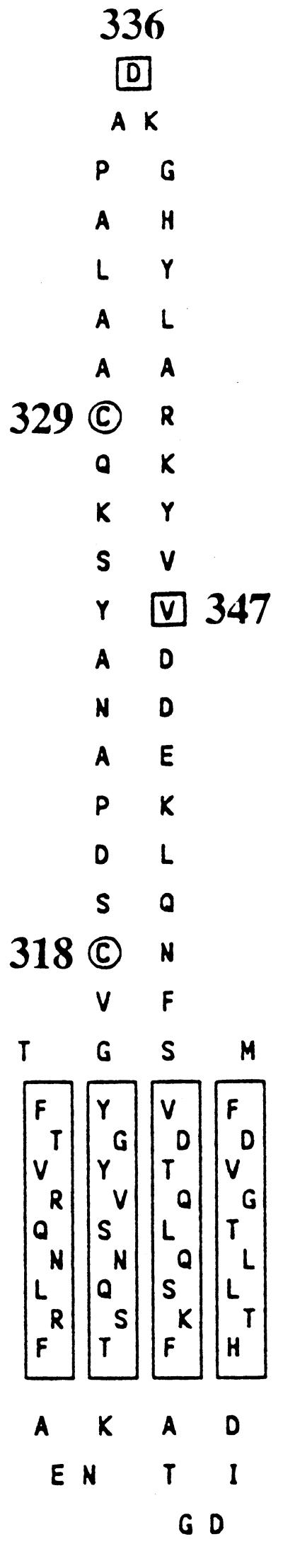
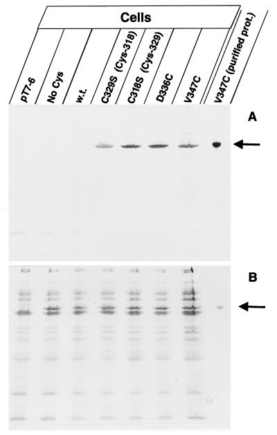
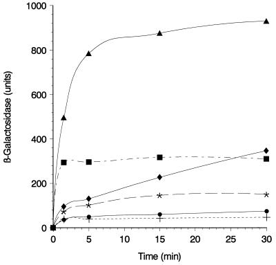
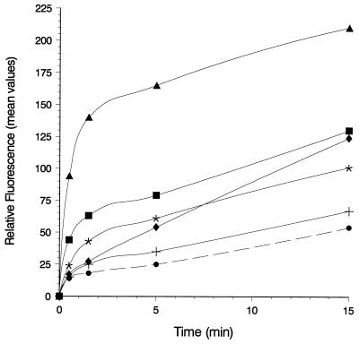
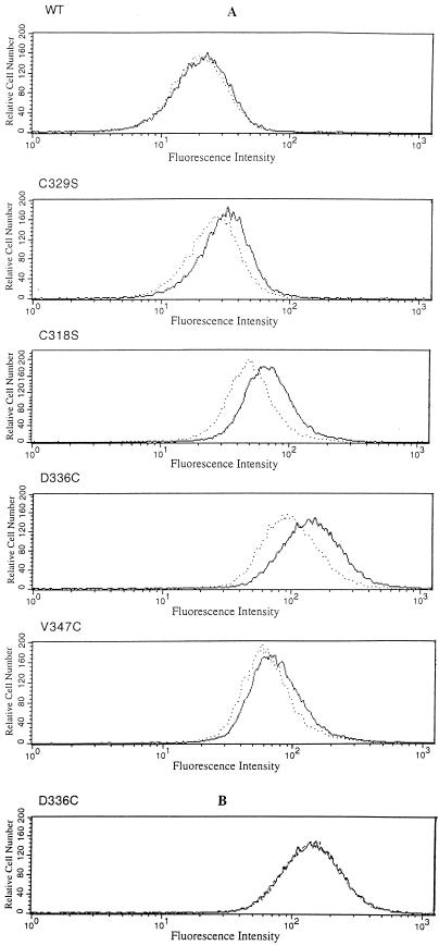
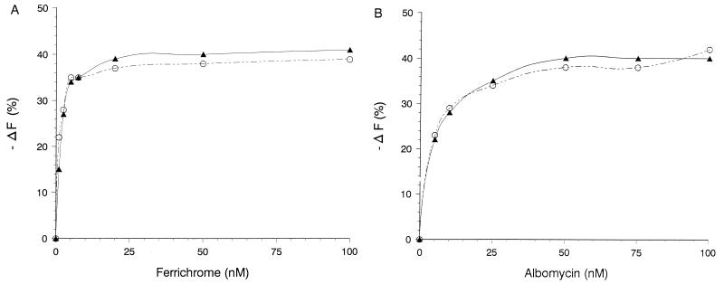
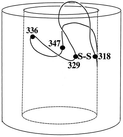
The FhuA protein of Escherichia coli K-12 transports ferrichrome, the antibiotic albomycin, colicin M, and microcin 25 across the outer membrane and serves as a receptor for the phages T1, T5, phi80, and UC-1. FhuA is activated by the electrochemical potential of the cytoplasmic membrane, which probably opens a channel in FhuA. It is thought that the proteins TonB, ExbB, and ExbD function as a coupling device between the cytoplasmic membrane and the outer membrane. Excision of 34 residues from FhuA, tentatively designated the gating loop, converts FhuA into a permanently open channel. FhuA contains two disulfide bridges, one in the gating loop and one close to the C-terminal end. Reduction of the disulfide bridges results in a low in vivo reaction of the cysteines in the gating loop and no reaction of the C-terminal cysteines with biotin-maleimide, as determined by streptavidin-beta-galactosidase bound to biotin. In this study we show that a cysteine residue introduced into the gating loop by replacement of Asp-336 displayed a rather high reactivity and was used to monitor structural changes in FhuA upon binding of ferrichrome. Flow cytometric analysis revealed fluorescence quenching by ferrichrome and albomycin of fluorescein-maleimide bound to FhuA. Ferrichrome did not inhibit Cys-336 labeling. In contrast, labeling of Cys-347, obtained by replacing Val-347 in the gating loop, was inhibited by ferrichrome, but ferrichrome quenching was negligible. It is concluded that binding of ferrichrome causes a conformational change of the gating loop and that Cys-347 is part of or close to the ferrichrome binding site. Fluorescence quenching was independent of the TonB activity. The newly introduced cysteines and the replacement of the existing cysteines by serine did not alter sensitivity of cells to the FhuA ligands tested (T5, phi80, T1, colicin M, and albomycin) and fully supported growth on ferrichrome as the sole iron source. Since cells of E. coli K-12 display no reactivity to thiol reagents, newly introduced cysteines can be used to determine surface-exposed regions of outer membrane proteins and to monitor conformational changes during their function.


Figures

FIG. 1
FIG. 1
Amino acid sequence of the gating loop of FhuA in the outer membrane of E. coli, localized at the cell surface by determination of the proteolysis within genetically inserted peptides in cells and spheroplasts (21). The model illustrates the sites of the natural cysteines present at positions 318 and 329 in FhuA (○) and those of the newly inserted cysteines at sites 336 and 347 (□). The model does not imply that the surface loops (top) and periplasmic turns (bottom) extend from the outer membrane as drawn; rather, they are predicted to be highly folded (Fig. 8).
FIG. 2
FIG. 2
Specific labeling of the FhuA single-cysteine mutants with B-M in live cells (A). Cells of E. coli UL3 fhuA were transformed with one of the following plasmids: pT7-6 (vector without fhuA), pfhuA9 [FhuA(C318S C329S C692S C698S); No Cys], pfhuA8 [FhuA(wild type); w.t.], pfhuA5 [FhuA(C329S) Cys-318], pfhuA4 [FhuA(C318S) Cys-329], pfhuA6 [FhuA(D336C)], and pfhuA7 [FhuA(V347C)], as indicated. Cells were incubated for 30 min with 0.5 mM B-M at 30°C, and the proteins of whole cells were separated by SDS-PAGE. In addition, isolated FhuA(V347C) protein was heated for 3 min in 4% SDS and subsequently labeled with B-M (purified prot.). The proteins were separated by SDS-PAGE, blotted onto a nitrocellulose membrane, and stained with Ponceau-S-Red (B) and, after destaining and incubation with streptavidin-β-galactosidase, stained with X-Gal (A). The FhuA band is indicated by an arrow.
FIG. 3
FIG. 3
Time course of specific labeling of gating loop cysteines with B-M in intact cells. Exponentially growing cells of E. coli UL3 fhuA transformed with plasmids pT7-6 (+), pfhuA8 [FhuA(wild type)] (•), pfhuA5 [FhuA(C329S) Cys-318] (∗), pfhuA4 [FhuA(C318S) Cys-329] (⧫), pfhuA7 [FhuA(V347C)] (▪), and pfhuA6 [FhuA(D336C)] (▴) were harvested, washed with PBS, and incubated with B-M (0.5 mM) for 1.5, 5, 15, and 30 min at 30°C. Labeling was interrupted by the addition of 0.02 M dithiothreitol. Cells were washed, treated with 2% bovine serum albumin, incubated with S-G, and washed again, and β-galactosidase activity was measured with o-nitrophenyl-β-d-galactoside (10) and related to the cell density (5% standard deviation).
FIG. 4
FIG. 4
Labeling of the gating loop cysteines with F-M (A). Cells of E. coli UL3 fhuA were transformed with one of the following plasmids: pT7-6 (vector without fhuA), pfhuA9 [FhuA(C318S C329S C692S C698S); No Cys], pfhuA8 [FhuA(wild type); w.t.], pfhuA5 [FhuA(C329S) Cys-318], pfhuA4 [FhuA(C318S) Cys-329], pfhuA6 [FhuA(D336C)], and pfhuA7 [FhuA(V347C)], as indicated. Cells were incubated for 15 min with 0.5 mM F-M at 30°C in darkness, and the proteins of whole cells were separated by SDS-PAGE. In addition, purified protein of FhuA(V347C) was heated for 3 min in 4% SDS and subsequently labeled with F-M (purified prot.). Proteins were separated by SDS-PAGE, illuminated at 302 nm, and photographed with a Cybertech video camera (A). The proteins were then stained with Serva R to show that equal amounts were applied to the lanes (B).
FIG. 5
FIG. 5
Flow cytometry of E. coli UL3 fhuA transformed with plasmids pT7-6 (vector without fhuA) (+), pfhuA8 [FhuA (wild type)] (•), pfhuA5 [FhuA(C329S) Cys-318] (∗), pfhuA4 [FhuA(C318S) Cys-329] (⧫), pfhuA7 [FhuA(V347C)] (▪), and pfhuA6 [FhuA(D336C)] (▴) after labeling with 0.5 mM F-M for 0.5, 1.5, 5, or 15 min at 30°C. The reaction was stopped by adding 20 mM DTT. The fluorescence values showed a standard deviation of 10%.
FIG. 6
FIG. 6
Fluorescence quenching of F–M-labeled cells by ferrichrome. E. coli UL3 fhuA transformed with plasmid pfhuA8 [FhuA(wild type)], pfhuA5 [FhuA (C329S) Cys-318], pfhuA4 [FhuA(C318S) Cys-329], pfhuA6 [FhuA (D336C)], or pfhuA7 [FhuA(V347C)] was labeled with F-M. The fluorescence was determined by flow cytometry prior to (−) and after (…) the addition of 10 nM ferrichrome (A) or 1 μM ferrioxamine B {pfhuA6 [FhuA(D336C)]} (B).
FIG. 7
FIG. 7
Fluorescence quenching of F–M-labeled TonB+ versus TonB cells by ferrichrome (A) and albomycin (B). Cells of E. coli UL3 fhuA (▴) and E. coli HK99 fhuA tonB (○), each transformed with pfhuA6 [FhuA(D336C)], were labeled for 5 min with 0.5 mM F-M and then incubated with ferrichrome (A) and albomycin (B) at the concentrations indicated. Fluorescence was determined by flow cytometry.
FIG. 8
FIG. 8
Illustration of the natural disulfide bridge between residues 318 and 329 and of the cysteine residues introduced at sites 336 and 347 of the FhuA gating loop. The model takes into account the relatively high reactivity of Cys-336 by localizing it close to the cell surface, the lower reactivity of Cys-347, and the low reactivity of Cys-318 and Cys-329. The ferrichrome binding site is close to Cys-347. This model is not intended to predict the conformation of the gating loop.

References

    1. Bardwell J C A, McGovern K, Beckwith J. Identification of a protein required for disulfide bond formation in vivo. Cell. 1991;67:581–589. - PubMed
    1. Bassford P J, Jr, Kadner R J, Schnaitman C A. Functional stability of the bfe and tonB gene products in Escherichia coli. J Bacteriol. 1977;130:750–758. - PMC - PubMed
    1. Bayer E A, Safars M, Wilchek M. Selective labeling of sulfhydryls and disulfides on blot transfers using avidin-biotin technology: studies on purified proteins and erythrocyte membranes. Anal Biochem. 1987;161:262–271. - PubMed
    1. Bayer E A, Zalis M G, Wilchek M. 3-(N-Maleimido-propionyl) biocytin: a versatile thiol-specific biotinylating reagent. Anal Biochem. 1985;149:529–536. - PubMed
    1. Bonhivers M, Ghazi A, Boulanger P, Lettelier L. FhuA, a transporter of the Escherichia coli outer membrane, is converted into a channel upon binding of bacteriophage T5. EMBO J. 1996;15:1850–1856. - PMC - PubMed

Publication types

MeSH terms

LinkOut - more resources