Comparative Study
. 1998 Feb;4(2):187-193.
doi: 10.1038/nm0298-187.

Absence of neurological deficits following extensive demyelination in a class I-deficient murine model of multiple sclerosis

Affiliations
Comparative Study

Absence of neurological deficits following extensive demyelination in a class I-deficient murine model of multiple sclerosis

Cynthia Rivera-Quiñones et al. Nat Med. 1998 Feb.

Abstract

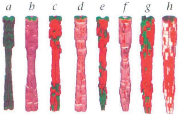
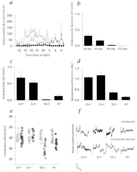
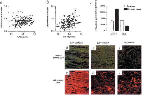
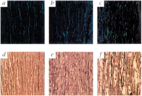
Demyelination alone has been considered sufficient for development of neurological deficits following central nervous system (CNS) disease. However, extensive demyelination is not always associated with clinical deficits in patients with multiple sclerosis (MS), the most common primary demyelinating disease in humans. We used the Theiler's murine encephalomyelitis virus model of demyelination to investigate the role of major histocompatibility complex (MHC) class I and class II gene products in the development of functional and neurophysiological deficits following demyelination. We measured spontaneous clinical activity by two independent assays and recorded hind-limb motor-evoked potentials in infected class I-deficient and class II-deficient mice of an identical genetic background as well as in highly susceptible SJL/J mice. The results show that despite a similar distribution and extent of demyelinated lesions in all mice, only class I-deficient mice were functionally normal. We propose that the mechanism by which demyelinated class I-deficient mice maintain neurologic function results from increased sodium channel densities and the relative preservation of axons. These findings are the first to implicate a role for MHC class I in the development of neurological deficits following demyelination.


Figures

Fig. 1
Fig. 1
Three-dimensional reconstruction of demyelinated lesions from chronically infected nonmutant H–2b (β2m+/+), class I-deficient (β2m−/−), SJL/J and class II-deficient (Ab°) mice. The extent and distribution of demyelination is similar in β2m−/− (c and d), SJL/J (e and f) and class II-deficient (Ab°) (g and h) mice. β2m+/+ mice (a and b) show no demyelination. Green, gray matter; white, white matter; red, demyelinated lesions.
Fig. 2
Fig. 2
Functional status of mice as measured by the Digiscan activity monitoring system, the activity wheel and conduction velocities in hind-limb motor-evoked potentials. Vertical activity was monitored hourly (over 3 days) for all groups of mice. a, The progressive decrease in nocturnal vertical activity from 0 to 180 days after infection is illustrated for susceptible SJL/J mice. b, A time course for SJL/J mice revealed a decrease in the ratio of nocturnal vertical activity as early as 60 days after infection when compared with nonmutant H–2b (β2m+/+) mice. This activity was completely abolished by 270 days after infection. c, At 180 days after infection there was no difference observed between the mean nocturnal vertical activity ratios for infected nonmutant H–2b (β2m+/+) and class I-deficient (α2m−/−) mice, indicating that hind-limb motor abnormalities were minimal in these mice. In contrast the ratios of mean nocturnal vertical activity for 180-day-infected SJL/J and 100-day-infected class II-deficient (Ab°) mice were significantly lower than those of nonmutant H–2b (β2m+/+) and class I-deficient β2m−/− mice. d, Spontaneous activity measured by the activity wheel. The ratios of mean revolutions per day revealed the same pattern of normal activity in class I-deficient (β2m−/−) mice when compared with infected H–2b (β2m+/+) mice, whereas infected SJL/J and class II-deficient (Ab°) mice displayed significantly decreased activities. e, Conduction velocities of 180-day-infected β2m−/− mice did not differ significantly from those of uninfected control β2m−/− mice or uninfected and 180-day-infected β2m+/+ mice. Conduction velocities for 180-day-infected SJL/J mice were slower when compared with uninfected SJL/J mice, 180-day-infected β2m+/+ mice and 180-day-infected β2m−/− mice. Conduction velocities for 100-day-infected, class II-deficient mice were also slower as compared with uninfected class II-deficient mice, 180-day-infected β2m+/+ and 180-day-infected β2m−/− mice. There were no differences observed between the mean conduction velocities for any of the uninfected controls. Uninfected, open symbols; chronically infected, shaded symbols. f, Representative tracings of recorded motor-evoked potentials from β2m+/+, β2m−/−, SJL/J and class II-deficient mice. Recordings from 180-day-infected β2m+/+ and β2m−/− mice are similar to their littermate uninfected controls. Recordings from 180-day-infected SJL/J and 100-day-infected class II-deficient mice showed prolonged latencies, dispersion of potentials and smaller amplitudes when compared with their uninfected controls. The arrow represents the point used to calculate latency to the beginning of the potential.
Fig. 3
Fig. 3
Sodium channel distributions in uninfected (filled triangle) and chronically infected (open circle with dot) class I-deficient (β2m−/−) and SJL/J mice. a, A significant increase was observed in the sodium channel intensities of 323-day-infected β2m−/− mice when compared with their uninfected counterparts. b, In contrast, there was a significant decrease in sodium channel intensities of 270-day-infected SJL/J mice as compared with uninfected controls. a and b, A positive linear correlation (thickened gray lines, chronically infected groups; thin black lines, uninfected groups) exists between sodium channel and corresponding myelin proteolipid protein (PLP) intensities for uninfected SJL/J (r = 0.50), infected SJL/J (r = 0.20) and uninfected β2m−/− mice (r = 0.47). However, there is no positive linear correlation between sodium channel and PLP intensities for chronically infected β2m−/− mice (a), which can be explained by regions of high sodium channel intensities in areas of low PLP intensities. c, [3H]Saxitoxin autoradiography showed a 1.7-fold increase in the sodium channel grain density of 323-day-infected β2m−/− mice when compared with that of uninfected controls. In contrast, there was a 50% decrease in the sodium channel grain density of 270-day-infected SJL/J mice when compared with their uninfected controls. White bars, uninfected; black bars, chronically infected. Preincubation with 200-fold excess of unlabeled tetrodotoxin blocked approximately 50% of the [3H]saxitoxin labeling in an uninfected β2m+/+ mouse (data not shown). Representative examples of sodium channel staining (d, e and f) and PLP-myelin staining (g, h and I) in an uninfected β2m−/− mouse (d and g), a 323-day-infected β2m−/− mouse (e and h) and a 270-day-infected SJL/J mouse (f and i). Images for the sodium channel stain and their respective PLP stained counterparts were taken from the same field. A normal distribution of sodium channel (d) and myelin (g) is shown for an uninfected β2m−/− mouse. (Uninfected nonmutant mice had a comparable staining pattern.) Note the elevated and homogenous distribution of sodium channel staining (e) in an area of relative myelin loss (h) from an uninfected β2m−/− mouse. Note the severe loss of sodium channel intensity (f) in a similar area of low myelin (i) in a chronically infected SJL/J mouse. Even though the degree of myelin loss is similar in infected β2m−/− (h) and SJL/J (i) mice, there is more sodium channel expression in β2m−/− (e) compared with the SJL/J (f) mouse. Regions of negative staining were determined from the corresponding ultraviolet images to be cellular nuclei. Green, sodium channels; red, PLP.
Fig. 4
Fig. 4
Representative examples of axons in the spinal cord white matter of chronically infected class I-deficient (β2m−/−) and SJL/J mice and uninfected class I-deficient and nonmutant H–2b (β2m+/+) mice. Representative examples of antineurofilament (a-c) and Bielschowski (d-f) staining are shown for an uninfected β2m+/+ mouse (a), an uninfected β2m−/− mouse (d), a chronically infected (323 days postinfection) class I-deficient mouse (b and e) and a chronically infected (270 days) SJL/J mouse (c and f). A normal distribution of axons is shown for an uninfected β2m+/+ mouse (a) and an uninfected β2m−/− mouse (d). The staining pattern in uninfected animals of all groups is comparable. Axons are relatively well preserved in chronically infected β2m−/− mice despite the presence of inflammatory cells and demyelination. Both the neurofilament (b) and the Bielschowski (e) stains show that the axons in β2m−/− mice are neither significantly disrupted nor degenerating. In contrast the axons in chronically infected SJL/J mice are severely disrupted. The neurofilament stain (c) shows a significant loss of immunostainable material, whereas the Bielschowski stain (f) shows swelling axons (red arrows) which are actively undergoing degeneration.

Similar articles

Cited by

References

    1. Rodriguez M, Oleszak E, Leibowitz J. Theiler’s murine encephalomyelitis: A model of demyelination and persistence of virus. Crit. Rev. Immunol. 1987;7:325–365. - PubMed
    1. Fiette L, Aubert C, Brahic M, Rossi CP. Theiler’s virus infection of β2-microglobulin-deficient mice. J. Virol. 1993;67:589–592. - PMC - PubMed
    1. Pullen LC, Miller SD, Dal Canto MC, Kim BS. Class I-deficient resistant mice intracerebrally inoculated with Theiler’s virus show an increased T cell response to viral antigens and susceptibility to demyelination. Eur. J. Immunol. 1993;23:2287–2293. - PubMed
    1. Rodriguez M, et al. Abrogation of resistance to Theiler’s-induced demyelination in H–2b mice deficient in β2-microglobulin. J. Immunol. 1993;151:266–276. - PubMed
    1. Njenga MK, et al. Theiler’s virus persistence and demyelination in major histocompatibility complex class II-deficient mice. J. Virol. 1996;70:1729–1737. - PMC - PubMed

Publication types

Substances