. 1997 Dec;41(6):740-7.
doi: 10.1136/gut.41.6.740.

Helicobacter pylori gastritis and epithelial cell proliferation in patients with reflux oesophagitis after treatment with lansoprazole

Affiliations

Helicobacter pylori gastritis and epithelial cell proliferation in patients with reflux oesophagitis after treatment with lansoprazole

A E Berstad et al. Gut. 1997 Dec.

Abstract

Background: Helicobacter pylori gastritis may spread proximally in the stomach during profound acid inhibition.

Aims: To examine histological gastric body changes and epithelial cell proliferation before and after treatment with lansoprazole.

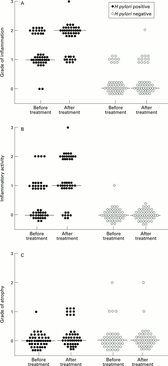
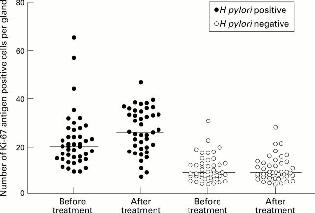
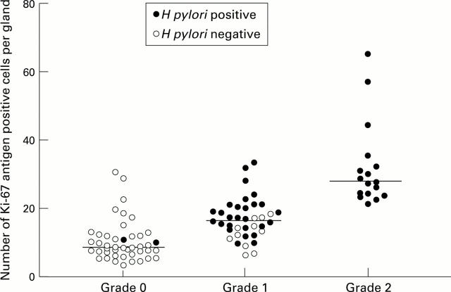
Patients and methods: Patients diagnosed as having reflux oesophagitis grade 1 or 2 were enrolled and treated for 12 weeks with lansoprazole (30 mg every morning). After 12 weeks, 103 of the 118 patients appeared endoscopically healed and were asymptomatic; they then received maintenance treatment with 15 or 30 mg lansoprazole daily. Biopsy specimens obtained from similar sites before and after treatment, were available from 90 patients after a median of 64 weeks (range 15-73 weeks). Epithelial cell proliferation was determined by the number of Ki-67 antigen positive cells per gland.

Results: Of these 90 patients, 44 (49%) were found to be infected with H pylori. Their median inflammation score had increased from grade 1 before to grade 2 after treatment (p < 0.0001). Initially, the number of Ki-67 antigen positive cells per gland was significantly higher in the H pylori infected than in the uninfected group and increased further after treatment (p < 0.0001). In uninfected patients, no significant change in inflammation or proliferation occurred during treatment.

Conclusions: A marked increase in body gastritis was observed in H pylori infected individuals during long term treatment with the proton pump inhibitor lansoprazole. Epithelial cell proliferation and atrophy also increased in infected but not in uninfected patients.


Figures

Figure 1
Figure 1
: Scatter diagrams depicting (A) grade of corpus inflammation, (B) inflammatory activity, and (C) atrophy in H pylori infected and uninfected patients before and after treatment. Median scores indicated by horizontal lines.
Figure 2
Figure 2
: Effect of lansoprazole treatment on histology of gastric body mucosa (H&E staining) in H pylori infected patients. Few leucocytes are present within the lamina propria (grade 1 inflammation, grade 0 activity) in one patient before treatment (A); after 64 weeks of treatment, intensified inflammation (grade 2 inflammation, grade 1 activity) is evident (B). In another patient, neutrophils are absent before treatment (C); after 48 weeks of treatment, numerous neutrophils appear in the isthmus region and within the epithelium (D). In a third patient, moderate (grade 2) inflammation but no glandular atrophy is present before treatment (E); after 65 weeks of treatment, mild to moderate loss of glands (grade 1 atrophy) is evident together with persistent inflammation (F). Original magnifications: A and B, ×250; C and D, ×400; E and F, ×160.
Figure 3
Figure 3
: Immunostaining of Ki-67 nuclear antigen positive cells in the body isthmus zone of a patient without (A) or with (B) gastritis before lansoprazole treatment. Positive nuclei are stained brown. Immunoperoxidase counterstained with haematoxylin. Original magnifications: A, ×1000; B, ×400.
Figure 4
Figure 4
: Scatter diagram depicting gastric body epithelial cell proliferation in H pylori infected and uninfected patients before and after lansoprazole treatment. Data based on immunostaining for Ki-67 nuclear antigen. Median values indicated by horizontal lines.
Figure 5
Figure 5
: Scatter diagram depicting Ki-67 nuclear antigen positive cells per gastric gland in relation to grade of inflammation at the start of the study in H pylori infected and uninfected patients as indicated (Spearman's r=0.72, p<0.0001). Median values indicated by horizontal lines.

References

    1. Histopathology. 1993 Jun;22(6):543-7 - PubMed
    1. Lab Invest. 1993 Jun;68(6):629-36 - PubMed
    1. J Clin Pathol. 1994 Feb;47(2):122-5 - PubMed
    1. Ann Intern Med. 1994 Aug 1;121(3):161-7 - PubMed
    1. Scand J Gastroenterol. 1994 Nov;29(11):995-1000 - PubMed

Publication types

MeSH terms