. 1998 Feb 16;187(4):537-46.
doi: 10.1084/jem.187.4.537.

An interleukin (IL)-10/IL-12 immunoregulatory circuit controls susceptibility to autoimmune disease

Affiliations

An interleukin (IL)-10/IL-12 immunoregulatory circuit controls susceptibility to autoimmune disease

B M Segal et al. J Exp Med. .

Abstract

Cells of the innate immune system secrete cytokines early in immune responses that guide maturing T helper (Th) cells along appropriate lineages. This study investigates the role of cytokine networks, bridging the innate and acquired immune systems, in the pathogenesis of an organ specific autoimmune disease. Experimental allergic encephalomyelitis (EAE), a demyelinating disease of the central nervous system, is widely used as an animal model for multiple sclerosis. We demonstrate that interleukin (IL)-12 is essential for the generation of the autoreactive Th1 cells that induce EAE, both in the presence and absence of interferon gamma. The disease-promoting effects of IL-12 are antagonized by IL-10 produced by an antigen nonspecific CD4+ T cell which, in turn, is regulated by the endogenous production of IL-12. This unique immunoregulatory circuit appears to play a critical role in controlling Th cell differentiation and provides a mechanism by which microbial triggers of the innate immune system can modulate autoimmune disease.


Figures
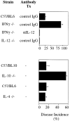
Figure 1
Figure 1
(A) Induction of EAE in both wild-type and IFN-γ−/− mice is dependent on the presence of IL-12. C57BL/6 wild-type (n = 66), IFN-γ−/− (n = 89) and IL-12−/− (n = 33) mice were immunized with bovine MBP (400 μg) emulsified in CFA (1:1) on days 0 and 7. IFN-γ−/− mice were injected intraperitoneally with 0.5 mg of neutralizing anti–IL-12 antibody (n = 30) or control IgG (n = 59) on days 0, 3, 6, and 12. Mice were examined for signs of neurologic impairment from days 0 to 50. Mice with clinical scores of 1 (indicating a limp tail) or higher were considered symptomatic. Data was pooled from five independent experiments; standard deviation reflects the variability between individual experiments. (B) IL-10−/− mice, but not IL-4−/− mice, exhibit heightened susceptibility to EAE. C57BL/10 wild-type (n = 32) and C57BL/10 IL-10−/− (n = 39) mice, and C57BL/6 wild-type (n = 20) and C57BL/6 IL-4−/− (n = 15) mice, were immunized with MBP and examined for clinical signs according to the protocol described above. Data was pooled from three independent experiments.
Figure 2
Figure 2
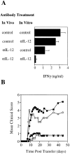
The ability of MBP-reactive LN cells to produce IFN-γ and transfer disease is compromised after neutralization of IL-12 during antigen priming. (A) SJL mice were immunized with MBP87–106 (100 μg) in CFA and injected intraperitoneally with 0.5 mg of either control IgG or goat anti–mouse IL-12 on days 0, 3, and 6. Draining LN were harvested on day 10 and LN cells were cultured in the presence of MBP87–106 (50 μg/ ml) with anti–IL-12 (10 μg/ml) or control IgG (10 μg/ml). 96 h later the cells were washed extensively and restimulated for 48 h with antigen and irradiated syngeneic splenocytes to measure IFN-γ production. The values indicated represent the means and standard deviations of five independent experiments using a total of 25–30 donor mice/ group. (B) Cells (5 × 10⁷) from the four groups described in A (exposed to control IgG only [filled squares]; anti–IL-12 in vitro [open circles]; anti–IL-12 in vivo [filled circles]; and anti–IL-12 both in vivo and in vitro [open squares]) were injected intraperitoneally into naive syngeneic recipients and recipient animals were evaluated for neurological impairment. The incidence of disease was 90.5, 80, 51, and 29%, respectively. Results were pooled from five independent experiments with a total of 25–35 recipient mice in each group.
Figure 3
Figure 3
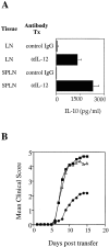
Systemic administration of anti–IL-12 elicits IL-10 production by lymphoid tissue from MBP-primed mice. The protection from EAE mediated by anti–IL-12 is largely reversed by neutralization of IL-10. (A) Spleens and draining LN were removed from SJL mice that had been immunized with MBP87–106 10 d earlier and treated with either anti–IL-12 or control IgG according to the protocol described in Fig. 2. Splenocytes and LN cells were stimulated with antigen and supernatants were assayed for IL-10 production at 96 h. Results represent the means and standard deviations of six independent experiments in which cells were pooled from 4–5 mice per group per experiment. (B) Draining LN were removed from MBP87–106 primed SJL mice that had been injected with control IgG (filled squares), anti–IL-12 (filled circles) or a combination of anti–IL-12 and anti–IL-10 (open triangles) according to the schedule described in Fig. 2. LN cells from each group were stimulated with MBP87–106 for 96 h, in the presence (open triangles) or absence (filled symbols) of anti–IL-10, washed extensively, and then injected (5 × 10⁷) into naive syngeneic recipients. The incidence of disease was 87, 58, and 95% among recipients of control IgG–, anti–IL-12–, and anti–IL-12/anti–IL-10–treated splenocytes, respectively. The results shown in the figure are mean clinical scores obtained from three independent experiments with a total of 20–30 recipient mice per experimental group.
Figure 4
Figure 4
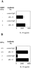
Suppression of IL-10 production by endogenous IL-12 is antigen and IFN-γ independent. (A) Spleens were harvested from naive or MBP87–106-primed SJL mice that had been injected with anti–IL-12 or control IgG on days −10, −6, and −3 before killing. Supernatants were assayed for IL-10 after 96 h of culture. Results represent the means of four independent experiments with groups consisting of 5–7 mice in each experiment. Standard deviations reflect the variability between individual experiments. (B) Naive C57BL/ 6 wild-type and IFN-γ−/− mice were treated as in A. Results represent the means and standard deviations of two independent experiments in which spleens were pooled from four mice in each group.
Figure 5
Figure 5
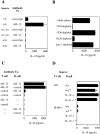
After neutralization of IL-12, IL-10 is produced by antigen nonspecific CD4+ T cells in collaboration with B cells. Spleens were harvested from unimmunized mice that had been injected intraperitoneally with 0.5 mg of control IgG or anti–IL-12 on days −10, −6, and −3 before killing. Supernatants were collected for assay of IL-10 after 96–120 h of culture. (A) IL-10 production by splenocytes from BALB/c wild-type (w/t), nu/nu, and SCID mice at 96 h. Results represent means and standard deviations of two independent experiments in which spleens were pooled from 3–5 mice in each group in each experiment. (B) Splenocytes from anti–IL-12 treated SJL mice (n = 5–6) were depleted of various subpopulations using sheep antifluorescein biomag beads and FITC-conjugated mAbs specific for the cell surface markers indicated. Recovered cells (4 × 10⁶ cells/ml) from each group were cultured and supernatants assayed for IL-10 after 120 h. In each case, the recovered cells were 99– 100% free of the depleted population as determined by flow cytometry using PE-conjugated mAbs (not shown). Results represent means and standard deviations of three independent experiments. Whole spleen and subpopulation preparations derived from control IgG–treated donors failed to produce detectable IL-10 (not shown). (C) CD4+ T cells and B220+ B cells purified from spleens of anti–IL-12 or control IgG–treated SJL mice (n = 4–7/group) were cocultured (2 × 10⁵ of each in 200 μl) in 96-well microtiter plates. Supernatants were collected at 120 h to quantify IL-10 levels. Results represent means and standard deviations of four independent experiments. Levels of purity ranged from 90 to 95% for CD4+ T cells and 93-98% for B220+ B cells as assessed by flow cytometry (not shown). (D) CD4+ T cells from spleens of anti–IL-12–treated C57BL/10 wild-type or IL-10−/− mice were cocultured with B220+ B cells purified from spleens of either anti–IL-12 or control IgG–treated donors (2 × 10⁵ of each in 200 μl). An asterisk (*) denotes B cells taken from anti–IL-12–treated mice. Supernatants were collected at 144 h to quantify IL-10 levels. Results represent means and standard deviations of two independent experiments. Levels of purity ranged from 92 to 94% for CD4+ T cells and from 95 to 99% for B220+ B cells. Three to five donor mice were used for each group in each experiment. CD4+ T cells purified from control IgG–treated donors failed to produce IL-10 irrespective of the B cells with which they were combined (not shown).
Figure 6
Figure 6
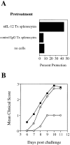
Splenocytes from anti–IL-12-treated naive mice directly suppress the induction of EAE. SJL mice were injected with splenocytes (1 × 10⁸) pooled from 5–7 anti–IL-12– treated or control IgG–treated syngeneic donors, or they were left untreated. Mice from all three groups were subsequently immunized with bovine MBP in CFA 1 and 7 d later. (A) Results represent the percent of mice that remained healthy (clinical score of 0) over the 30 d period between the second immunization and killing. The experiment shown consisted of 10 recipients of anti–IL-12–treated splenocytes, 14 recipients of control IgG–treated splenocytes, and 26 mice that were not pretreated. (B) The mice described in A were rated daily for signs of neurologic impairment according to the scale used in Fig. 2.

References

    1. Mosmann TR, Coffman RL. Th1 and Th2 cells: different patterns of lymphokine secretion lead to different functional properties. Annu Rev Immunol. 1989;7:145–173. - PubMed
    1. Sher A, Coffman RL. Regulation of autoimmunity to parasites and T cell-derived cytokines. Annu Rev Immunol. 1992;10:385–409. - PubMed
    1. Liblau RS, Singer SM, McDevitt HO. Th1 and Th2 CD4+ T cells in the pathogenesis of organ-specific autoimmune diseases. Immunol Today. 1995;16:34–38. - PubMed
    1. Zamvil SS, Steinman L. The T lymphocyte in experimental allergic encephalomyelitis. Annu Rev Immunol. 1990;8:579–621. - PubMed
    1. Bergman B, Haskins K. Islet-specific T-cell clones from the NOD mouse respond to β-granule antigen. Diabetes. 1994;43:197–203. - PubMed

Publication types

MeSH terms